FANCD2 Antibody (1290D) - BSA Free
Novus Biologicals | Catalog # NBP2-54808
Recombinant Monoclonal Antibody
Key Product Details
Validated by
Knockout/Knockdown
Species Reactivity
Human
Applications
Knockout Validated, Immunohistochemistry, Immunohistochemistry-Paraffin, Western Blot, Flow Cytometry, Flow (Intracellular), Simple Western
Label
Unconjugated
Antibody Source
Recombinant Monoclonal Rabbit IgG Clone # 1290D expressed in HEK293
Format
BSA Free
Loading...
Product Specifications
Immunogen
This FANCD2 Antibody (1290D) was developed against human FANCD2 fusion protein (N-terminal fragment). [Swiss-Prot #Q9BXW9]
Clonality
Monoclonal
Host
Rabbit
Isotype
IgG
Theoretical MW
164.1 kDa.
Disclaimer note: The observed molecular weight of the protein may vary from the listed predicted molecular weight due to post translational modifications, post translation cleavages, relative charges, and other experimental factors.
Disclaimer note: The observed molecular weight of the protein may vary from the listed predicted molecular weight due to post translational modifications, post translation cleavages, relative charges, and other experimental factors.
Scientific Data Images for FANCD2 Antibody (1290D) - BSA Free
![Knockout Validated: FANCD2 Antibody (1290D) - BSA Free [NBP2-54808]](https://resources.rndsystems.com/images/products/FANCD2-Antibody-1290D-Knockout-Validated-NBP2-54808-img0003.jpg "Western Blot: FANCD2 Antibody (1290D) - BSA Free [NBP2-54808]")
Western Blot: FANCD2 Antibody (1290D) - BSA Free [NBP2-54808]
Western Blot: FANCD2 Antibody (1290D) [NBP2-54808] - Western blot shows lysates of HeLa human cervical epithelial carcinoma parental cell line and FANCD2 knockout (KO) HeLa cell line. PVDF membrane was probed with 0.1 ug/mL of Rabbit Monoclonal (Catalog # NBP2-54808) followed by HRP-conjugated Anti-Rabbit IgG Secondary Antibody (Catalog #HAF008). Specific band was detected for FANCD2 at approximately 150 kDa (as indicated) in the parental HeLa cell line, but is not detectable in the knockout HeLa cell line. This experiment was conducted under reducing conditions.![Simple Western: FANCD2 Antibody (1290D)BSA Free [NBP2-54808]](https://resources.rndsystems.com/images/products/FANCD2-Antibody-1290D-Simple-Western-NBP2-54808-img0002.jpg "Simple Western: FANCD2 Antibody (1290D)BSA Free [NBP2-54808]")
Simple Western: FANCD2 Antibody (1290D)BSA Free [NBP2-54808]
Simple Western: FANCD2 Antibody (1290D) [NBP2-54808] - Detection of Human FANCD2 by Simple Western using FANCD2 Antibody (1290D) in HeLa and K562 cell lysate.![Immunohistochemistry: FANCD2 Antibody (1290D) - BSA Free [NBP2-54808]](https://resources.rndsystems.com/images/products/FANCD2-Antibody-1290D-Immunohistochemistry-NBP2-54808-img0004.jpg "Immunohistochemistry: FANCD2 Antibody (1290D) - BSA Free [NBP2-54808]")
Immunohistochemistry: FANCD2 Antibody (1290D) - BSA Free [NBP2-54808]
Immunohistochemistry: FANCD2 Antibody (1290D) [NBP2-54808] - FANCD2 was detected in immersion fixed paraffin-embedded sections of human tonsil using Rabbit Anti-Human FANCD2 Antibody (1290D) (Catalog # NBP2-54808) at 5 ug/mL for 1 hour at room temperature followed by incubation with the Anti-Rabbit IgG VisUCyte(TM) HRP Polymer Antibody (Catalog # VC003). Tissue was stained using DAB (brown) and counterstained with hematoxylin (blue). Specific staining was localized to nuclei.![Flow (Intracellular): FANCD2 Antibody (1290D) - BSA Free [NBP2-54808]](https://resources.rndsystems.com/images/products/FANCD2-Antibody-1290D-Flow-Intracellular-NBP2-54808-img0005.jpg "Flow (Intracellular): FANCD2 Antibody (1290D) - BSA Free [NBP2-54808]")
Flow (Intracellular): FANCD2 Antibody (1290D) - BSA Free [NBP2-54808]
Flow (Intracellular): FANCD2 Antibody (1290D) [NBP2-54808] - An intracellular stain was performed on HeLa Cells with FANCD2 Antibody (1290D) (blue) and a matched isotype control MAB1050 (orange). Cells were fixed with 4% paraformaldehyde, following fixation, cells were permeabilized with 0.1% saponin. Cells were incubated in an antibody dilution of 1 ug/mL for 30 minutes at room temperature, followed by rabbit IgG APC-conjugated secondary antibody (F0111, R&D Systems).![Western Blot: FANCD2 Antibody (1290D)BSA Free [NBP2-54808]](https://resources.rndsystems.com/images/products/FANCD2-Antibody-1290D-Western-Blot-NBP2-54808-img0006.jpg "Western Blot: FANCD2 Antibody (1290D)BSA Free [NBP2-54808]")
Western Blot: FANCD2 Antibody (1290D)BSA Free [NBP2-54808]
Western Blot: FANCD2 Antibody (1290D) [NBP2-54808] - Total protein from HeLa and K562 cells (Molecular weight: 164.1 KDa) was separated on a 7.5% gel by SDS-PAGE, transferred to PVDF membrane and blocked in 5% non-fat milk in TBST. The membrane was probed with 0.1 ug/ml FANCD2 Antibody (1290D) (catalog number NBP2-54808) in blocking buffer and detected with an anti-rabbit HRP secondary antibody using chemiluminescence.Applications for FANCD2 Antibody (1290D) - BSA Free
Application
Recommended Usage
Flow Cytometry
1 ug/ml
Immunohistochemistry
5-10 ug/ml
Immunohistochemistry-Paraffin
5-10 ug/ml
Simple Western
1 ug/ml
Western Blot
0.1 - 0.5 ug/ml
Reviewed Applications
Read 1 review rated 4 using NBP2-54808 in the following applications:
Flow Cytometry Panel Builder
Bio-Techne Knows Flow Cytometry
Save time and reduce costly mistakes by quickly finding compatible reagents using the Panel Builder Tool.
Advanced Features
- Spectra Viewer - Custom analysis of spectra from multiple fluorochromes
- Spillover Popups - Visualize the spectra of individual fluorochromes
- Antigen Density Selector - Match fluorochrome brightness with antigen density

Formulation, Preparation, and Storage
Purification
Protein A or G purified from cell culture supernatant
Formulation
PBS
Format
BSA Free
Preservative
0.02% Sodium Azide
Concentration
1.0 mg/ml
Shipping
The product is shipped with polar packs. Upon receipt, store it immediately at the temperature recommended below.
Stability & Storage
Store at 4C short term. Aliquot and store at -20C long term. Avoid freeze-thaw cycles.
Background: FANCD2
References
1. Bi, J., Areecheewakul, S., Li, Y., Yang, S., Zhang, Y., Ebeid, K.,... Meng, X. (2019). MTDH/AEG-1 downregulation using pristimerin-loaded nanoparticles inhibits Fanconi anemia proteins and increases sensitivity to platinum-based chemotherapy. Gynecol Oncol, 155(2), 349-358. doi:10.1016/j.ygyno.2019.08.014
2. Balcerek, J., Jiang, J., Li, Y., Jiang, Q., Holdreith, N., Singh, B.,... Tong, W. (2018). Lnk/Sh2b3 deficiency restores hematopoietic stem cell function and genome integrity in Fancd2 deficient Fanconi anemia. Nat Commun, 9(1), 3915. doi:10.1038/s41467-018-06380-1
Long Name
Fanconi Anemia, Complementation Group D2
Alternate Names
FA4, FACD, FAD, FAD2, FANCD
Gene Symbol
FANCD2
Additional FANCD2 Products
Product Documents for FANCD2 Antibody (1290D) - BSA Free
Certificate of Analysis
To download a Certificate of Analysis, please enter a lot or batch number in the search box below.
Product Specific Notices for FANCD2 Antibody (1290D) - BSA Free
This product is for research use only and is not approved for use in humans or in clinical diagnosis. Primary Antibodies are guaranteed for 1 year from date of receipt.
Customer Reviews for FANCD2 Antibody (1290D) - BSA Free (1)
4 out of 5
1 Customer Rating
Have you used FANCD2 Antibody (1290D) - BSA Free?
Submit a review and receive an Amazon gift card!
$25/€18/£15/$25CAN/¥2500 Yen for a review with an image
$10/€7/£6/$10CAN/¥1110 Yen for a review without an image
Submit a review
Customer Images
Showing
1
-
1 of
1 review
Showing All
Filter By:
-
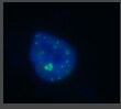
Application: ImmunofluorescenceSample Tested: HumanSpecies: HumanVerified Customer | Posted 09/08/2017D2 Foci PD20 ce;;s

There are no reviews that match your criteria.
Protocols
Find general support by application which include: protocols, troubleshooting, illustrated assays, videos and webinars.
- 7-Amino Actinomycin D (7-AAD) Cell Viability Flow Cytometry Protocol
- Antigen Retrieval Protocol (PIER)
- Antigen Retrieval for Frozen Sections Protocol
- Appropriate Fixation of IHC/ICC Samples
- Cellular Response to Hypoxia Protocols
- Chromogenic IHC Staining of Formalin-Fixed Paraffin-Embedded (FFPE) Tissue Protocol
- Chromogenic Immunohistochemistry Staining of Frozen Tissue
- ClariTSA™ Fluorophore Kits
- Detection & Visualization of Antibody Binding
- Extracellular Membrane Flow Cytometry Protocol
- Flow Cytometry Protocol for Cell Surface Markers
- Flow Cytometry Protocol for Staining Membrane Associated Proteins
- Flow Cytometry Staining Protocols
- Flow Cytometry Troubleshooting Guide
- Fluorescent IHC Staining of Frozen Tissue Protocol
- Graphic Protocol for Heat-induced Epitope Retrieval
- Graphic Protocol for the Preparation and Fluorescent IHC Staining of Frozen Tissue Sections
- Graphic Protocol for the Preparation and Fluorescent IHC Staining of Paraffin-embedded Tissue Sections
- Graphic Protocol for the Preparation of Gelatin-coated Slides for Histological Tissue Sections
- IHC Sample Preparation (Frozen sections vs Paraffin)
- Immunofluorescent IHC Staining of Formalin-Fixed Paraffin-Embedded (FFPE) Tissue Protocol
- Immunohistochemistry (IHC) and Immunocytochemistry (ICC) Protocols
- Immunohistochemistry Frozen Troubleshooting
- Immunohistochemistry Paraffin Troubleshooting
- Intracellular Flow Cytometry Protocol Using Alcohol (Methanol)
- Intracellular Flow Cytometry Protocol Using Detergents
- Intracellular Nuclear Staining Flow Cytometry Protocol Using Detergents
- Intracellular Staining Flow Cytometry Protocol Using Alcohol Permeabilization
- Intracellular Staining Flow Cytometry Protocol Using Detergents to Permeabilize Cells
- Preparing Samples for IHC/ICC Experiments
- Preventing Non-Specific Staining (Non-Specific Binding)
- Primary Antibody Selection & Optimization
- Propidium Iodide Cell Viability Flow Cytometry Protocol
- Protocol for Heat-Induced Epitope Retrieval (HIER)
- Protocol for Liperfluo
- Protocol for Making a 4% Formaldehyde Solution in PBS
- Protocol for VisUCyte™ HRP Polymer Detection Reagent
- Protocol for the Characterization of Human Th22 Cells
- Protocol for the Characterization of Human Th9 Cells
- Protocol for the Preparation & Fixation of Cells on Coverslips
- Protocol for the Preparation and Chromogenic IHC Staining of Frozen Tissue Sections
- Protocol for the Preparation and Chromogenic IHC Staining of Frozen Tissue Sections - Graphic
- Protocol for the Preparation and Chromogenic IHC Staining of Paraffin-embedded Tissue Sections
- Protocol for the Preparation and Chromogenic IHC Staining of Paraffin-embedded Tissue Sections - Graphic
- Protocol for the Preparation and Fluorescent IHC Staining of Frozen Tissue Sections
- Protocol for the Preparation and Fluorescent IHC Staining of Paraffin-embedded Tissue Sections
- Protocol for the Preparation of Gelatin-coated Slides for Histological Tissue Sections
- Protocol: Annexin V and PI Staining by Flow Cytometry
- Protocol: Annexin V and PI Staining for Apoptosis by Flow Cytometry
- R&D Systems Quality Control Western Blot Protocol
- TUNEL and Active Caspase-3 Detection by IHC/ICC Protocol
- The Importance of IHC/ICC Controls
- Troubleshooting Guide: Fluorokine Flow Cytometry Kits
- Troubleshooting Guide: Immunohistochemistry
- Troubleshooting Guide: Western Blot Figures
- Western Blot Conditions
- Western Blot Protocol
- Western Blot Protocol for Cell Lysates
- Western Blot Troubleshooting
- Western Blot Troubleshooting Guide
- View all Protocols, Troubleshooting, Illustrated assays and Webinars
Loading...