FANCD2 Antibody - BSA Free
Novus Biologicals | Catalog # NB100-182
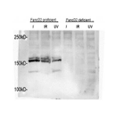
![Knockdown Validated: FANCD2 Antibody - BSA Free [NB100-182]](https://resources.rndsystems.com/images/products/FANCD2-Antibody-Knockdown-Validated-NB100-182-img0040.jpg "Western Blot: FANCD2 Antibody - BSA Free [NB100-182]")
Key Product Details
Validated by
Knockout/Knockdown, Biological Validation
Species Reactivity
Validated:
Human, Mouse, Rat, Avian, Canine, Kangaroo, Primate, Zebrafish
Cited:
Human, Mouse, Avian, Canine, Fish - Danio rerio (Zebrafish), Primate
Applications
Validated:
Knockout Validated, Immunohistochemistry, Immunohistochemistry-Paraffin, Western Blot, Immunoblotting, Flow Cytometry, Immunocytochemistry/ Immunofluorescence, Simple Western, Immunoprecipitation, Chromatin Immunoprecipitation, Chromatin Immunoprecipitation (ChIP), RNA Inhibition, Knockdown Validated
Cited:
Knockout Validated, Immunohistochemistry-Paraffin, Western Blot, Immunofluorescence, Immunocytochemistry/ Immunofluorescence, Immunoprecipitation, Chromatin Immunoprecipitation, Chemotaxis, IF/IHC, RNA Inhibition, Knockdown Validated
Label
Unconjugated
Antibody Source
Polyclonal Rabbit IgG
Format
BSA Free
Loading...
Product Specifications
Immunogen
This FANCD2 Antibody was developed against human FANCD2 fusion protein (N-terminal fragment). [Swiss-Prot #Q9BXW9]
Reactivity Notes
Primate reactivity reported in literature (PMID: 21421661). Canine reactivity reported in literature (PMID: 27257868). Zebrafish reactivity reported in scientific literature (PMID: 30540754). Rat reactivity reported in multiple pieces of scientific literature. Kangaroo reactivity reported in scientific literature (PMID: 24982423).
Clonality
Polyclonal
Host
Rabbit
Isotype
IgG
Theoretical MW
164.1 kDa.
Disclaimer note: The observed molecular weight of the protein may vary from the listed predicted molecular weight due to post translational modifications, post translation cleavages, relative charges, and other experimental factors.
Disclaimer note: The observed molecular weight of the protein may vary from the listed predicted molecular weight due to post translational modifications, post translation cleavages, relative charges, and other experimental factors.
Scientific Data Images for FANCD2 Antibody - BSA Free
![Knockdown Validated: FANCD2 Antibody - BSA Free [NB100-182]](https://resources.rndsystems.com/images/products/FANCD2-Antibody-Knockdown-Validated-NB100-182-img0035.jpg "Immunocytochemistry/ Immunofluorescence: FANCD2 Antibody - BSA Free [NB100-182]")
![Western Blot: FANCD2 AntibodyBSA Free [NB100-182]](https://resources.rndsystems.com/images/products/FANCD2-Antibody-Western-Blot-NB100-182-img0039.jpg "Western Blot: FANCD2 AntibodyBSA Free [NB100-182]")
![Simple Western: FANCD2 AntibodyBSA Free [NB100-182]](https://resources.rndsystems.com/images/products/FANCD2-Antibody-Simple-Western-NB100-182-img0014.jpg "Simple Western: FANCD2 AntibodyBSA Free [NB100-182]")
Simple Western: FANCD2 AntibodyBSA Free [NB100-182]
Simple Western: FANCD2 Antibody - BSA Free [NB100-182] - FANCD2 Antibody [NB100-182] - Simple Western lane view shows a specific band for FANCD2 in 0.1 mg/ml of HeLa lysate using FANCD2 Antibody. This experiment was performed under reducing conditions using the 12-230 kDa separation system.![Immunocytochemistry/ Immunofluorescence: FANCD2 Antibody - BSA Free [NB100-182]](https://resources.rndsystems.com/images/products/FANCD2-Antibody-Immunocytochemistry-Immunofluorescence-NB100-182-img0025.jpg "Immunocytochemistry/ Immunofluorescence: FANCD2 Antibody - BSA Free [NB100-182]")
Immunocytochemistry/ Immunofluorescence: FANCD2 Antibody - BSA Free [NB100-182]
Immunocytochemistry/Immunofluorescence: FANCD2 Antibody - BSA Free [NB100-182] - FANCD2 Antibody [NB100-182] - Analysis using the Biotin conjugate of FANCD2 Antibody. FANCD2 colocalizes in vivo with another protein in SiHa cells after cell exposure to IR. Proliferating SiHa cells were exposed to 10 Gy of IR and double color immunofluorescence staining was performed after 8 h. Images were captured in a Kodak digital image system on a Leica fluorescence microscope.![Western Blot: FANCD2 AntibodyBSA Free [NB100-182]](https://resources.rndsystems.com/images/products/FANCD2-Antibody-Western-Blot-NB100-182-img0037.jpg "Western Blot: FANCD2 AntibodyBSA Free [NB100-182]")
![Western Blot: FANCD2 AntibodyBSA Free [NB100-182]](https://resources.rndsystems.com/images/products/FANCD2-Antibody-Western-Blot-NB100-182-img0044.jpg "Western Blot: FANCD2 AntibodyBSA Free [NB100-182]")
Western Blot: FANCD2 AntibodyBSA Free [NB100-182]
FANCD2-Antibody-Western-Blot-NB100-182-img0044.jpg![Immunocytochemistry/ Immunofluorescence: FANCD2 Antibody - BSA Free [NB100-182]](https://resources.rndsystems.com/images/products/FANCD2-Antibody-Immunocytochemistry-Immunofluorescence-NB100-182-img0045.jpg "Immunocytochemistry/ Immunofluorescence: FANCD2 Antibody - BSA Free [NB100-182]")
![Immunohistochemistry: FANCD2 Antibody - BSA Free [NB100-182]](https://resources.rndsystems.com/images/products/FANCD2-Antibody-Immunohistochemistry-NB100-182-img0046.jpg "Immunohistochemistry: FANCD2 Antibody - BSA Free [NB100-182]")
Immunohistochemistry: FANCD2 Antibody - BSA Free [NB100-182]
FANCD2-Antibody-Immunohistochemistry-NB100-182-img0046.jpg![Western Blot: FANCD2 AntibodyBSA Free [NB100-182]](https://resources.rndsystems.com/images/products/FANCD2-Antibody-Western-Blot-NB100-182-img0029.jpg "Western Blot: FANCD2 AntibodyBSA Free [NB100-182]")
Western Blot: FANCD2 AntibodyBSA Free [NB100-182]
Western Blot: FANCD2 Antibody [NB100-182] - Analysis of FANCD2 (Molecular weight: 164.1 KDa) using the HRP conjugate of FANCD2 Antibody (lot C) in HeLa WCE.![Western Blot: FANCD2 AntibodyBSA Free [NB100-182]](https://resources.rndsystems.com/images/products/FANCD2-Antibody-Western-Blot-NB100-182-img0043.jpg "Western Blot: FANCD2 AntibodyBSA Free [NB100-182]")
Western Blot: FANCD2 AntibodyBSA Free [NB100-182]
FANCD2-Antibody-Western-Blot-NB100-182-img0043.jpg![Immunocytochemistry/ Immunofluorescence: FANCD2 Antibody - BSA Free [NB100-182]](https://resources.rndsystems.com/images/products/FANCD2-Antibody-Immunocytochemistry-Immunofluorescence-NB100-182-img0031.jpg "Immunocytochemistry/ Immunofluorescence: FANCD2 Antibody - BSA Free [NB100-182]")
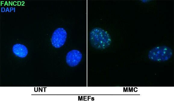
Immunocytochemistry/ Immunofluorescence: FANCD2 Antibody - BSA Free [NB100-182]
Immunocytochemistry/Immunofluorescence: FANCD2 Antibody [NB100-182] - Mouse Embryonic Fibroblasts Untreated and treated with Mitomycin C for 24hr with FANCD2 Antibody diluted 1:500 in 15%BCS+0.3% Triton X 100 in PBS. This image was submitted via customer Review.![Immunohistochemistry: FANCD2 Antibody - BSA Free [NB100-182]](https://resources.rndsystems.com/images/products/FANCD2-Antibody-Immunohistochemistry-NB100-182-img0041.jpg "Immunohistochemistry: FANCD2 Antibody - BSA Free [NB100-182]")
Immunohistochemistry: FANCD2 Antibody - BSA Free [NB100-182]
Immunohistochemistry: FANCD2 Antibody [NB100-182] - Staining of human prostate, glandular epithelium using FANCD2 Antibody. Image using the Biotin format of this antibody.
Immunohistochemistry: FANCD2 Antibody - BSA Free [NB100-182] -
Representative images of the cellular response to RS (induced by a low dose of hydroxyurea; HU) in U2OS cells, as determined by IF staining for gamma H2AX and FANCD2A selected cell is defined by the yellow arrow, and is enlarged in the bottom panel. Scale bars are indicated.
FANCD2 in A431 Human Cell Line.
FANCD2 was detected in immersion fixed A431 human skin carcinoma cell line using Rabbit anti-FANCD2 Antigen Affinity Purified Polyclonal Antibody conjugated to DyLight 550 (Catalog # NB100-182R) (red) at 5 µg/mL overnight at 4C. Cells were counterstained with DAPI (blue). Cells were imaged using a 100X objective and digitally deconvolved.
Western Blot: FANCD2 Antibody - BSA Free [NB100-182] -
Western Blot: FANCD2 Antibody - BSA Free [NB100-182] - Western blot analysis of FANCD2 & ERCC1 expression in lung cancer cell H1299 following treatment with veliparib. PAR protein level was reduced at 6 h post veliparib 5 μM exposure & maintained at low levels through 48 h in the H1299 cells. However, FANCD2 & ERCC1 protein expression raised simultaneously in these cells post treatment with veliparib. The data suggest that veliparib induces compensatory activation of alternative DNA-repair pathways. C is control (without treatment). Image collected & cropped by CiteAb from the following publication (http://journal.frontiersin.org/article/10.3389/fonc.2014.00368/abstract), licensed under a CC-BY license. Not internally tested by Novus Biologicals.
Immunocytochemistry/ Immunofluorescence: FANCD2 Antibody - BSA Free [NB100-182] -
Immunocytochemistry/ Immunofluorescence: FANCD2 Antibody - BSA Free [NB100-182] - Workflow for MS-based quantification of CFS associated proteins. (A) Experimental workflow for SILAC-based quantitative MS identification of CFS associated proteins. (B) Flow cytometry analysis of cell cycle distribution of SILAC labeled cells used for enrichment of CFSs as illustrated in (A). (C) IF of FANCD2 & EdU incorporation to assess formation of FANCD2 foci at late replicating regions with & without APH treatments. Cells were synchronized as in (A) & (B). Image collected & cropped by CiteAb from the following publication (https://pubmed.ncbi.nlm.nih.gov/31180492), licensed under a CC-BY license. Not internally tested by Novus Biologicals.
Western Blot: FANCD2 Antibody - BSA Free [NB100-182] -
Western Blot: FANCD2 Antibody - BSA Free [NB100-182] - The response to replication stress is a defining feature for Ewing sarcoma compared to the PHATE_1-high developmental context. (A) Western blot showing expression of Fanconi Anemia proteins FANCA, FANCD2, & FANCI in TC32 (Ewing sarcoma cell line) compared to IMR90 (fibroblast cell line); (B) Bar-plot showing the impact of FANC genes knockdown on cell viability in TC32 compared to IMR90; (C) Western blot showing the expression of FEN1 in Ewing sarcoma cell lines TC32, EWS502, & CHLA10 compared to IMR90; (D) Line plot showing the viability of Ewing sarcoma cells compared to IMR90 with increasing doses of FEN1 inhibitor. (** p ≤ 0.01; *** p ≤ 0.001; **** p ≤ 0.0001). Image collected & cropped by CiteAb from the following publication (https://pubmed.ncbi.nlm.nih.gov/32290418), licensed under a CC-BY license. Not internally tested by Novus Biologicals.
Western Blot: FANCD2 Antibody - BSA Free [NB100-182] -
Western Blot: FANCD2 Antibody - BSA Free [NB100-182] - Activation of FANCD2 monoubiquitination following treatment with the EZH2 inhibitor EPZ-6438(A-C) MCF10A (A), HCT116 p53+/+ & p53-/- (B), & U2OS (C) cells were treated with the indicated concentrations of the EZH2-specific inhibitor EPZ-6438 for 24 h (A & B) or 24, 48, & 72 h (C). Whole-cell lysates were prepared & immunoblotted with the indicated antibodies. L.E., light exposure; D.E., dark exposure; L:S Ratio, ratio of monoubiquitinated to nonubiquitinated FANCI; or FANCD2 RBI, relative band intensity. Image collected & cropped by CiteAb from the following publication (https://pubmed.ncbi.nlm.nih.gov/29100324), licensed under a CC-BY license. Not internally tested by Novus Biologicals.
Western Blot: FANCD2 Antibody - BSA Free [NB100-182] -
Western Blot: FANCD2 Antibody - BSA Free [NB100-182] - Inhibition of class I & II HDACs attenuates FANCD2 & FANCI monoubiquitination in BJ-TERT cells(A) & (B), HeLa cells were pre-treated with the indicated concentrations of trichostatin A (TSA) (A) or vorinostat (SAHA) (B) for 4 h, followed by co-incubation with (+) & without (-) 200 nM MMC for a further 20 h. Whole-cell lysates were prepared & immunoblotted with anti-FANCD2, anti-FANCI, anti-CHK1 pS345, anti-CHK2 pT68, anti-H4K16ac, & anti-alpha -Tubulin antibodies. (C) & (D), BJ-TERT cells were treated identically to that described for HeLa cells above. L:S Ratio, ratio of monoubiquitinated to nonubiquitinated FANCD2 or FANCI. Image collected & cropped by CiteAb from the following publication (https://pubmed.ncbi.nlm.nih.gov/29100324), licensed under a CC-BY license. Not internally tested by Novus Biologicals.
Immunocytochemistry/ Immunofluorescence: FANCD2 Antibody - BSA Free [NB100-182] -
Immunocytochemistry/ Immunofluorescence: FANCD2 Antibody - BSA Free [NB100-182] - Representative images of the cellular response to RS (induced by a low dose of hydroxyurea; HU) in U2OS cells, as determined by IF staining for gamma H2AX & FANCD2A selected cell is defined by the yellow arrow, & is enlarged in the bottom panel. Scale bars are indicated. Image collected & cropped by CiteAb from the following publication (https://www.oncotarget.com/lookup/doi/10.18632/oncotarget.16940), licensed under a CC-BY license. Not internally tested by Novus Biologicals.
Western Blot: FANCD2 Antibody - BSA Free [NB100-182] -
Western Blot: FANCD2 Antibody - BSA Free [NB100-182] - Functional evaluation of FANCBWT & mutant cDNA. Proband fibroblasts, FANCB mutant (null) fibroblasts & BJ control fibroblasts were HPV16 E6E7 transformed. Either empty vector, wild‐type FANCBcDNA, or mutant FANCBcDNA (p.A319*) was introduced into proband fibroblasts & FANCB mutant (null) fibroblasts. After puromycin selection, cells were cultured with or without MMC 1 μM for 24 hr, after which cells were harvested for the FANCD2 & HA western blot assays Image collected & cropped by CiteAb from the following publication (https://pubmed.ncbi.nlm.nih.gov/29193904), licensed under a CC-BY license. Not internally tested by Novus Biologicals.
Western Blot: FANCD2 Antibody - BSA Free [NB100-182] -
Western Blot: FANCD2 Antibody - BSA Free [NB100-182] - BRD4770-induced activation of the FA pathway may occur via inhibition of the PRC2 complex(A) U2OS cells were incubated in the absence (NT) or presence of BRD4770, DZNep, UNC0646, & DZNep & UNC0646 combined (4 μM each) for 24 h. Whole-cell lysates were prepared & immunoblotted with anti-FANCD2, anti-FANCI, anti-EZH2, & anti-alpha -Tubulin antibodies. (B) U2OS cells were incubated in the absence or presence of 2, 5, & 10 μM BRD4770 for 24, 48, or 72 h. Whole-cell lysates were prepared & immunoblotted with anti-FANCD2, anti-FANCI, anti-CHK1 pS345, anti-EZH2, anti-H3K27me3, & anti-alpha -Tubulin antibodies. L:S Ratio, ratio of monoubiquitinated to nonubiquitinated FANCI; or FANCD2 RBI, relative band intensity. Image collected & cropped by CiteAb from the following publication (https://pubmed.ncbi.nlm.nih.gov/29100324), licensed under a CC-BY license. Not internally tested by Novus Biologicals.
Immunocytochemistry/ Immunofluorescence: FANCD2 Antibody - BSA Free [NB100-182] -
Immunocytochemistry/ Immunofluorescence: FANCD2 Antibody - BSA Free [NB100-182] - Chemical library screening for small molecules that inhibit the Fanconi anemia pathway. (A) Schematic of the screening for small molecules that inhibit IR-induced FANCD2 foci formation. (B) Representative photomicrographs of EGFP-FANCD2 foci in PD20F-EGFP-FANCD2 cells untreated & treated with the indicated compounds at the indicated concentration. The cells were treated with compounds immediately before irradiation (15 Gy), & fixed after 12 hours. (C) FANCD2 foci in HeLa cells untreated & treated with the indicated compounds at the indicated concentration. The cells were fixed 8 hours after irradiation (10 Gy) & immunostained with anti-FANCD2 antibody. Scale bar = 20 μm. Image collected & cropped by CiteAb from the following publication (https://molecular-cancer.biomedcentral.com/articles/10.1186/1476-4598-1…), licensed under a CC-BY license. Not internally tested by Novus Biologicals.
Western Blot: FANCD2 Antibody - BSA Free [NB100-182] -
Western Blot: FANCD2 Antibody - BSA Free [NB100-182] - AMPK‐activating AICAR treatment activates FANCD2, a pivotal molecule of Fanconi anemia DNA damage signaling pathway. (A) AICAR treatment induces FANCD2 monoubiquitination in transformed normal fibroblasts (GM00637I). GM00637I cells were treated with 1 mm 2‐deoxyglucose, 0.25 mM AICAR, or 1 mm phenformin for 24 h. Lysates were subjected to western blotting with anti‐FANCD2, phospho‐AMPK alpha 1 (T172), & AMPK alpha & beta ‐actin. In FANCD2 blots, the position of monoubiquitinated FANCD2 (Ub‐FANCD2) is indicated by an arrow. (B) AICAR treatment induces formation of FANCD2 nuclear foci in GM00637I fibroblasts. Cells grown on coverslips in 12‐well plates were treated with 0.25 mm AICAR for 24 h. Cells were immunostained with FANCD2 antibody & Alexa 488‐conjugated anti‐rabbit secondary antibody. FANCD2 foci were visualized by confocal microscopy. Representative images are shown at the top. The number of foci per cell was counted & plotted for ≥ 20 cells (bottom panel). The values represent the mean ± SEM (Student's t‐test, ***P < 0.001). (C) AICAR treatment induces FANCD2 monoubiquitination in Caki‐1 cells. Caki‐1 cells were treated with 0.25 mm AICAR for 24 h & monoubiquitination of FANCD2 was monitored as in (A). Image collected & cropped by CiteAb from the following publication (https://pubmed.ncbi.nlm.nih.gov/28174693), licensed under a CC-BY license. Not internally tested by Novus Biologicals.
Western Blot: FANCD2 Antibody - BSA Free [NB100-182] -
Western Blot: FANCD2 Antibody - BSA Free [NB100-182] - BRD4770-induced activation of the FA pathway does not occur via the direct induction of DNA damage or altered cell cycle progression(A) BJ-TERT cells were incubated with (+) & without (-) 200 nM mitomycin C (MMC) in the absence (NT) or presence of 10 μM BRD4770 for 24 h. Cells were fixed & stained with mouse monoclonal anti-gamma H2AX antibody & counterstained with DAPI, & the number of nuclei with >5 gamma H2AX foci were scored. At least 300 nuclei were scored for each treatment & this experiment was performed three times with similar results. Error bars represent the standard errors of the means from three independent experiments. (B) BJ-TERT cells were incubated with (+) & without (-) 0.4 μM etoposide (VP-16), in the absence or presence of 10 μM BRD4770, 5 μM GSK-J1 & 2.5 μM BIX01294, for 24 h. Whole-cell lysates were immunoblotted with anti-gamma H2AX & anti-PCNA (loading control) antibodies. (C) U2OS cells were incubated in the absence (NT) or presence of 200 nM MMC or 5 & 10 μM BRD4770 for 24 h. Whole-cell lysates were prepared & immunoblotted with anti-FANCD2, anti-FANCA, anti-UBE2T, anti-USP1, anti-CHK1 pS345, anti-RPA, anti-RPA pS4/8, & anti-alpha -Tubulin antibodies. L:S Ratio, ratio of monoubiquitinated to nonubiquitinated FANCD2; RBI, relative band intensity. (D) U2OS cells were incubated in the absence or presence of 2, 5, & 10 μM BRD4770 for 24, 48, or 72 h. Cells were fixed in ice-cold ethanol, stained with propidium iodide, & analyzed by flow cytometry. Cell cycle stage distributions were determined using FlowJo v10.2. Image collected & cropped by CiteAb from the following publication (https://pubmed.ncbi.nlm.nih.gov/29100324), licensed under a CC-BY license. Not internally tested by Novus Biologicals.
Western Blot: FANCD2 Antibody - BSA Free [NB100-182] -
Western Blot: FANCD2 Antibody - BSA Free [NB100-182] - AICAR‐induced FANCD2 monoubiquitination is dependent on AMPK. (A) Inhibitor of AMPK blocks AICAR‐induced FANCD2 monoubiquitination in GM00637I normal fibroblasts. Cells were pretreated with 5 μm of Compound C (an AMPK inhibitor) 1 h before treatment with 0.25 mm AICAR for 24 h. Cell lysates were subjected to immunoblotting with FANCD2 to visualize monoubiquitinated FANCD2 (Ub‐FANCD2). (B) AMPK inhibitor abrogates AICAR‐induced FANCD2 nuclear foci formation in GM00637I fibroblasts. GM006387I cells grown on coverslips were pretreated with 5 μm Compound C 1 h prior to 0.25 mm AICAR treatment for 24 h. FANCD2 foci were visualized by immunofluorescence staining & confocal microscopy. Representative images are shown at the top. The number of foci per cell was counted & plotted for ≥ 20 cells (bottom panel). The values represent the mean ± SEM (Student's t‐test, *P < 0.05; ***P < 0.001). (C) Knockdown of AMPK alpha 1 inhibits AICAR‐induced FANCD2 monoubiquitination in Caki‐1 cells. Caki‐1 cells were transfected with siRNAs (siControl or siAMPK#8) & after 48 h, AICAR was treated for 24 h. FANCD2 monoubiquitination was monitored by immunoblotting. Image collected & cropped by CiteAb from the following publication (https://pubmed.ncbi.nlm.nih.gov/28174693), licensed under a CC-BY license. Not internally tested by Novus Biologicals.
Western Blot: FANCD2 Antibody - BSA Free [NB100-182] -
Western Blot: FANCD2 Antibody - BSA Free [NB100-182] - LNK depletion restores FA-like human progenitor cells. a depicts a schematic overview of isolation of primary human HSPCs for lentiviral transduction followed by CFC assay. b UCB-derived CD34 + cells were sequentially infected with lentiviruses expressing shRNA to Luciferase (Luc) or FANCD2 (D2) with GFP marker, followed by shRNA to Luc or LNK with mCherry marker. GFP + mCherry + cells were then sorted & plated onto semi-solid methylcellulose media. Colony forming progenitor numbers are shown with a two-tailed Student's t-test. Representative data from two independent repeats are shown. c TF-1 cells with shRNA-mediated depletion of LNK or FANCD2 or CRISPR/Cas9-mediated depletion of LNK were analyzed for depletion efficiency by western blot. Luc, luciferase. gRNA, guide RNA Image collected & cropped by CiteAb from the following publication (https://pubmed.ncbi.nlm.nih.gov/30254368), licensed under a CC-BY license. Not internally tested by Novus Biologicals.
Western Blot: FANCD2 Antibody - BSA Free [NB100-182] -
Western Blot: FANCD2 Antibody - BSA Free [NB100-182] - SAN1 functions independently of the FA pathway & does not affect FA pathway activation. a, b CSAs of HeLa WT & SAN1−/− cells treated with scrambled ctrl siRNA or FANCD2 siRNA, in response to Cisplatin & MMC (N = 3). Statistical significance determined by two-way ANOVA. Error bars denote s.e.m. *p < 0.05, **p < 0.01, ***p < 0.001, ****p < 0.0001. c Immunoblot showing siRNA knockdown of FANCD2 in HeLa WT & SAN1−/− cells. d IF staining of FANCD2 foci in HeLa WT cells & SAN1−/− cells treated with 0.045 μM MMC. e Immunoblot of FANCD2 showing mono-ubiquitylation in HeLa WT & SAN1−/− cells treated with vehicle or 0.045 μM MMC. f, g CSAs of HeLa WT & SAN1−/− cells treated with ctrl or SNM1A siRNA & exposed to Cisplatin or MMC. Statistical significance was determined by two-way ANOVA test. h Immunoblot of SNM1A in HeLa WT & SAN1−/− cells treated with ctrl or SNM1A siRNA Image collected & cropped by CiteAb from the following publication (https://pubmed.ncbi.nlm.nih.gov/29968717), licensed under a CC-BY license. Not internally tested by Novus Biologicals.
Immunocytochemistry/ Immunofluorescence: FANCD2 Antibody - BSA Free [NB100-182] -
Immunocytochemistry/ Immunofluorescence: FANCD2 Antibody - BSA Free [NB100-182] - SAN1 functions independently of the FA pathway & does not affect FA pathway activation. a, b CSAs of HeLa WT & SAN1−/− cells treated with scrambled ctrl siRNA or FANCD2 siRNA, in response to Cisplatin & MMC (N = 3). Statistical significance determined by two-way ANOVA. Error bars denote s.e.m. *p < 0.05, **p < 0.01, ***p < 0.001, ****p < 0.0001. c Immunoblot showing siRNA knockdown of FANCD2 in HeLa WT & SAN1−/− cells. d IF staining of FANCD2 foci in HeLa WT cells & SAN1−/− cells treated with 0.045 μM MMC. e Immunoblot of FANCD2 showing mono-ubiquitylation in HeLa WT & SAN1−/− cells treated with vehicle or 0.045 μM MMC. f, g CSAs of HeLa WT & SAN1−/− cells treated with ctrl or SNM1A siRNA & exposed to Cisplatin or MMC. Statistical significance was determined by two-way ANOVA test. h Immunoblot of SNM1A in HeLa WT & SAN1−/− cells treated with ctrl or SNM1A siRNA Image collected & cropped by CiteAb from the following publication (https://pubmed.ncbi.nlm.nih.gov/29968717), licensed under a CC-BY license. Not internally tested by Novus Biologicals.
Immunocytochemistry/ Immunofluorescence: FANCD2 Antibody - BSA Free [NB100-182] -
Immunocytochemistry/ Immunofluorescence: FANCD2 Antibody - BSA Free [NB100-182] - The FANCD2 NLS is required for the nuclear localization of a subset of FANCI.(A) FA-D2 patient cells or FA-D2 cells stably expressing FANCD2-WT were incubated in the absence (NT) or presence of MMC for 24 h, fixed, stained with rabbit polyclonal anti-FANCD2 or anti-FANCI antibody & counterstained with phalloidin & DAPI. AF-488, Alexa Fluor 488. (B) FA-D2 cells stably expressing LacZ, FANCD2-WT, FANCD2-∆N57, FANCD2-∆N100, & FANCD2-3N were incubated in the absence (NT) or presence of MMC for 24 h, fixed, & stained with rabbit polyclonal anti-FANCI antibody, & counterstained with phalloidin & DAPI. At least 300 cells were scored for cytoplasmic (Cyto.), nuclear (Nucl.), & both cytoplasmic & nuclear (Both) localization of FANCI. (C) COS-7 cells were transiently transfected with no DNA, FANCI-GFP, FANCI-GFP plus FANCD2-V5-WT, or FANCI-GFP plus FANCD2-V5-∆N57. Whole-cell lysates were immunoprecipitated with anti-V5 or anti-GFP antibodies & immune complexes immunoblotted with anti-GFP & anti-V5 antibodies. Image collected & cropped by CiteAb from the following publication (https://pubmed.ncbi.nlm.nih.gov/24278431), licensed under a CC-BY license. Not internally tested by Novus Biologicals.
Western Blot: FANCD2 Antibody - BSA Free [NB100-182] -
Western Blot: FANCD2 Antibody - BSA Free [NB100-182] - Functional assessment of Fanconi anemia pathway. (a) The experimental scheme for MMC treatment. Twenty‐four hours after plating, cells were cultured with or without MMC 1 μM for an additional 24 hr, after which the cells were harvested for western blot or immunostaining. (b) Western blot with FANCD2 antibody of BJ, proband fibroblasts, FANCB mutant (null) fibroblasts & FANCD2 mutant (null) fibroblasts. (c) Western blot with FANCD2 antibody of non‐FA control lymphoblasts (LCL), proband LCL‐A‐2017, proband LCL‐B‐2017, FANCB mutant (null) LCL, & FANCD2 mutant (null) LCL. (d) Representative figures of FANCD2 foci formation in the indicated cells. (e) Quantification of FANCD2 foci formation following treatment with or without 1 μM MMC. Experiments were performed in triplicate. One hundred cells were counted for each experiment Image collected & cropped by CiteAb from the following publication (https://pubmed.ncbi.nlm.nih.gov/29193904), licensed under a CC-BY license. Not internally tested by Novus Biologicals.
Western Blot: FANCD2 Antibody - BSA Free [NB100-182] -
Western Blot: FANCD2 Antibody - BSA Free [NB100-182] - The FAN1 PIP-box motif is not required for FAN1 foci formation upon exposure to MMC. a Total cell extracts & chromatin-enriched fractions of U2OS cells expressing the indicated eGFP-FAN1 variants, treated or mock-treated with MMC (150 ng/ml, 24 h), were analysed by immunoblotting using the indicated antibodies. A representative blot of three independent experiments is shown. b Cells as in a were immunostained with anti-FANCD2 antibody. Representative images are shown. Scale bar: 25 μm. c, d Quantification of eGFP-FAN1 foci count (c) & the sum of their intensities (d) was obtained from QIBC analysis of b. Median levels are indicated by black bars. Statistical analyses were carried out using unpaired, two-tailed t-tests. P values expressed as ***(P < 0.01) were considered significant, n = 3. e Total cell extracts derived from cells as in a, treated or mock-treated with MMC (150 ng/ml, 24 h), were incubated with anti-eGFP affinity resin. Inputs & immunoprecipitates were analysed by immunoblotting with the indicated antibodies. A representative blot of two independent experiments is shown Image collected & cropped by CiteAb from the following publication (https://pubmed.ncbi.nlm.nih.gov/29051491), licensed under a CC-BY license. Not internally tested by Novus Biologicals.
Immunocytochemistry/ Immunofluorescence: FANCD2 Antibody - BSA Free [NB100-182] -
Immunocytochemistry/ Immunofluorescence: FANCD2 Antibody - BSA Free [NB100-182] - Functional assessment of Fanconi anemia pathway. (a) The experimental scheme for MMC treatment. Twenty‐four hours after plating, cells were cultured with or without MMC 1 μM for an additional 24 hr, after which the cells were harvested for western blot or immunostaining. (b) Western blot with FANCD2 antibody of BJ, proband fibroblasts, FANCB mutant (null) fibroblasts & FANCD2 mutant (null) fibroblasts. (c) Western blot with FANCD2 antibody of non‐FA control lymphoblasts (LCL), proband LCL‐A‐2017, proband LCL‐B‐2017, FANCB mutant (null) LCL, & FANCD2 mutant (null) LCL. (d) Representative figures of FANCD2 foci formation in the indicated cells. (e) Quantification of FANCD2 foci formation following treatment with or without 1 μM MMC. Experiments were performed in triplicate. One hundred cells were counted for each experiment Image collected & cropped by CiteAb from the following publication (https://pubmed.ncbi.nlm.nih.gov/29193904), licensed under a CC-BY license. Not internally tested by Novus Biologicals.
Immunocytochemistry/ Immunofluorescence: FANCD2 Antibody - BSA Free [NB100-182] -
Immunocytochemistry/ Immunofluorescence: FANCD2 Antibody - BSA Free [NB100-182] - DNA damage & cell survivalas a function of FANCD2 status. (a) Representative images of the formation of gamma H2AX foci in PD20 versus wild-typecomplemented cells 30 minutes after completion of H2O2 treatment. (b) Quantification of gamma H2AX foci response. Data represent means with upper standard error based on two independent experiments. (c) G2-type chromosomal aberrations are expressed as breaks per cell as a function of increasing H2O2 concentration in FANCD2-deficient andwild-type complemented PD20 cells. Data represent means with SEM based on at least three repeat experiments. (d) Apoptosis induction by H2O2 (50 μM) in cells with or without wild-type FANCD2 using fluorescence microscopy to assess apoptotic morphology by DAPI staining & flow cytometric analysis for sub-G1 DNA content. Representative experiments based on theapoptotic response at 24 hours are shown (similar results were obtained at 48 hours & with 25 μM H2O2). Image collected & cropped by CiteAb from the following publication (https://pubmed.ncbi.nlm.nih.gov/18483568), licensed under a CC-BY license. Not internally tested by Novus Biologicals.
Western Blot: FANCD2 Antibody - BSA Free [NB100-182] -
Western Blot: FANCD2 Antibody - BSA Free [NB100-182] - The FANCD2 NLS is required for efficient FANCD2 & FANCI monoubiquitination & chromatin association.(A) FA-D2 cells stably expressing LacZ, FANCD2-WT, FANCD2-K561R, FANCD2-∆N57, FANCD2-∆N100 & FANCD2-3N were incubated in the absence & presence of 250 nM MMC for 18 h, & whole-cell lysates were immunoblotted with antibodies to FANCD2, V5, FANCI & alpha -tubulin. The FANCD2 & FANCI L/S ratios are the ratios of monoubiquitinated to nonubiquitinated protein, & were calculated by measuring protein band intensities using ImageJ image processing & analysis software (http://rsb.info.nih.gov/ij/). (B & C) FA-D2 cells stably expressing FANCD2-WT, FANCD2-∆N57, FANCD2-∆N100 & FANCD2-3N were treated as above & cell pellets were fractionated into soluble (S) & chromatin-associated (C) fractions. Fractions were immunoblotted with antibodies against V5, FANCI, alpha -tubulin & H2A. W, unfractionated whole cell extract. Image collected & cropped by CiteAb from the following publication (https://pubmed.ncbi.nlm.nih.gov/24278431), licensed under a CC-BY license. Not internally tested by Novus Biologicals.
Western Blot: FANCD2 Antibody - BSA Free [NB100-182] -
Western Blot: FANCD2 Antibody - BSA Free [NB100-182] - AICAR‐induced FANCD2 monoubiquitination requires FANCA. (A) AICAR‐induced FANCD2 monoubiquitination does not occur in transformed FANCA−/− fibroblasts originated from a patient with defective FANCA. Transformed FANCA−/− (GM06914B) or normal fibroblasts (GM00637I) were treated with 0.25 mm AICAR for 24 h, & monoubiquitinated FANCD2 (Ub‐FANCD2) was visualized by immunoblotting. (B) AICAR‐induced FANCD2 nuclear foci formation is abrogated in FANCA−/− fibroblasts. FANCD2 nuclear foci were visualized by immunofluorescence staining & confocal microscopy. (C) Knockdown of FANCA abolishes FANCD2 monoubiquitination after AICAR treatment in Caki‐1 cells. Caki‐1 cells were transfected with FANCA‐targeting siRNAs (siFANCA#5 or siFANCA#6), treated with AICAR & subjected to FANCD2 immunoblotting. Image collected & cropped by CiteAb from the following publication (https://pubmed.ncbi.nlm.nih.gov/28174693), licensed under a CC-BY license. Not internally tested by Novus Biologicals.
Western Blot: FANCD2 Antibody - BSA Free [NB100-182] -
Western Blot: FANCD2 Antibody - BSA Free [NB100-182] - Acetaldehyde toxicity to human FANCD2‐deleted human cells & FANCD2 ubiquitylation in BRCA2‐deleted cellsA, BHuman DLD1 cells in which FANCD2 was deleted with CRISPR/Cas9 & control cells were incubated with the indicated concentrations of cisplatin (A) or acetaldehyde (B) for 6 days before processing for dose‐dependent viability assays. Graphs are representative of two independent experiments, each performed in triplicate. Error bars represent SD of triplicate values obtained from a single experiment. Inset, Western blot detection of FANCD2 expression. SMC1 was used as a loading control.CBRCA2‐proficient (+BRCA2) or BRCA2‐deficient (−BRCA2) DLD1 cells were incubated with 4 mM acetaldehyde for 48 h before being processed for immunoblotting as indicated.DH1299 cells expressing a DOX‐inducible BRCA2 shRNA were grown in the presence or absence of DOX & transfected with control or FANCD2 siRNA before being processed for immunoblotting as indicated. DOX, doxycycline. Image collected & cropped by CiteAb from the following publication (https://pubmed.ncbi.nlm.nih.gov/28729482), licensed under a CC-BY license. Not internally tested by Novus Biologicals.
Western Blot: FANCD2 Antibody - BSA Free [NB100-182] -
Western Blot: FANCD2 Antibody - BSA Free [NB100-182] - Functional assessment of Fanconi anemia pathway. (a) The experimental scheme for MMC treatment. Twenty‐four hours after plating, cells were cultured with or without MMC 1 μM for an additional 24 hr, after which the cells were harvested for western blot or immunostaining. (b) Western blot with FANCD2 antibody of BJ, proband fibroblasts, FANCB mutant (null) fibroblasts & FANCD2 mutant (null) fibroblasts. (c) Western blot with FANCD2 antibody of non‐FA control lymphoblasts (LCL), proband LCL‐A‐2017, proband LCL‐B‐2017, FANCB mutant (null) LCL, & FANCD2 mutant (null) LCL. (d) Representative figures of FANCD2 foci formation in the indicated cells. (e) Quantification of FANCD2 foci formation following treatment with or without 1 μM MMC. Experiments were performed in triplicate. One hundred cells were counted for each experiment Image collected & cropped by CiteAb from the following publication (https://pubmed.ncbi.nlm.nih.gov/29193904), licensed under a CC-BY license. Not internally tested by Novus Biologicals.
Western Blot: FANCD2 Antibody - BSA Free [NB100-182] -
Western Blot: FANCD2 Antibody - BSA Free [NB100-182] - SAN1 functions independently of the FA pathway & does not affect FA pathway activation. a, b CSAs of HeLa WT & SAN1−/− cells treated with scrambled ctrl siRNA or FANCD2 siRNA, in response to Cisplatin & MMC (N = 3). Statistical significance determined by two-way ANOVA. Error bars denote s.e.m. *p < 0.05, **p < 0.01, ***p < 0.001, ****p < 0.0001. c Immunoblot showing siRNA knockdown of FANCD2 in HeLa WT & SAN1−/− cells. d IF staining of FANCD2 foci in HeLa WT cells & SAN1−/− cells treated with 0.045 μM MMC. e Immunoblot of FANCD2 showing mono-ubiquitylation in HeLa WT & SAN1−/− cells treated with vehicle or 0.045 μM MMC. f, g CSAs of HeLa WT & SAN1−/− cells treated with ctrl or SNM1A siRNA & exposed to Cisplatin or MMC. Statistical significance was determined by two-way ANOVA test. h Immunoblot of SNM1A in HeLa WT & SAN1−/− cells treated with ctrl or SNM1A siRNA Image collected & cropped by CiteAb from the following publication (https://pubmed.ncbi.nlm.nih.gov/29968717), licensed under a CC-BY license. Not internally tested by Novus Biologicals.
Western Blot: FANCD2 Antibody - BSA Free [NB100-182] -
Western Blot: FANCD2 Antibody - BSA Free [NB100-182] - AICAR‐induced FANCD2 monoubiquitination requires FANCA. (A) AICAR‐induced FANCD2 monoubiquitination does not occur in transformed FANCA−/− fibroblasts originated from a patient with defective FANCA. Transformed FANCA−/− (GM06914B) or normal fibroblasts (GM00637I) were treated with 0.25 mm AICAR for 24 h, & monoubiquitinated FANCD2 (Ub‐FANCD2) was visualized by immunoblotting. (B) AICAR‐induced FANCD2 nuclear foci formation is abrogated in FANCA−/− fibroblasts. FANCD2 nuclear foci were visualized by immunofluorescence staining & confocal microscopy. (C) Knockdown of FANCA abolishes FANCD2 monoubiquitination after AICAR treatment in Caki‐1 cells. Caki‐1 cells were transfected with FANCA‐targeting siRNAs (siFANCA#5 or siFANCA#6), treated with AICAR & subjected to FANCD2 immunoblotting. Image collected & cropped by CiteAb from the following publication (https://pubmed.ncbi.nlm.nih.gov/28174693), licensed under a CC-BY license. Not internally tested by Novus Biologicals.
Immunocytochemistry/ Immunofluorescence: FANCD2 Antibody - BSA Free [NB100-182] -
Immunocytochemistry/ Immunofluorescence: FANCD2 Antibody - BSA Free [NB100-182] - BRCA2-deficient cells accumulate single-stranded DNA lesions in G2. a gamma H2AX foci analysis in early mitotic cells. Cells were untreated, or treated with RO-3306 (10 µM, 24 h) to delay mitotic entry, & released for 1 h before analysis of gamma H2AX & FANCD2 foci pairs in early mitotic cells. Representative images are shown (left). Analysis is by an unpaired two-tailed t-test. n = 3. Scale bars 10 µm. b–g Cells treated with the indicated siRNAs or mirin (50 µM, 5 h) were incubated with EdU for 30 min & then analyzed for gamma H2AX & RPA foci in G2 cells (EdU–, 2N DNA content). Representative deconvolved images are shown (b). Quantification of gamma H2AX foci (top, c–g) & RPA+ gamma H2AX foci (bottom, c–g) for BRCA2-deficient cells (c), cells transfected with siRNAs (d, RAD51; e SMARCAL1; f EXO1 & DNA2), & cells treated with mirin (g) are shown. n ≥ 3. Scale bars 10 µm. h, i Cells were incubated with EdU as in b before analysis of gamma H2AX & pCHK2-T68 (h) or pATM-S1981 (i) foci in G2 cells. Representative deconvolved images with magnified inset highlighting foci co-localization are shown (left in each panel). n ≥ 3. Scale bars 10 µm. Error bars s.d. ns, not significant; *p < 0.05; **p < 0.01; ***p < 0.001; ****p < 0.0001 (unpaired two-tailed t-test) Image collected & cropped by CiteAb from the following publication (https://www.nature.com/articles/s41467-017-00634-0), licensed under a CC-BY license. Not internally tested by Novus Biologicals.
Western Blot: FANCD2 Antibody - BSA Free [NB100-182] -
Western Blot: FANCD2 Antibody - BSA Free [NB100-182] - SAN1 functions independently of the FA pathway & does not affect FA pathway activation. a, b CSAs of HeLa WT & SAN1−/− cells treated with scrambled ctrl siRNA or FANCD2 siRNA, in response to Cisplatin & MMC (N = 3). Statistical significance determined by two-way ANOVA. Error bars denote s.e.m. *p < 0.05, **p < 0.01, ***p < 0.001, ****p < 0.0001. c Immunoblot showing siRNA knockdown of FANCD2 in HeLa WT & SAN1−/− cells. d IF staining of FANCD2 foci in HeLa WT cells & SAN1−/− cells treated with 0.045 μM MMC. e Immunoblot of FANCD2 showing mono-ubiquitylation in HeLa WT & SAN1−/− cells treated with vehicle or 0.045 μM MMC. f, g CSAs of HeLa WT & SAN1−/− cells treated with ctrl or SNM1A siRNA & exposed to Cisplatin or MMC. Statistical significance was determined by two-way ANOVA test. h Immunoblot of SNM1A in HeLa WT & SAN1−/− cells treated with ctrl or SNM1A siRNA Image collected & cropped by CiteAb from the following publication (https://pubmed.ncbi.nlm.nih.gov/29968717), licensed under a CC-BY license. Not internally tested by Novus Biologicals.
Immunocytochemistry/ Immunofluorescence: FANCD2 Antibody - BSA Free [NB100-182] -
Immunocytochemistry/ Immunofluorescence: FANCD2 Antibody - BSA Free [NB100-182] - BRCA2 deficiency causes DNA under replication that results in abnormal mitoses. a–cBRCA2∆Ex3-4/– cells released for 22 h from serum starvation to increase mitotic cells, incubated w/ EdU for 1 h, & then analyzed for mitotic DNA synthesis. Early mitotic cells defined as being in prophase, prometaphase,/metaphase analyzed for EdU foci that co-localize w/ FANCD2 foci pairs. a Representative images of mitotic DNA synthesis. Scale bars 10 µm. b Percent early mitotic cells containing EdU foci, analyzed by an unpaired two-tailed t-test. n = 3. c FANCD2 foci pairs w//w/out EdU foci co-localization. Graphs represent pooled results of 3 independent experiments, each analyzed by a two-tailed Mann–Whitney test. Median FANCD2 foci pair number, red bars. Image collected & cropped by CiteAb from following publication (https://www.nature.com/articles/s41467-017-00634-0), licensed under a CC-BY license. Not internally tested by Novus Biologicals.
Western Blot: FANCD2 Antibody - BSA Free [NB100-182] -
Western Blot: FANCD2 Antibody - BSA Free [NB100-182] - The FANCD2 NLS is required for efficient FANCD2 & FANCI monoubiquitination & chromatin association.(A) FA-D2 cells stably expressing LacZ, FANCD2-WT, FANCD2-K561R, FANCD2-∆N57, FANCD2-∆N100 & FANCD2-3N were incubated in the absence & presence of 250 nM MMC for 18 h, & whole-cell lysates were immunoblotted with antibodies to FANCD2, V5, FANCI & alpha -tubulin. The FANCD2 & FANCI L/S ratios are the ratios of monoubiquitinated to nonubiquitinated protein, & were calculated by measuring protein band intensities using ImageJ image processing & analysis software (http://rsb.info.nih.gov/ij/). (B & C) FA-D2 cells stably expressing FANCD2-WT, FANCD2-∆N57, FANCD2-∆N100 & FANCD2-3N were treated as above & cell pellets were fractionated into soluble (S) & chromatin-associated (C) fractions. Fractions were immunoblotted with antibodies against V5, FANCI, alpha -tubulin & H2A. W, unfractionated whole cell extract. Image collected & cropped by CiteAb from the following publication (https://pubmed.ncbi.nlm.nih.gov/24278431), licensed under a CC-BY license. Not internally tested by Novus Biologicals.
Immunocytochemistry/ Immunofluorescence: FANCD2 Antibody - BSA Free [NB100-182] -
Immunocytochemistry/ Immunofluorescence: FANCD2 Antibody - BSA Free [NB100-182] - Detection of FANCD2 foci formation in human lung tumors by the FATSI staining analysis. The paraffin-embedded lung tumor tissues sections were deparaffinized & rehydrated. The tissue sections were incubated with a primary antibody cocktail of rabbit polyclonal FANCD2 antibody (Novus Biologicals, Littleton, CO, USA) at a dilution of 1:1000 & a monoclonal anti-Ki67 mouse antibody (Dako, Carpenteria, CA, USA) at a dilution of 1:150 for 1 h at room temperature. Sections then were incubated with a secondary antibody cocktail containing FITC conjugated anti-rabbit IgG & Alexafluor 594 donkey anti-mouse secondary for 1 h at room temperature. The sections were mounted on glass slides using a 4′ 6-diamidino-2-phenylindole (DAPI)-containing embedding medium (Vysis Dapi 1, Abbott Laboratories, Downers Grove, IL, USA). The slides were analyzed under a fluorescence microscope. (A) FANCD2 foci positive NSCL tumor, & (B) FANCD2 foci negative NSCL tumor. Magnification: 1000×. Image collected & cropped by CiteAb from the following publication (http://journal.frontiersin.org/article/10.3389/fonc.2014.00368/abstract), licensed under a CC-BY license. Not internally tested by Novus Biologicals.
Immunocytochemistry/ Immunofluorescence: FANCD2 Antibody - BSA Free [NB100-182] -
Immunocytochemistry/ Immunofluorescence: FANCD2 Antibody - BSA Free [NB100-182] - Normal DNA damage responses in FANCD2-deficient cells treated with hydrogen peroxide (H2O2). (a) Illustration of RAD51 foci formation in PD20 & PD20-wtD2 cells 5 hours after exposure to MMC (0.25 μg/mL for 1 hour) or H2O2 (25 μM for 2 hours). (b) Induction of RAD51 foci formation above background levels by MMC or H2O2. Because equal drug concentrations were used, rather than isoeffective concentrations with regard to cell survival, the extent of foci induction between FANCD-deficient & -complemented cells is not directly comparable. Data represent means with upperstandard error based on three independent repeats. (c) DNA synthesis measured by BrdU pulse labeling in cells treated with ionizing radiation (IR, 8 Gy) or H2O2 (25 μM). Data represent means with upper standard error based on twoindependent experiments. (d) Cell cycle distribution of propidium-iodide cell populations by flow cytometry. A representative experiment is shown. Percentages of cells in the G1, S, & G2 (and M) phases of the cell cycle are indicated. Image collected & cropped by CiteAb from the following publication (https://pubmed.ncbi.nlm.nih.gov/18483568), licensed under a CC-BY license. Not internally tested by Novus Biologicals.
Western Blot: FANCD2 Antibody - BSA Free [NB100-182] -
Western Blot: FANCD2 Antibody - BSA Free [NB100-182] - Dicer inhibition prevents FANCD2 foci formation after replication stress without affecting FANCD2 mono-ubiquitylation.(A) HCT116 cells were treated with control or Dicer siRNA & then with aphidicolin to induce replicative stress. They were then stained for FANCD2 & analyzed by immunofluorescence microscopy. (B) Quantification of the experiments in panel (A) showing the percentage of FANCD2 positive cells. Error bars represent the SD of three independent experiments. To score the FANCD2 positive cells a threshold value was calculated on the base of the average number of foci in control cells for each replicate, using Image J. Unpaired t-test: *p < 0.05. (C) FANCD2 foci formation in WT & Dicer Ex5/Ex5 cells after replication stress induced by aphidicolin. (D) Quantification of the experiments in panel (C) showing the percentage of FANCD2 positive cells. Error bars represent the SD of three independent experiments. Unpaired t-test: **p < 0.005. (E) Western blotting showing the levels of FANCD2 mono-ubiquitylation (L-FANCD2) after siRNA-mediated inhibition of Dicer, in the presence or absence of replication stress induced by aphidicolin. Vinculin was used as a loading control. (F) Western blotting showing FANCD2 mono-ubiquitylation levels after Drosha inhibition by siRNA. (G) FANCD2 mono-ubiquitylation levels in WT & Dicer Ex5/Ex5 cells assayed by western blotting. Image collected & cropped by CiteAb from the following publication (https://pubmed.ncbi.nlm.nih.gov/31320994), licensed under a CC-BY license. Not internally tested by Novus Biologicals.
Western Blot: FANCD2 Antibody - BSA Free [NB100-182] -
Western Blot: FANCD2 Antibody - BSA Free [NB100-182] - Acetaldehyde toxicity to human FANCD2‐deleted human cells & FANCD2 ubiquitylation in BRCA2‐deleted cellsA, BHuman DLD1 cells in which FANCD2 was deleted with CRISPR/Cas9 & control cells were incubated with the indicated concentrations of cisplatin (A) or acetaldehyde (B) for 6 days before processing for dose‐dependent viability assays. Graphs are representative of two independent experiments, each performed in triplicate. Error bars represent SD of triplicate values obtained from a single experiment. Inset, Western blot detection of FANCD2 expression. SMC1 was used as a loading control.CBRCA2‐proficient (+BRCA2) or BRCA2‐deficient (−BRCA2) DLD1 cells were incubated with 4 mM acetaldehyde for 48 h before being processed for immunoblotting as indicated.DH1299 cells expressing a DOX‐inducible BRCA2 shRNA were grown in the presence or absence of DOX & transfected with control or FANCD2 siRNA before being processed for immunoblotting as indicated. DOX, doxycycline. Image collected & cropped by CiteAb from the following publication (https://pubmed.ncbi.nlm.nih.gov/28729482), licensed under a CC-BY license. Not internally tested by Novus Biologicals.
Western Blot: FANCD2 Antibody - BSA Free [NB100-182] -
Western Blot: FANCD2 Antibody - BSA Free [NB100-182] - The FAN1 PIP-box motif is not required for FAN1 foci formation upon exposure to MMC. a Total cell extracts & chromatin-enriched fractions of U2OS cells expressing the indicated eGFP-FAN1 variants, treated or mock-treated with MMC (150 ng/ml, 24 h), were analysed by immunoblotting using the indicated antibodies. A representative blot of three independent experiments is shown. b Cells as in a were immunostained with anti-FANCD2 antibody. Representative images are shown. Scale bar: 25 μm. c, d Quantification of eGFP-FAN1 foci count (c) & the sum of their intensities (d) was obtained from QIBC analysis of b. Median levels are indicated by black bars. Statistical analyses were carried out using unpaired, two-tailed t-tests. P values expressed as ***(P < 0.01) were considered significant, n = 3. e Total cell extracts derived from cells as in a, treated or mock-treated with MMC (150 ng/ml, 24 h), were incubated with anti-eGFP affinity resin. Inputs & immunoprecipitates were analysed by immunoblotting with the indicated antibodies. A representative blot of two independent experiments is shown Image collected & cropped by CiteAb from the following publication (https://pubmed.ncbi.nlm.nih.gov/29051491), licensed under a CC-BY license. Not internally tested by Novus Biologicals.
Western Blot: FANCD2 Antibody - BSA Free [NB100-182] -
Western Blot: FANCD2 Antibody - BSA Free [NB100-182] - AMPK‐activating AICAR treatment activates FANCD2, a pivotal molecule of Fanconi anemia DNA damage signaling pathway. (A) AICAR treatment induces FANCD2 monoubiquitination in transformed normal fibroblasts (GM00637I). GM00637I cells were treated with 1 mm 2‐deoxyglucose, 0.25 mM AICAR, or 1 mm phenformin for 24 h. Lysates were subjected to western blotting with anti‐FANCD2, phospho‐AMPK alpha 1 (T172), & AMPK alpha & beta ‐actin. In FANCD2 blots, the position of monoubiquitinated FANCD2 (Ub‐FANCD2) is indicated by an arrow. (B) AICAR treatment induces formation of FANCD2 nuclear foci in GM00637I fibroblasts. Cells grown on coverslips in 12‐well plates were treated with 0.25 mm AICAR for 24 h. Cells were immunostained with FANCD2 antibody & Alexa 488‐conjugated anti‐rabbit secondary antibody. FANCD2 foci were visualized by confocal microscopy. Representative images are shown at the top. The number of foci per cell was counted & plotted for ≥ 20 cells (bottom panel). The values represent the mean ± SEM (Student's t‐test, ***P < 0.001). (C) AICAR treatment induces FANCD2 monoubiquitination in Caki‐1 cells. Caki‐1 cells were treated with 0.25 mm AICAR for 24 h & monoubiquitination of FANCD2 was monitored as in (A). Image collected & cropped by CiteAb from the following publication (https://pubmed.ncbi.nlm.nih.gov/28174693), licensed under a CC-BY license. Not internally tested by Novus Biologicals.
Western Blot: FANCD2 Antibody - BSA Free [NB100-182] -
Western Blot: FANCD2 Antibody - BSA Free [NB100-182] - AICAR‐induced FANCD2 monoubiquitination is dependent on AMPK. (A) Inhibitor of AMPK blocks AICAR‐induced FANCD2 monoubiquitination in GM00637I normal fibroblasts. Cells were pretreated with 5 μm of Compound C (an AMPK inhibitor) 1 h before treatment with 0.25 mm AICAR for 24 h. Cell lysates were subjected to immunoblotting with FANCD2 to visualize monoubiquitinated FANCD2 (Ub‐FANCD2). (B) AMPK inhibitor abrogates AICAR‐induced FANCD2 nuclear foci formation in GM00637I fibroblasts. GM006387I cells grown on coverslips were pretreated with 5 μm Compound C 1 h prior to 0.25 mm AICAR treatment for 24 h. FANCD2 foci were visualized by immunofluorescence staining & confocal microscopy. Representative images are shown at the top. The number of foci per cell was counted & plotted for ≥ 20 cells (bottom panel). The values represent the mean ± SEM (Student's t‐test, *P < 0.05; ***P < 0.001). (C) Knockdown of AMPK alpha 1 inhibits AICAR‐induced FANCD2 monoubiquitination in Caki‐1 cells. Caki‐1 cells were transfected with siRNAs (siControl or siAMPK#8) & after 48 h, AICAR was treated for 24 h. FANCD2 monoubiquitination was monitored by immunoblotting. Image collected & cropped by CiteAb from the following publication (https://pubmed.ncbi.nlm.nih.gov/28174693), licensed under a CC-BY license. Not internally tested by Novus Biologicals.
Western Blot: FANCD2 Antibody - BSA Free [NB100-182] -
Western Blot: FANCD2 Antibody - BSA Free [NB100-182] - Acetaldehyde toxicity to human FANCD2‐deleted human cells & FANCD2 ubiquitylation in BRCA2‐deleted cellsA, BHuman DLD1 cells in which FANCD2 was deleted with CRISPR/Cas9 & control cells were incubated with the indicated concentrations of cisplatin (A) or acetaldehyde (B) for 6 days before processing for dose‐dependent viability assays. Graphs are representative of two independent experiments, each performed in triplicate. Error bars represent SD of triplicate values obtained from a single experiment. Inset, Western blot detection of FANCD2 expression. SMC1 was used as a loading control.CBRCA2‐proficient (+BRCA2) or BRCA2‐deficient (−BRCA2) DLD1 cells were incubated with 4 mM acetaldehyde for 48 h before being processed for immunoblotting as indicated.DH1299 cells expressing a DOX‐inducible BRCA2 shRNA were grown in the presence or absence of DOX & transfected with control or FANCD2 siRNA before being processed for immunoblotting as indicated. DOX, doxycycline. Image collected & cropped by CiteAb from the following publication (https://pubmed.ncbi.nlm.nih.gov/28729482), licensed under a CC-BY license. Not internally tested by Novus Biologicals.
Detection of FANCD2 in A431 Human Cell Line by Flow Cytometry.
An intracellular stain was performed on A431 human skin carcinoma cell line using Rabbit anti-FANCD2 Affinity Purified Polyclonal Antibody conjugated to DyLight 550 (Catalog # NB100-182R, blue histogram) or matched control antibody (Catalog # NBP2-24981R, orange histogram) at 2.5 µg/mL for 30 minutes at RT.
FANCD2 in U-2 OS Human Cell Line.
FANCD2 was detected in immersion fixed U-2 OS human osteosarcoma cell line using Rabbit anti-FANCD2 Antigen Affinity Purified Polyclonal Antibody conjugated to Biotin (Catalog # NB100-182B) at 5 µg/mL overnight at 4C. Cells were stained using Streptavidin conjugated to DyLight 550 (red) and counterstained with DAPI (blue). Cells were imaged using a 100X objective and digitally deconvolved.
FANCD2 in U-2 OS Human Cell Line.
FANCD2 was detected in immersion fixed U-2 OS human osteosarcoma cell line using Rabbit anti-FANCD2 Antigen Affinity Purified Polyclonal Antibody conjugated to DyLight 550 (Catalog # NB100-182R) (red) at 5 µg/mL overnight at 4C. Cells were counterstained with DAPI (blue). Cells were imaged using a 100X objective and digitally deconvolved.
Immunohistochemistry-Paraffin: FANCD2 Antibody - BSA Free [NB100-182] -
Detection of FANCD2 foci formation in human lung tumors by the FATSI staining analysis. The paraffin-embedded lung tumor tissues sections were deparaffinized and rehydrated. The tissue sections were incubated with a primary antibody cocktail of rabbit polyclonal FANCD2 antibody (Novus Biologicals, Littleton, CO, USA) at a dilution of 1:1000 and a monoclonal anti-Ki67 mouse antibody (Dako, Carpenteria, CA, USA) at a dilution of 1:150 for 1 h at room temperature. Sections then were incubated with a secondary antibody cocktail containing FITC conjugated anti-rabbit IgG and Alexafluor 594 donkey anti-mouse secondary for 1 h at room temperature. The sections were mounted on glass slides using a 4′ 6-diamidino-2-phenylindole (DAPI)-containing embedding medium (Vysis Dapi 1, Abbott Laboratories, Downers Grove, IL, USA). The slides were analyzed under a fluorescence microscope. (A) FANCD2 foci positive NSCL tumor, and (B) FANCD2 foci negative NSCL tumor. Magnification: 1000×. Image collected and cropped by CiteAb from the following open publication (https://pubmed.ncbi.nlm.nih.gov/25566506), licensed under a CC-BY license. Not internally tested by Novus Biologicals.
Immunohistochemistry-Paraffin: FANCD2 Antibody - BSA Free [NB100-182] -
Detection of FANCD2 foci formation in human lung tumors by the FATSI staining analysis. The paraffin-embedded lung tumor tissues sections were deparaffinized and rehydrated. The tissue sections were incubated with a primary antibody cocktail of rabbit polyclonal FANCD2 antibody (Novus Biologicals, Littleton, CO, USA) at a dilution of 1:1000 and a monoclonal anti-Ki67 mouse antibody (Dako, Carpenteria, CA, USA) at a dilution of 1:150 for 1 h at room temperature. Sections then were incubated with a secondary antibody cocktail containing FITC conjugated anti-rabbit IgG and Alexafluor 594 donkey anti-mouse secondary for 1 h at room temperature. The sections were mounted on glass slides using a 4′ 6-diamidino-2-phenylindole (DAPI)-containing embedding medium (Vysis Dapi 1, Abbott Laboratories, Downers Grove, IL, USA). The slides were analyzed under a fluorescence microscope. (A) FANCD2 foci positive NSCL tumor, and (B) FANCD2 foci negative NSCL tumor. Magnification: 1000×. Image collected and cropped by CiteAb from the following open publication (https://pubmed.ncbi.nlm.nih.gov/25566506), licensed under a CC-BY license. Not internally tested by Novus Biologicals.Applications for FANCD2 Antibody - BSA Free
Application
Recommended Usage
Chromatin Immunoprecipitation
reported in scientific literature (PMID 28196964)
Chromatin Immunoprecipitation (ChIP)
reported in scientific literature (PMID 28196964)
Flow Cytometry
2 - 5 ug/ml
Immunoblotting
reported in multiple pieces of scientific literature
Immunocytochemistry/ Immunofluorescence
5 ug/ml
Immunohistochemistry
2.5-5.0 ug/ml
Immunohistochemistry-Paraffin
2.5-5.0 ug/ml
Immunoprecipitation
1:10-1:500
RNA Inhibition
reported in scientific literature (PMID 27694619)
Simple Western
1:25
Western Blot
1 - 2 ug/ml
Application Notes
By Western blot, this antibody should recognize a band at ~166 kDa (post-translationally modified form). Additional bands may be seen at lower molecular weights. For immunofluorescence, it has been tested in human MMC and IR treated MEF cells. In Simple Western only 10 - 15 uL of the recommended dilution is used per data point.
See Simple Western Antibody Database for Simple Western validation: Tested in HeLa lysate 0.1 mg/mL, separated by Size, antibody dilution of 1:25, apparent MW was 160 kDa. Separated by Size-Wes, Sally Sue/Peggy Sue.
See Simple Western Antibody Database for Simple Western validation: Tested in HeLa lysate 0.1 mg/mL, separated by Size, antibody dilution of 1:25, apparent MW was 160 kDa. Separated by Size-Wes, Sally Sue/Peggy Sue.
Reviewed Applications
Read 8 reviews rated 4.5 using NB100-182 in the following applications:
Flow Cytometry Panel Builder
Bio-Techne Knows Flow Cytometry
Save time and reduce costly mistakes by quickly finding compatible reagents using the Panel Builder Tool.
Advanced Features
- Spectra Viewer - Custom analysis of spectra from multiple fluorochromes
- Spillover Popups - Visualize the spectra of individual fluorochromes
- Antigen Density Selector - Match fluorochrome brightness with antigen density

Formulation, Preparation, and Storage
Purification
Immunogen affinity purified
Formulation
PBS
Format
BSA Free
Preservative
0.05% Sodium Azide
Concentration
1.0 mg/ml
Shipping
The product is shipped with polar packs. Upon receipt, store it immediately at the temperature recommended below.
Stability & Storage
Aliquot and store at -20C or -80C. Avoid freeze-thaw cycles.
Background: FANCD2
References
1. Bi, J., Areecheewakul, S., Li, Y., Yang, S., Zhang, Y., Ebeid, K.,... Meng, X. (2019). MTDH/AEG-1 downregulation using pristimerin-loaded nanoparticles inhibits Fanconi anemia proteins and increases sensitivity to platinum-based chemotherapy. Gynecol Oncol, 155(2), 349-358. doi:10.1016/j.ygyno.2019.08.014
2. Balcerek, J., Jiang, J., Li, Y., Jiang, Q., Holdreith, N., Singh, B.,... Tong, W. (2018). Lnk/Sh2b3 deficiency restores hematopoietic stem cell function and genome integrity in Fancd2 deficient Fanconi anemia. Nat Commun, 9(1), 3915. doi:10.1038/s41467-018-06380-1
Long Name
Fanconi Anemia, Complementation Group D2
Alternate Names
FA4, FACD, FAD, FAD2, FANCD
Gene Symbol
FANCD2
Additional FANCD2 Products
Product Documents for FANCD2 Antibody - BSA Free
Certificate of Analysis
To download a Certificate of Analysis, please enter a lot or batch number in the search box below.
Product Specific Notices for FANCD2 Antibody - BSA Free
This product is for research use only and is not approved for use in humans or in clinical diagnosis. Primary Antibodies are guaranteed for 1 year from date of receipt.
Citations for FANCD2 Antibody - BSA Free
 Powered by Bioz
Customer Reviews for FANCD2 Antibody - BSA Free (8)
4.5 out of 5
8 Customer Ratings
Have you used FANCD2 Antibody - BSA Free?
Submit a review and receive an Amazon gift card!
$25/€18/£15/$25CAN/¥2500 Yen for a review with an image
$10/€7/£6/$10CAN/¥1110 Yen for a review without an image
Submit a review
Customer Images
Showing
1
-
5 of
8 reviews
Showing All
Filter By:
-
Application: IF experimentSample Tested: Mouse embryonic fibroblastsSpecies: MouseVerified Customer | Posted 01/21/2020Mouse Embryonic Fibroblasts Untreated and treated with Mitomycin C for 24hr Antibody Conc. Used 1:500 in 15%BCS+0.3% Triton X 100 in PBS

-
Application: Western BlotSample Tested: Human Cell linesSpecies: humam cell linesVerified Customer | Posted 01/22/2018
-
Application: Western BlotSample Tested: 20 ug whole cell lysateSpecies: HumanVerified Customer | Posted 12/15/2017The WB was performed using 3%-8% Gel.
-
Application: Western BlotSample Tested: See PMID 24124520Species: OtherVerified Customer | Posted 12/12/2014
-
Application: ImmunofluorescenceSample Tested: See PMID 24124520Species: OtherVerified Customer | Posted 12/12/2014
-
Application: Western BlotSample Tested: See PMID 23318456Species: HumanVerified Customer | Posted 12/12/2014
-
Application: Western BlotSample Tested: Human Cell LysateSpecies: HumanVerified Customer | Posted 08/31/2011
-
Application: Western BlotSample Tested: PD20 cell line (corrected with FancD2)- whole cell extract, Sample Amount: 20 ug / laneSpecies: HumanVerified Customer | Posted 02/11/2011
There are no reviews that match your criteria.
Protocols
Find general support by application which include: protocols, troubleshooting, illustrated assays, videos and webinars.
- 7-Amino Actinomycin D (7-AAD) Cell Viability Flow Cytometry Protocol
- Antigen Retrieval Protocol (PIER)
- Antigen Retrieval for Frozen Sections Protocol
- Appropriate Fixation of IHC/ICC Samples
- Cellular Response to Hypoxia Protocols
- ChIP Protocol Video
- Chromatin Immunoprecipitation (ChIP) Protocol
- Chromatin Immunoprecipitation Protocol
- Chromogenic IHC Staining of Formalin-Fixed Paraffin-Embedded (FFPE) Tissue Protocol
- Chromogenic Immunohistochemistry Staining of Frozen Tissue
- ClariTSA™ Fluorophore Kits
- Detection & Visualization of Antibody Binding
- Extracellular Membrane Flow Cytometry Protocol
- Flow Cytometry Protocol for Cell Surface Markers
- Flow Cytometry Protocol for Staining Membrane Associated Proteins
- Flow Cytometry Staining Protocols
- Flow Cytometry Troubleshooting Guide
- Fluorescent IHC Staining of Frozen Tissue Protocol
- Graphic Protocol for Heat-induced Epitope Retrieval
- Graphic Protocol for the Preparation and Fluorescent IHC Staining of Frozen Tissue Sections
- Graphic Protocol for the Preparation and Fluorescent IHC Staining of Paraffin-embedded Tissue Sections
- Graphic Protocol for the Preparation of Gelatin-coated Slides for Histological Tissue Sections
- ICC Cell Smear Protocol for Suspension Cells
- ICC Immunocytochemistry Protocol Videos
- ICC for Adherent Cells
- IHC Sample Preparation (Frozen sections vs Paraffin)
- Immunocytochemistry (ICC) Protocol
- Immunocytochemistry Troubleshooting
- Immunofluorescence of Organoids Embedded in Cultrex Basement Membrane Extract
- Immunofluorescent IHC Staining of Formalin-Fixed Paraffin-Embedded (FFPE) Tissue Protocol
- Immunohistochemistry (IHC) and Immunocytochemistry (ICC) Protocols
- Immunohistochemistry Frozen Troubleshooting
- Immunohistochemistry Paraffin Troubleshooting
- Immunoprecipitation Protocol
- Intracellular Flow Cytometry Protocol Using Alcohol (Methanol)
- Intracellular Flow Cytometry Protocol Using Detergents
- Intracellular Nuclear Staining Flow Cytometry Protocol Using Detergents
- Intracellular Staining Flow Cytometry Protocol Using Alcohol Permeabilization
- Intracellular Staining Flow Cytometry Protocol Using Detergents to Permeabilize Cells
- Preparing Samples for IHC/ICC Experiments
- Preventing Non-Specific Staining (Non-Specific Binding)
- Primary Antibody Selection & Optimization
- Propidium Iodide Cell Viability Flow Cytometry Protocol
- Protocol for Heat-Induced Epitope Retrieval (HIER)
- Protocol for Liperfluo
- Protocol for Making a 4% Formaldehyde Solution in PBS
- Protocol for VisUCyte™ HRP Polymer Detection Reagent
- Protocol for the Characterization of Human Th22 Cells
- Protocol for the Characterization of Human Th9 Cells
- Protocol for the Fluorescent ICC Staining of Cell Smears - Graphic
- Protocol for the Fluorescent ICC Staining of Cultured Cells on Coverslips - Graphic
- Protocol for the Preparation & Fixation of Cells on Coverslips
- Protocol for the Preparation and Chromogenic IHC Staining of Frozen Tissue Sections
- Protocol for the Preparation and Chromogenic IHC Staining of Frozen Tissue Sections - Graphic
- Protocol for the Preparation and Chromogenic IHC Staining of Paraffin-embedded Tissue Sections
- Protocol for the Preparation and Chromogenic IHC Staining of Paraffin-embedded Tissue Sections - Graphic
- Protocol for the Preparation and Fluorescent ICC Staining of Cells on Coverslips
- Protocol for the Preparation and Fluorescent ICC Staining of Non-adherent Cells
- Protocol for the Preparation and Fluorescent ICC Staining of Stem Cells on Coverslips
- Protocol for the Preparation and Fluorescent IHC Staining of Frozen Tissue Sections
- Protocol for the Preparation and Fluorescent IHC Staining of Paraffin-embedded Tissue Sections
- Protocol for the Preparation of Gelatin-coated Slides for Histological Tissue Sections
- Protocol for the Preparation of a Cell Smear for Non-adherent Cell ICC - Graphic
- Protocol: Annexin V and PI Staining by Flow Cytometry
- Protocol: Annexin V and PI Staining for Apoptosis by Flow Cytometry
- R&D Systems Quality Control Western Blot Protocol
- TUNEL and Active Caspase-3 Detection by IHC/ICC Protocol
- The Importance of IHC/ICC Controls
- Troubleshooting Guide: Fluorokine Flow Cytometry Kits
- Troubleshooting Guide: Immunohistochemistry
- Troubleshooting Guide: Western Blot Figures
- Western Blot Conditions
- Western Blot Protocol
- Western Blot Protocol for Cell Lysates
- Western Blot Troubleshooting
- Western Blot Troubleshooting Guide
- View all Protocols, Troubleshooting, Illustrated assays and Webinars
FAQs for FANCD2 Antibody - BSA Free
Showing
1
-
3 of
3 FAQs
Showing All
-
Q: We would like to know if the antibody is specific for isoform a, 1471aa (Q9BXW9) or isoform b, 1451aa (Q9BXW9-2)?
A: This antibody has been generated using human FANCD2 fusion protein (N-terminal fragment) as an immunogen. Because the isoforms a and b of FANCD2 are different in their c-terminal sequence, this antibody should not be able to differentiate the isoforms.
-
Q: Where does the FANCD2 Ab specifically recognizes on the protein? I see that it recognizes the N-terminus but could you tell me which AA range exactly?
A:
The amino acid range for this antibody corresponds to amino acids 11-230 [Swiss-Prot #Q9BXW9].
-
Q: Which clone is used to produce this antibody? I have some suspicion that it might be the same as the one from Abcam (ab2187). As I want to have two different staining of FANCD2, I would like to be sure that it’s not the same antibody.
A: These are polyclonal antibodies, so there will not be a clone ID associated with them. However, I can tell you that NB100-182 is the same as ab2187.
-
Q: We would like to know if the antibody is specific for isoform a, 1471aa (Q9BXW9) or isoform b, 1451aa (Q9BXW9-2)?
A: This antibody has been generated using human FANCD2 fusion protein (N-terminal fragment) as an immunogen. Because the isoforms a and b of FANCD2 are different in their c-terminal sequence, this antibody should not be able to differentiate the isoforms.
-
Q: Where does the FANCD2 Ab specifically recognizes on the protein? I see that it recognizes the N-terminus but could you tell me which AA range exactly?
A:
The amino acid range for this antibody corresponds to amino acids 11-230 [Swiss-Prot #Q9BXW9].
-
Q: Which clone is used to produce this antibody? I have some suspicion that it might be the same as the one from Abcam (ab2187). As I want to have two different staining of FANCD2, I would like to be sure that it’s not the same antibody.
A: These are polyclonal antibodies, so there will not be a clone ID associated with them. However, I can tell you that NB100-182 is the same as ab2187.
Loading...