FANCM Antibody (CV5.1) - BSA Free
Novus Biologicals | Catalog # NBP2-50418
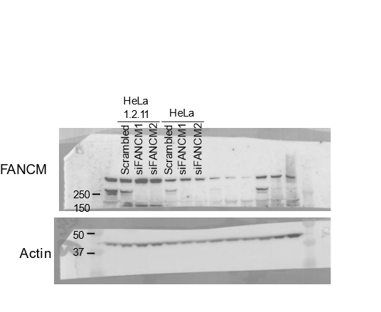
![Knockdown Validated: FANCM Antibody (CV5.1) - BSA Free [NBP2-50418]](https://resources.rndsystems.com/images/products/FANCM-Antibody-CV5-1-Knockdown-Validated-NBP2-50418-img0001.jpg "Western Blot: FANCM Antibody (CV5.1) - BSA Free [NBP2-50418]")
Key Product Details
Validated by
Knockout/Knockdown
Species Reactivity
Validated:
Human
Cited:
Human
Applications
Validated:
Immunohistochemistry, Immunohistochemistry-Paraffin, Western Blot, Immunocytochemistry/ Immunofluorescence, Immunoprecipitation, Knockdown Validated
Cited:
Immunohistochemistry-Paraffin, Western Blot
Label
Unconjugated
Antibody Source
Monoclonal Mouse IgG1 kappa Clone # CV5.1
Format
BSA Free
Loading...
Product Specifications
Immunogen
His-tagged denatured FANCM (aa 1507-1679) made in E.coli
Clonality
Monoclonal
Host
Mouse
Isotype
IgG1 kappa
Scientific Data Images for FANCM Antibody (CV5.1) - BSA Free
![Immunohistochemistry: FANCM Antibody (CV5.1) - BSA Free [NBP2-50418]](https://resources.rndsystems.com/images/products/FANCM-Antibody-CV5-1-Immunohistochemistry-NBP2-50418-img0002.jpg "Immunohistochemistry: FANCM Antibody (CV5.1) - BSA Free [NBP2-50418]")
Immunohistochemistry: FANCM Antibody (CV5.1) - BSA Free [NBP2-50418]
FANCM-Antibody-CV5-1-Immunohistochemistry-NBP2-50418-img0002.jpg - BSA Free [NBP2-50418] -")
Immunohistochemistry: FANCM Antibody (CV5.1) - BSA Free [NBP2-50418] -
FANCM expression in human fetal ovaries.(A) Relative FANCM mRNA abundance was measured by RT-qPCR in human fetal ovaries from 5 to 32 weeks post-fertilization (wpf). (B) Germ cells (D2-40+) & somatic cells (D2-40-) were sorted from three ovaries ranging from 8 to 12 wpf & FANCM expression was measured. ACTB was used to normalize FANCM expression in all samples. Dots represent different ovaries & the mean is indicated by the line. (C) Immunohistochemistry of FANCM in human fetal & adult ovaries. Fetal ovaries at 8 & 22 wpf & adult ovaries were studied. FANCM positive cells appear in yellow/brown color (monoclonal FANCM CV5.1 antibody, Novus Biologicals, Abingdon, UK). Ovarian sections were counterstained with hematoxylin (blue staining). Oo, oogonia; Pa, oocyte at the pachytene stage of meiosis I, D, oocyte at the diplotene stage of meiosis I; Pr, oocyte in primordial follicle. (D) Co-staining in 22 wpf ovaries, for FANCM (purple) & DDX4 (brown) confirmed the germ cell identity of FANCM-positive cells (left). Successive staining for FANCM & SYCP3 in the same section (panels a & b). Negative control performed with non-immune mouse IgG (right). Scale bar: 10 μm. Image collected & cropped by CiteAb from the following publication (https://pubmed.ncbi.nlm.nih.gov/29231814), licensed under a CC-BY license. Not internally tested by Novus Biologicals.Applications for FANCM Antibody (CV5.1) - BSA Free
Application
Recommended Usage
Immunocytochemistry/ Immunofluorescence
1:100
Immunoprecipitation
2 - 4 ug/ml lysate
Western Blot
1:1000
Application Notes
Positive control(s): 293 cell extract. Use in Immunohistochemistry-paraffin reported in scientific literature (PMID: 29231814).
Reviewed Applications
Read 1 review rated 4 using NBP2-50418 in the following applications:
Formulation, Preparation, and Storage
Purification
Protein A purified
Formulation
PBS
Format
BSA Free
Preservative
0.02% Sodium Azide
Concentration
1 mg/ml
Shipping
The product is shipped with polar packs. Upon receipt, store it immediately at the temperature recommended below.
Stability & Storage
Store at 4C short term. Aliquot and store at -20C long term. Avoid freeze-thaw cycles.
Background: FANCM
Alternate Names
ATP-dependent RNA helicase FANCM, EC 3.6.1, FAAP250EC 3.6.4.13, Fanconi anemia, complementation group M, Fanconi anemia-associated polypeptide of 250 kDa, KIAA1596Fanconi anemia group M protein, MGC176453, Protein FACM, Protein Hef ortholog
Gene Symbol
FANCM
Additional FANCM Products
Product Documents for FANCM Antibody (CV5.1) - BSA Free
Certificate of Analysis
To download a Certificate of Analysis, please enter a lot or batch number in the search box below.
Product Specific Notices for FANCM Antibody (CV5.1) - BSA Free
This product is for research use only and is not approved for use in humans or in clinical diagnosis. Primary Antibodies are guaranteed for 1 year from date of receipt.
Citations for FANCM Antibody (CV5.1) - BSA Free
 Powered by Bioz
Customer Reviews for FANCM Antibody (CV5.1) - BSA Free (1)
4 out of 5
1 Customer Rating
Have you used FANCM Antibody (CV5.1) - BSA Free?
Submit a review and receive an Amazon gift card!
$25/€18/£15/$25CAN/¥2500 Yen for a review with an image
$10/€7/£6/$10CAN/¥1110 Yen for a review without an image
Submit a review
Customer Images
Showing
1
-
1 of
1 review
Showing All
Filter By:
-
Application: Western BlotSample Tested: Hela whole cell lysateSpecies: HumanVerified Customer | Posted 10/07/2020Uncropped Western blot showing FANCM depletion using two different siRNA's (Life Technologies - siRNA ID s33619, s33621 respectively). Note that there is a prominent non-specific band at about 350-450kD.Cells were lysed using RIPA buffer supplemented with protease inhibitor cocktail. 20ug protein per lane. PVDF membrane was blocked with 5% milk in PBST. Diluted in 0.5% milk in PBST. Band signal requires requires femtomolar ECL reagent in order to detect in under 3 minutes.

There are no reviews that match your criteria.
Protocols
Find general support by application which include: protocols, troubleshooting, illustrated assays, videos and webinars.
- Antigen Retrieval Protocol (PIER)
- Antigen Retrieval for Frozen Sections Protocol
- Appropriate Fixation of IHC/ICC Samples
- Cellular Response to Hypoxia Protocols
- Chromogenic IHC Staining of Formalin-Fixed Paraffin-Embedded (FFPE) Tissue Protocol
- Chromogenic Immunohistochemistry Staining of Frozen Tissue
- ClariTSA™ Fluorophore Kits
- Detection & Visualization of Antibody Binding
- Fluorescent IHC Staining of Frozen Tissue Protocol
- Graphic Protocol for Heat-induced Epitope Retrieval
- Graphic Protocol for the Preparation and Fluorescent IHC Staining of Frozen Tissue Sections
- Graphic Protocol for the Preparation and Fluorescent IHC Staining of Paraffin-embedded Tissue Sections
- Graphic Protocol for the Preparation of Gelatin-coated Slides for Histological Tissue Sections
- ICC Cell Smear Protocol for Suspension Cells
- ICC Immunocytochemistry Protocol Videos
- ICC for Adherent Cells
- IHC Sample Preparation (Frozen sections vs Paraffin)
- Immunocytochemistry (ICC) Protocol
- Immunocytochemistry Troubleshooting
- Immunofluorescence of Organoids Embedded in Cultrex Basement Membrane Extract
- Immunofluorescent IHC Staining of Formalin-Fixed Paraffin-Embedded (FFPE) Tissue Protocol
- Immunohistochemistry (IHC) and Immunocytochemistry (ICC) Protocols
- Immunohistochemistry Frozen Troubleshooting
- Immunohistochemistry Paraffin Troubleshooting
- Immunoprecipitation Protocol
- Preparing Samples for IHC/ICC Experiments
- Preventing Non-Specific Staining (Non-Specific Binding)
- Primary Antibody Selection & Optimization
- Protocol for Heat-Induced Epitope Retrieval (HIER)
- Protocol for Making a 4% Formaldehyde Solution in PBS
- Protocol for VisUCyte™ HRP Polymer Detection Reagent
- Protocol for the Fluorescent ICC Staining of Cell Smears - Graphic
- Protocol for the Fluorescent ICC Staining of Cultured Cells on Coverslips - Graphic
- Protocol for the Preparation & Fixation of Cells on Coverslips
- Protocol for the Preparation and Chromogenic IHC Staining of Frozen Tissue Sections
- Protocol for the Preparation and Chromogenic IHC Staining of Frozen Tissue Sections - Graphic
- Protocol for the Preparation and Chromogenic IHC Staining of Paraffin-embedded Tissue Sections
- Protocol for the Preparation and Chromogenic IHC Staining of Paraffin-embedded Tissue Sections - Graphic
- Protocol for the Preparation and Fluorescent ICC Staining of Cells on Coverslips
- Protocol for the Preparation and Fluorescent ICC Staining of Non-adherent Cells
- Protocol for the Preparation and Fluorescent ICC Staining of Stem Cells on Coverslips
- Protocol for the Preparation and Fluorescent IHC Staining of Frozen Tissue Sections
- Protocol for the Preparation and Fluorescent IHC Staining of Paraffin-embedded Tissue Sections
- Protocol for the Preparation of Gelatin-coated Slides for Histological Tissue Sections
- Protocol for the Preparation of a Cell Smear for Non-adherent Cell ICC - Graphic
- R&D Systems Quality Control Western Blot Protocol
- TUNEL and Active Caspase-3 Detection by IHC/ICC Protocol
- The Importance of IHC/ICC Controls
- Troubleshooting Guide: Immunohistochemistry
- Troubleshooting Guide: Western Blot Figures
- Western Blot Conditions
- Western Blot Protocol
- Western Blot Protocol for Cell Lysates
- Western Blot Troubleshooting
- Western Blot Troubleshooting Guide
- View all Protocols, Troubleshooting, Illustrated assays and Webinars
Loading...