FFAR4/GPR120 Antibody - BSA Free
Novus Biologicals | Catalog # NBP1-00858
![Knockout Validated: FFAR4/GPR120 Antibody - BSA Free [NBP1-00858]](https://resources.rndsystems.com/images/products/FFAR4-GPR120-Antibody-Knockout-Validated-NBP1-00858-img0010.jpg "Western Blot: FFAR4/GPR120 Antibody - BSA Free [NBP1-00858]")
Key Product Details
Validated by
Knockout/Knockdown
Species Reactivity
Validated:
Human, Mouse, Rat
Cited:
Human, Mouse, Rat
Applications
Validated:
Knockout Validated, Immunohistochemistry, Immunohistochemistry-Paraffin, Western Blot, Flow Cytometry, Immunocytochemistry/ Immunofluorescence, Knockdown Validated
Cited:
Immunohistochemistry-Paraffin, Western Blot, Flow Cytometry, Immunocytochemistry/ Immunofluorescence, IF/IHC
Label
Unconjugated
Antibody Source
Polyclonal Rabbit IgG
Format
BSA Free
Loading...
Product Specifications
Immunogen
A synthetic peptide made to an internal portion of the human GPR120 protein (between residues 200-300) [UniProt Q5NUL3]
Localization
FFAR4/GPR120 is a transmembrane protein with extracellular-helical-cytoplasmic domains
Clonality
Polyclonal
Host
Rabbit
Isotype
IgG
Scientific Data Images for FFAR4/GPR120 Antibody - BSA Free
![Immunohistochemistry-Paraffin: FFAR4/GPR120 Antibody - BSA Free [NBP1-00858]](https://resources.rndsystems.com/images/products/FFAR4-GPR120-Antibody-Immunohistochemistry-Paraffin-NBP1-00858-img0005.jpg "Immunohistochemistry-Paraffin: FFAR4/GPR120 Antibody - BSA Free [NBP1-00858]")
Immunohistochemistry-Paraffin: FFAR4/GPR120 Antibody - BSA Free [NBP1-00858]
Immunohistochemistry-Paraffin: FFAR4/GPR120 Antibody [NBP1-00858] - IHC analysis of formalin fixed paraffin embedded tissue section of mouse intestine using FFAR4/GPR120 antibody at 1:200 dilution. The intestinal epithelial cells showed an expected cytoplasmic staining (immunogen of NBP1-00858 corresponds to the cytoplasmic domain of GPR120 protein ), while some cells especially the goblet cells population depicted nuclear positivity also. The observed nuclear signal may be justified on the fact that - some GPCRs are known for nuclear translocation upon binding to extracellular or endogenous/non-secreted ligands, wherein they impact the trancriptional regulation via GPCR-heterotrimeric G-protein-effector complexes.![Western Blot: FFAR4/GPR120 AntibodyBSA Free [NBP1-00858]](https://resources.rndsystems.com/images/products/FFAR4-GPR120-Antibody-Western-Blot-NBP1-00858-img0007.jpg "Western Blot: FFAR4/GPR120 AntibodyBSA Free [NBP1-00858]")
Western Blot: FFAR4/GPR120 AntibodyBSA Free [NBP1-00858]
Western Blot: FFAR4/GPR120 Antibody [NBP1-00858] - Analysis of GPR120 (V259) antibody in extracts from LOVO cells.![Western Blot: FFAR4/GPR120 AntibodyBSA Free [NBP1-00858]](https://resources.rndsystems.com/images/products/FFAR4-GPR120-Antibody-Western-Blot-NBP1-00858-img0008.jpg "Western Blot: FFAR4/GPR120 AntibodyBSA Free [NBP1-00858]")
Western Blot: FFAR4/GPR120 AntibodyBSA Free [NBP1-00858]
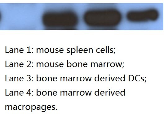
Western Blot: FFAR4/GPR120 Antibody [NBP1-00858] - GPR120 expression in mouse spleen cells (1), bone marrow (2), bone marrow derived DC (3) and macrophages (4). This image was submitted through a verified customer review.![Immunocytochemistry/ Immunofluorescence: FFAR4/GPR120 Antibody - BSA Free [NBP1-00858]](https://resources.rndsystems.com/images/products/FFAR4-GPR120-Antibody-Immunocytochemistry-Immunofluorescence-NBP1-00858-img0006.jpg "Immunocytochemistry/ Immunofluorescence: FFAR4/GPR120 Antibody - BSA Free [NBP1-00858]")
Immunocytochemistry/ Immunofluorescence: FFAR4/GPR120 Antibody - BSA Free [NBP1-00858]
Immunocytochemistry/Immunofluorescence: FFAR4/GPR120 Antibody [NBP1-00858] - GPR120 antibody was tested at 1:50 in HeLa cells with Dylight 488 (green). Nuclei and alpha-tubulin were counterstained with DAPI (blue) and Dylight 550 (red). Image objective 40x.![Immunohistochemistry-Paraffin: FFAR4/GPR120 Antibody - BSA Free [NBP1-00858]](https://resources.rndsystems.com/images/products/FFAR4-GPR120-Antibody-Immunohistochemistry-Paraffin-NBP1-00858-img0003.jpg "Immunohistochemistry-Paraffin: FFAR4/GPR120 Antibody - BSA Free [NBP1-00858]")
Immunohistochemistry-Paraffin: FFAR4/GPR120 Antibody - BSA Free [NBP1-00858]
Immunohistochemistry-Paraffin: FFAR4/GPR120 Antibody [NBP1-00858] - IHC analysis of formalin fixed paraffin embedded tissue section of mouse intestine using FFAR4/GPR120 antibody at 1:200 dilution. The intestinal epithelial cells showed an expected cytoplasmic staining (immunogen of NBP1-00858 corresponds to the cytoplasmic domain of GPR120 protein), while some cells especially the goblet cells population depicted nuclear positivity also. The observed nuclear signal may be justified on the fact that - some GPCRs are known for nuclear translocation upon binding to extracellular or endogenous/non-secreted ligands, wherein they impact the trancriptional regulation via GPCR-heterotrimeric G-protein-effector complexes.
Western Blot: FFAR4/GPR120 Antibody - BSA Free [NBP1-00858] -
Western Blot: FFAR4/GPR120 Antibody - BSA Free [NBP1-00858] - The combination of RA & omega -3 PUFAs induces G alpha q-P38 activation through RAR alpha & GPR40(A): Cells were treated with RA(20μM) + EPA(80μM) with or without GPR120-knockdown for 15 min. Cell extracts were prepared & subjected to western blotting analysis. (B): Cells were treated with RA(20μM) + EPA(80μM) with or without GPR40-knockdown for 15 min. Cell extracts were prepared & subjected to western blotting analysis. (C): Cells were treated with RA(20μM) + EPA(80μM) with or without RAR alpha -knockdown for 15 min. Cell extracts were prepared & subjected to western blotting analysis. (D): Cells were treated with RA(20μM) + EPA(80μM) with or without RAR beta -knockdown for 15 min. Cell extracts were prepared & subjected to western blotting analysis. (E): Cells were treated with RA(20μM) + EPA(80μM) with or without RAR gamma -knockdown for 15 min. Cell extracts were prepared & subjected to western blotting analysis. (F): MCF-7 cells were treated with RA(20μM) + EPA(80μM) & their extracts fractionated using an iodixanol density gradient, as described in the Materials & Methods section. Each fraction was subjected to SDS-PAGE & immunoblot analysis using antibodies against the indicated proteins. (G): MCF-7 cells were pretreated with the indicated concentrations of methyl-beta -cyclodextrin (M beta CD) for 1 h, followed by 15 min treatment with RA(20μM) + EPA(80μM). Cell lysates were prepared & subjected to SDS-PAGE & immunoblot analysis. (H): MCF-7 cells were pretreated with the indicated concentrations of methyl-beta -cyclodextrin (M beta CD) for 1 h followed by 24h treatment with RA(20μM) + EPA(80μM), & then subjected to cell counts. Image collected & cropped by CiteAb from the following publication (https://www.oncotarget.com/lookup/doi/10.18632/oncotarget.22629), licensed under a CC-BY license. Not internally tested by Novus Biologicals.
Western Blot: FFAR4/GPR120 Antibody - BSA Free [NBP1-00858] -
Western Blot: FFAR4/GPR120 Antibody - BSA Free [NBP1-00858] - The expression of corin & ANP was increased in DHA-induced adipocytes. 3T3-L1 cells were exposed to DHA (100 μM) for 2 d in the presence of the differentiation medium. (A–C) The expression of corin & ANP in DHA-induced adipocytes was analyzed by qRT-PCR & Western blotting. The basal delta-Ct levels for tested genes are presented as Supplementary Table S2. ** p < 0.01. (D,E) The expression of corin & ANP was measured in the GPR120 deficient adipocytes treated with DHA by qRT-PCR & Western blotting. * p < 0.05, ** p < 0.01, *** p < 0.001. (F) 3T3-L1 cells were treated with 1 μM TUG-891, a potent GPR120 agonist for 24 h. The corin & ANP expression levels were analyzed by Western blotting. (G) The concentration of ANP was measured in the media derived from the DHA-induced adipocytes using ELISA. ** p < 0.01. The data are shown as the means ± standard deviations from three or more independent experiments. Image collected & cropped by CiteAb from the following publication (https://pubmed.ncbi.nlm.nih.gov/31817347), licensed under a CC-BY license. Not internally tested by Novus Biologicals.Applications for FFAR4/GPR120 Antibody - BSA Free
Application
Recommended Usage
Flow Cytometry
reported in scientific literature (PMID 34911928)
Immunocytochemistry/ Immunofluorescence
1:50-1:200
Immunohistochemistry
1:200
Immunohistochemistry-Paraffin
1:200
Western Blot
1:500-1:1000
Reviewed Applications
Read 1 review rated 5 using NBP1-00858 in the following applications:
Flow Cytometry Panel Builder
Bio-Techne Knows Flow Cytometry
Save time and reduce costly mistakes by quickly finding compatible reagents using the Panel Builder Tool.
Advanced Features
- Spectra Viewer - Custom analysis of spectra from multiple fluorochromes
- Spillover Popups - Visualize the spectra of individual fluorochromes
- Antigen Density Selector - Match fluorochrome brightness with antigen density

Formulation, Preparation, and Storage
Purification
Immunogen affinity purified
Formulation
PBS
Format
BSA Free
Preservative
0.02% Sodium Azide
Concentration
1.0 mg/ml
Shipping
The product is shipped with polar packs. Upon receipt, store it immediately at the temperature recommended below.
Stability & Storage
Store at 4C short term. Aliquot and store at -20C long term. Avoid freeze-thaw cycles.
Background: FFAR4/GPR120
Long Name
Free Fatty Acid Receptor 4
Alternate Names
GPR120, GPR129, GT01, O3FAR1, PGR4
Gene Symbol
FFAR4
UniProt
Additional FFAR4/GPR120 Products
Product Documents for FFAR4/GPR120 Antibody - BSA Free
Certificate of Analysis
To download a Certificate of Analysis, please enter a lot or batch number in the search box below.
Product Specific Notices for FFAR4/GPR120 Antibody - BSA Free
This product is for research use only and is not approved for use in humans or in clinical diagnosis. Primary Antibodies are guaranteed for 1 year from date of receipt.
Related Research Areas
Citations for FFAR4/GPR120 Antibody - BSA Free
 Powered by Bioz
Customer Reviews for FFAR4/GPR120 Antibody - BSA Free (1)
5 out of 5
1 Customer Rating
Have you used FFAR4/GPR120 Antibody - BSA Free?
Submit a review and receive an Amazon gift card!
$25/€18/£15/$25CAN/¥2500 Yen for a review with an image
$10/€7/£6/$10CAN/¥1110 Yen for a review without an image
Submit a review
Customer Images
Showing
1
-
1 of
1 review
Showing All
Filter By:
-
Application: Western BlotSample Tested: whole cell lysates from mouse bone marrow, spleen, bone marrow derived DCs/macrophages.Species: MouseVerified Customer | Posted 04/02/2015GPR120 expression in mouse spleen cells, bone marrow, bone marrow derived DC and macrophages

There are no reviews that match your criteria.
Protocols
View specific protocols for FFAR4/GPR120 Antibody - BSA Free (NBP1-00858):
Culture cells to appropriate density in 35 mm culture dishes or 6-well plates.
1. Remove culture medium and add 10% formalin to the dish. Fix at room temperature for 30 minutes.
2. Remove the formalin and add ice cold methanol. Incubate for 5-10 minutes.
3. Remove methanol and add washing solution (i.e. PBS). Be sure to not let the specimen dry out. Wash three times for 10 minutes.
4. To block nonspecific antibody binding incubate in 10% normal goat serum from 1 hour to overnight at room temperature.
5. Add primary antibody at appropriate dilution and incubate at room temperature from 2 hours to overnight at room temperature.
6. Remove primary antibody and replace with washing solution. Wash three times for 10 minutes.
7. Add secondary antibody at appropriate dilution. Incubate for 1 hour at room temperature.
8. Remove antibody and replace with wash solution, then wash for 10 minutes. Add Hoechst 33258 to wash solution at 1:25,0000 and incubate for 10 minutes. Wash a third time for 10 minutes.
9. Cells can be viewed directly after washing. The plates can also be stored in PBS containing Azide covered in Parafilm (TM). Cells can also be cover-slipped using Fluoromount, with appropriate sealing.
*The above information is only intended as a guide. The researcher should determine what protocol best meets their needs. Please follow safe laboratory procedures.
Western Blot Protocol
1. Perform SDS-PAGE using a 12% gel on samples to be analyzed, loading 25 ug of total protein per lane.
2. Transfer proteins to membrane according to the instructions provided by the manufacturer of the membrane and transfer apparatus.
3. Stain according to standard Ponceau S procedure (or similar product) to assess transfer success, and mark molecular weight standards where appropriate.
4. Rinse the blot.
5. Block the membrane using standard blocking buffer for at least 1 hour.
6. Wash the membrane in wash buffer three times for 10 minutes each.
7. Dilute anti-GPR120 primary antibody in blocking buffer and incubate 1 hour at room temperature.
8. Wash the membrane in wash buffer three times for 10 minutes each.
9. Apply the diluted HRP conjugated secondary antibody in blocking buffer (as per manufacturers instructions) and incubate 1 hour at room temperature.
10. Wash the blot in wash buffer three times for 10 minutes each (this step can be repeated as required to reduce background).
11. Apply the detection reagent of choice in accordance with the manufacturers instructions.
Note: Tween-20 can be added to the blocking or antibody dilution buffer at a final concentration of 0.05-0.2%.
Find general support by application which include: protocols, troubleshooting, illustrated assays, videos and webinars.
- 7-Amino Actinomycin D (7-AAD) Cell Viability Flow Cytometry Protocol
- Antigen Retrieval Protocol (PIER)
- Antigen Retrieval for Frozen Sections Protocol
- Appropriate Fixation of IHC/ICC Samples
- Cellular Response to Hypoxia Protocols
- Chromogenic IHC Staining of Formalin-Fixed Paraffin-Embedded (FFPE) Tissue Protocol
- Chromogenic Immunohistochemistry Staining of Frozen Tissue
- ClariTSA™ Fluorophore Kits
- Detection & Visualization of Antibody Binding
- Extracellular Membrane Flow Cytometry Protocol
- Flow Cytometry Protocol for Cell Surface Markers
- Flow Cytometry Protocol for Staining Membrane Associated Proteins
- Flow Cytometry Staining Protocols
- Flow Cytometry Troubleshooting Guide
- Fluorescent IHC Staining of Frozen Tissue Protocol
- Graphic Protocol for Heat-induced Epitope Retrieval
- Graphic Protocol for the Preparation and Fluorescent IHC Staining of Frozen Tissue Sections
- Graphic Protocol for the Preparation and Fluorescent IHC Staining of Paraffin-embedded Tissue Sections
- Graphic Protocol for the Preparation of Gelatin-coated Slides for Histological Tissue Sections
- ICC Cell Smear Protocol for Suspension Cells
- ICC Immunocytochemistry Protocol Videos
- ICC for Adherent Cells
- IHC Sample Preparation (Frozen sections vs Paraffin)
- Immunocytochemistry (ICC) Protocol
- Immunocytochemistry Troubleshooting
- Immunofluorescence of Organoids Embedded in Cultrex Basement Membrane Extract
- Immunofluorescent IHC Staining of Formalin-Fixed Paraffin-Embedded (FFPE) Tissue Protocol
- Immunohistochemistry (IHC) and Immunocytochemistry (ICC) Protocols
- Immunohistochemistry Frozen Troubleshooting
- Immunohistochemistry Paraffin Troubleshooting
- Intracellular Flow Cytometry Protocol Using Alcohol (Methanol)
- Intracellular Flow Cytometry Protocol Using Detergents
- Intracellular Nuclear Staining Flow Cytometry Protocol Using Detergents
- Intracellular Staining Flow Cytometry Protocol Using Alcohol Permeabilization
- Intracellular Staining Flow Cytometry Protocol Using Detergents to Permeabilize Cells
- Preparing Samples for IHC/ICC Experiments
- Preventing Non-Specific Staining (Non-Specific Binding)
- Primary Antibody Selection & Optimization
- Propidium Iodide Cell Viability Flow Cytometry Protocol
- Protocol for Heat-Induced Epitope Retrieval (HIER)
- Protocol for Liperfluo
- Protocol for Making a 4% Formaldehyde Solution in PBS
- Protocol for VisUCyte™ HRP Polymer Detection Reagent
- Protocol for the Characterization of Human Th22 Cells
- Protocol for the Characterization of Human Th9 Cells
- Protocol for the Fluorescent ICC Staining of Cell Smears - Graphic
- Protocol for the Fluorescent ICC Staining of Cultured Cells on Coverslips - Graphic
- Protocol for the Preparation & Fixation of Cells on Coverslips
- Protocol for the Preparation and Chromogenic IHC Staining of Frozen Tissue Sections
- Protocol for the Preparation and Chromogenic IHC Staining of Frozen Tissue Sections - Graphic
- Protocol for the Preparation and Chromogenic IHC Staining of Paraffin-embedded Tissue Sections
- Protocol for the Preparation and Chromogenic IHC Staining of Paraffin-embedded Tissue Sections - Graphic
- Protocol for the Preparation and Fluorescent ICC Staining of Cells on Coverslips
- Protocol for the Preparation and Fluorescent ICC Staining of Non-adherent Cells
- Protocol for the Preparation and Fluorescent ICC Staining of Stem Cells on Coverslips
- Protocol for the Preparation and Fluorescent IHC Staining of Frozen Tissue Sections
- Protocol for the Preparation and Fluorescent IHC Staining of Paraffin-embedded Tissue Sections
- Protocol for the Preparation of Gelatin-coated Slides for Histological Tissue Sections
- Protocol for the Preparation of a Cell Smear for Non-adherent Cell ICC - Graphic
- Protocol: Annexin V and PI Staining by Flow Cytometry
- Protocol: Annexin V and PI Staining for Apoptosis by Flow Cytometry
- R&D Systems Quality Control Western Blot Protocol
- TUNEL and Active Caspase-3 Detection by IHC/ICC Protocol
- The Importance of IHC/ICC Controls
- Troubleshooting Guide: Fluorokine Flow Cytometry Kits
- Troubleshooting Guide: Immunohistochemistry
- Troubleshooting Guide: Western Blot Figures
- Western Blot Conditions
- Western Blot Protocol
- Western Blot Protocol for Cell Lysates
- Western Blot Troubleshooting
- Western Blot Troubleshooting Guide
- View all Protocols, Troubleshooting, Illustrated assays and Webinars
Loading...