FGF-21 Antibody (OTI2F10)
Novus Biologicals | Catalog # NBP2-00645
Key Product Details
Species Reactivity
Human
Applications
Immunohistochemistry, Immunohistochemistry-Paraffin, Western Blot, Flow Cytometry, Immunocytochemistry/ Immunofluorescence
Label
Unconjugated
Antibody Source
Monoclonal Mouse IgG2A Clone # OTI2F10
Loading...
Product Specifications
Immunogen
Human recombinant protein fragment corresponding to amino acids 29-209 of human FGF21(NP_061986) produced in E.coli.
Clonality
Monoclonal
Host
Mouse
Isotype
IgG2A
Scientific Data Images for FGF-21 Antibody (OTI2F10)
![Western Blot: FGF-21 Antibody (OTI2F10) [NBP2-00645]](https://resources.rndsystems.com/images/products/FGF21-Antibody-2F10-Western-Blot-NBP2-00645-img0002.jpg "Western Blot: FGF-21 Antibody (OTI2F10) [NBP2-00645]")
Western Blot: FGF-21 Antibody (OTI2F10) [NBP2-00645]
Western Blot: FGF21 Antibody (2F10) [NBP2-00645] - HEK293T cells were transfected with the pCMV6-ENTRY control (Left lane) or pCMV6-ENTRY FGF21 (Right lane) cDNA for 48 hrs and lysed. Equivalent amounts of cell lysates (5 ug per lane) were separated by SDS-PAGE and immunoblotted with anti-FGF21.![Immunocytochemistry/ Immunofluorescence: FGF-21 Antibody (OTI2F10) [NBP2-00645]](https://resources.rndsystems.com/images/products/FGF-21-Antibody-2F10-Immunocytochemistry-Immunofluorescence-NBP2-00645-img0011.jpg "Immunocytochemistry/ Immunofluorescence: FGF-21 Antibody (OTI2F10) [NBP2-00645]")
Immunocytochemistry/ Immunofluorescence: FGF-21 Antibody (OTI2F10) [NBP2-00645]
Immunocytochemistry/Immunofluorescence: FGF-21 Antibody (2F10) [NBP2-00645] - Anti-FGF21 mouse monoclonal antibody immunofluorescent staining of COS7 cells transiently transfected by pCMV6-ENTRY FGF21(RC204538) at 1:100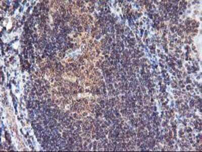
Immunohistochemistry-Paraffin: FGF21 Antibody (2F10) [NBP2-00645] Staining of paraffin-embedded Human tonsil using anti-FGF21 mouse monoclonal antibody.
![Flow Cytometry: FGF-21 Antibody (OTI2F10) [NBP2-00645]](https://resources.rndsystems.com/images/products/FGF-21-Antibody-2F10-Flow-Cytometry-NBP2-00645-img0013.jpg "Flow Cytometry: FGF-21 Antibody (OTI2F10) [NBP2-00645]")
Flow Cytometry: FGF-21 Antibody (OTI2F10) [NBP2-00645]
Flow Cytometry: FGF-21 Antibody (2F10) [NBP2-00645] - HEK293T cells transfected with either RC204538 overexpress plasmid (Red) or empty vector control plasmid(Blue) were immunostained by anti-FGF21 antibody and then analyzed by flow cytometry.![Immunohistochemistry-Paraffin: FGF-21 Antibody (OTI2F10) [NBP2-00645]](https://resources.rndsystems.com/images/products/FGF21-Antibody-2F10-Immunohistochemistry-Paraffin-NBP2-00645-img0003.jpg "Immunohistochemistry-Paraffin: FGF-21 Antibody (OTI2F10) [NBP2-00645]")
Immunohistochemistry-Paraffin: FGF-21 Antibody (OTI2F10) [NBP2-00645]
Immunohistochemistry-Paraffin: FGF21 Antibody (2F10) [NBP2-00645] - Staining of paraffin-embedded Adenocarcinoma of Human endometrium tissue using anti-FGF21 mouse monoclonal antibody.![Immunohistochemistry-Paraffin: FGF-21 Antibody (OTI2F10) [NBP2-00645]](https://resources.rndsystems.com/images/products/FGF21-Antibody-2F10-Immunohistochemistry-Paraffin-NBP2-00645-img0004.jpg "Immunohistochemistry-Paraffin: FGF-21 Antibody (OTI2F10) [NBP2-00645]")
Immunohistochemistry-Paraffin: FGF-21 Antibody (OTI2F10) [NBP2-00645]
Immunohistochemistry-Paraffin: FGF21 Antibody (2F10) [NBP2-00645] - Staining of paraffin-embedded Carcinoma of Human bladder tissue using anti-FGF21 mouse monoclonal antibody.![Immunohistochemistry-Paraffin: FGF-21 Antibody (OTI2F10) [NBP2-00645]](https://resources.rndsystems.com/images/products/FGF21-Antibody-2F10-Immunohistochemistry-Paraffin-NBP2-00645-img0005.jpg "Immunohistochemistry-Paraffin: FGF-21 Antibody (OTI2F10) [NBP2-00645]")
Immunohistochemistry-Paraffin: FGF-21 Antibody (OTI2F10) [NBP2-00645]
Immunohistochemistry-Paraffin: FGF21 Antibody (2F10) [NBP2-00645] - Staining of paraffin-embedded Carcinoma of Human liver tissue using anti-FGF21 mouse monoclonal antibody.![Immunohistochemistry-Paraffin: FGF-21 Antibody (OTI2F10) [NBP2-00645]](https://resources.rndsystems.com/images/products/FGF21-Antibody-2F10-Immunohistochemistry-Paraffin-NBP2-00645-img0006.jpg "Immunohistochemistry-Paraffin: FGF-21 Antibody (OTI2F10) [NBP2-00645]")
Immunohistochemistry-Paraffin: FGF-21 Antibody (OTI2F10) [NBP2-00645]
Immunohistochemistry-Paraffin: FGF21 Antibody (2F10) [NBP2-00645] - Staining of paraffin-embedded Carcinoma of Human lung tissue using anti-FGF21 mouse monoclonal antibody.![Immunohistochemistry-Paraffin: FGF-21 Antibody (OTI2F10) [NBP2-00645]](https://resources.rndsystems.com/images/products/FGF21-Antibody-2F10-Immunohistochemistry-Paraffin-NBP2-00645-img0007.jpg "Immunohistochemistry-Paraffin: FGF-21 Antibody (OTI2F10) [NBP2-00645]")
Immunohistochemistry-Paraffin: FGF-21 Antibody (OTI2F10) [NBP2-00645]
Immunohistochemistry-Paraffin: FGF21 Antibody (2F10) [NBP2-00645] - Staining of paraffin-embedded Carcinoma of Human prostate tissue using anti-FGF21 mouse monoclonal antibody.![Immunohistochemistry-Paraffin: FGF-21 Antibody (OTI2F10) [NBP2-00645]](https://resources.rndsystems.com/images/products/FGF21-Antibody-2F10-Immunohistochemistry-Paraffin-NBP2-00645-img0008.jpg "Immunohistochemistry-Paraffin: FGF-21 Antibody (OTI2F10) [NBP2-00645]")
Immunohistochemistry-Paraffin: FGF-21 Antibody (OTI2F10) [NBP2-00645]
Immunohistochemistry-Paraffin: FGF21 Antibody (2F10) [NBP2-00645] - Staining of paraffin-embedded Human breast tissue using anti-FGF21 mouse monoclonal antibody.![Immunohistochemistry-Paraffin: FGF-21 Antibody (OTI2F10) [NBP2-00645]](https://resources.rndsystems.com/images/products/FGF21-Antibody-2F10-Immunohistochemistry-Paraffin-NBP2-00645-img0009.jpg "Immunohistochemistry-Paraffin: FGF-21 Antibody (OTI2F10) [NBP2-00645]")
Immunohistochemistry-Paraffin: FGF-21 Antibody (OTI2F10) [NBP2-00645]
Immunohistochemistry-Paraffin: FGF21 Antibody (2F10) [NBP2-00645] - Staining of paraffin-embedded Human pancreas tissue using anti-FGF21 mouse monoclonal antibody.![Flow Cytometry: FGF-21 Antibody (OTI2F10) [NBP2-00645]](https://resources.rndsystems.com/images/products/FGF21-Antibody-2F10-Flow-Cytometry-NBP2-00645-img0001.jpg "Flow Cytometry: FGF-21 Antibody (OTI2F10) [NBP2-00645]")
Flow Cytometry: FGF-21 Antibody (OTI2F10) [NBP2-00645]
Flow Cytometry: FGF21 Antibody (2F10) [NBP2-00645] - Analysis of Jurkat cells, using anti-FGF21 antibody, (Red), compared to a nonspecific negative control antibody (Blue).Applications for FGF-21 Antibody (OTI2F10)
Application
Recommended Usage
Flow Cytometry
1:100
Immunocytochemistry/ Immunofluorescence
1:50-1:100
Immunohistochemistry
1:150
Immunohistochemistry-Paraffin
1:150
Western Blot
1:2000
Flow Cytometry Panel Builder
Bio-Techne Knows Flow Cytometry
Save time and reduce costly mistakes by quickly finding compatible reagents using the Panel Builder Tool.
Advanced Features
- Spectra Viewer - Custom analysis of spectra from multiple fluorochromes
- Spillover Popups - Visualize the spectra of individual fluorochromes
- Antigen Density Selector - Match fluorochrome brightness with antigen density

Formulation, Preparation, and Storage
Purification
Immunogen affinity purified
Formulation
PBS (pH 7.3), 1.0% BSA and 50% Glycerol
Preservative
0.02% Sodium Azide
Concentration
1.4 mg/ml
Shipping
The product is shipped with polar packs. Upon receipt, store it immediately at the temperature recommended below.
Stability & Storage
Store at -20C. Avoid freeze-thaw cycles.
Background: FGF-21
Long Name
Fibroblast Growth Factor 21
Alternate Names
FGF21
Entrez Gene IDs
26291 (Human)
Gene Symbol
FGF21
Additional FGF-21 Products
Product Documents for FGF-21 Antibody (OTI2F10)
Certificate of Analysis
To download a Certificate of Analysis, please enter a lot or batch number in the search box below.
Product Specific Notices for FGF-21 Antibody (OTI2F10)
This product is for research use only and is not approved for use in humans or in clinical diagnosis. Primary Antibodies are guaranteed for 1 year from date of receipt.
Related Research Areas
Customer Reviews for FGF-21 Antibody (OTI2F10)
There are currently no reviews for this product. Be the first to review FGF-21 Antibody (OTI2F10) and earn rewards!
Have you used FGF-21 Antibody (OTI2F10)?
Submit a review and receive an Amazon gift card!
$25/€18/£15/$25CAN/¥2500 Yen for a review with an image
$10/€7/£6/$10CAN/¥1110 Yen for a review without an image
Submit a review
Protocols
Find general support by application which include: protocols, troubleshooting, illustrated assays, videos and webinars.
- 7-Amino Actinomycin D (7-AAD) Cell Viability Flow Cytometry Protocol
- Antigen Retrieval Protocol (PIER)
- Antigen Retrieval for Frozen Sections Protocol
- Appropriate Fixation of IHC/ICC Samples
- Cellular Response to Hypoxia Protocols
- Chromogenic IHC Staining of Formalin-Fixed Paraffin-Embedded (FFPE) Tissue Protocol
- Chromogenic Immunohistochemistry Staining of Frozen Tissue
- ClariTSA™ Fluorophore Kits
- Detection & Visualization of Antibody Binding
- Extracellular Membrane Flow Cytometry Protocol
- Flow Cytometry Protocol for Cell Surface Markers
- Flow Cytometry Protocol for Staining Membrane Associated Proteins
- Flow Cytometry Staining Protocols
- Flow Cytometry Troubleshooting Guide
- Fluorescent IHC Staining of Frozen Tissue Protocol
- Graphic Protocol for Heat-induced Epitope Retrieval
- Graphic Protocol for the Preparation and Fluorescent IHC Staining of Frozen Tissue Sections
- Graphic Protocol for the Preparation and Fluorescent IHC Staining of Paraffin-embedded Tissue Sections
- Graphic Protocol for the Preparation of Gelatin-coated Slides for Histological Tissue Sections
- ICC Cell Smear Protocol for Suspension Cells
- ICC Immunocytochemistry Protocol Videos
- ICC for Adherent Cells
- IHC Sample Preparation (Frozen sections vs Paraffin)
- Immunocytochemistry (ICC) Protocol
- Immunocytochemistry Troubleshooting
- Immunofluorescence of Organoids Embedded in Cultrex Basement Membrane Extract
- Immunofluorescent IHC Staining of Formalin-Fixed Paraffin-Embedded (FFPE) Tissue Protocol
- Immunohistochemistry (IHC) and Immunocytochemistry (ICC) Protocols
- Immunohistochemistry Frozen Troubleshooting
- Immunohistochemistry Paraffin Troubleshooting
- Intracellular Flow Cytometry Protocol Using Alcohol (Methanol)
- Intracellular Flow Cytometry Protocol Using Detergents
- Intracellular Nuclear Staining Flow Cytometry Protocol Using Detergents
- Intracellular Staining Flow Cytometry Protocol Using Alcohol Permeabilization
- Intracellular Staining Flow Cytometry Protocol Using Detergents to Permeabilize Cells
- Preparing Samples for IHC/ICC Experiments
- Preventing Non-Specific Staining (Non-Specific Binding)
- Primary Antibody Selection & Optimization
- Propidium Iodide Cell Viability Flow Cytometry Protocol
- Protocol for Heat-Induced Epitope Retrieval (HIER)
- Protocol for Liperfluo
- Protocol for Making a 4% Formaldehyde Solution in PBS
- Protocol for VisUCyte™ HRP Polymer Detection Reagent
- Protocol for the Characterization of Human Th22 Cells
- Protocol for the Characterization of Human Th9 Cells
- Protocol for the Fluorescent ICC Staining of Cell Smears - Graphic
- Protocol for the Fluorescent ICC Staining of Cultured Cells on Coverslips - Graphic
- Protocol for the Preparation & Fixation of Cells on Coverslips
- Protocol for the Preparation and Chromogenic IHC Staining of Frozen Tissue Sections
- Protocol for the Preparation and Chromogenic IHC Staining of Frozen Tissue Sections - Graphic
- Protocol for the Preparation and Chromogenic IHC Staining of Paraffin-embedded Tissue Sections
- Protocol for the Preparation and Chromogenic IHC Staining of Paraffin-embedded Tissue Sections - Graphic
- Protocol for the Preparation and Fluorescent ICC Staining of Cells on Coverslips
- Protocol for the Preparation and Fluorescent ICC Staining of Non-adherent Cells
- Protocol for the Preparation and Fluorescent ICC Staining of Stem Cells on Coverslips
- Protocol for the Preparation and Fluorescent IHC Staining of Frozen Tissue Sections
- Protocol for the Preparation and Fluorescent IHC Staining of Paraffin-embedded Tissue Sections
- Protocol for the Preparation of Gelatin-coated Slides for Histological Tissue Sections
- Protocol for the Preparation of a Cell Smear for Non-adherent Cell ICC - Graphic
- Protocol: Annexin V and PI Staining by Flow Cytometry
- Protocol: Annexin V and PI Staining for Apoptosis by Flow Cytometry
- R&D Systems Quality Control Western Blot Protocol
- TUNEL and Active Caspase-3 Detection by IHC/ICC Protocol
- The Importance of IHC/ICC Controls
- Troubleshooting Guide: Fluorokine Flow Cytometry Kits
- Troubleshooting Guide: Immunohistochemistry
- Troubleshooting Guide: Western Blot Figures
- Western Blot Conditions
- Western Blot Protocol
- Western Blot Protocol for Cell Lysates
- Western Blot Troubleshooting
- Western Blot Troubleshooting Guide
- View all Protocols, Troubleshooting, Illustrated assays and Webinars
Loading...