FoxP3 Antibody - BSA Free
Novus Biologicals | Catalog # NB100-39002
![Immunohistochemistry-Paraffin: FoxP3 Antibody - BSA Free [NB100-39002]](https://resources.rndsystems.com/images/products/FoxP3-Antibody-Immunohistochemistry-Paraffin-NB100-39002-img0011.jpg "Immunohistochemistry-Paraffin: FoxP3 Antibody - BSA Free [NB100-39002]")
Key Product Details
Species Reactivity
Validated:
Cited:
Applications
Validated:
Cited:
Label
Antibody Source
Format
Product Specifications
Immunogen
Reactivity Notes
Localization
Clonality
Host
Isotype
Scientific Data Images for FoxP3 Antibody - BSA Free
Immunohistochemistry-Paraffin: FoxP3 Antibody - BSA Free [NB100-39002]
Immunohistochemistry-Paraffin: FoxP3 Antibody [NB100-39002] - Mouse spleen stained with FoxP3 antibody. IHC-P image submitted by a verified customer review.![Immunohistochemistry-Paraffin: FoxP3 Antibody - BSA Free [NB100-39002]](https://resources.rndsystems.com/images/products/FoxP3-Antibody-Immunohistochemistry-Paraffin-NB100-39002-img0025.jpg "Immunohistochemistry-Paraffin: FoxP3 Antibody - BSA Free [NB100-39002]")
Immunohistochemistry-Paraffin: FoxP3 Antibody - BSA Free [NB100-39002]
FoxP3-Antibody-Immunohistochemistry-Paraffin-NB100-39002-img0025.jpg![Immunohistochemistry: FoxP3 Antibody - BSA Free [NB100-39002]](https://resources.rndsystems.com/images/products/FoxP3-Antibody-Immunohistochemistry-NB100-39002-img0027.jpg "Immunohistochemistry: FoxP3 Antibody - BSA Free [NB100-39002]")
Immunohistochemistry: FoxP3 Antibody - BSA Free [NB100-39002]
FoxP3-Antibody-Immunohistochemistry-NB100-39002-img0027.jpg![Flow Cytometry: FoxP3 Antibody - BSA Free [NB100-39002]](https://resources.rndsystems.com/images/products/FoxP3-Antibody-Flow-Cytometry-NB100-39002-img0022.jpg "Flow Cytometry: FoxP3 Antibody - BSA Free [NB100-39002]")
Flow Cytometry: FoxP3 Antibody - BSA Free [NB100-39002]
Flow Cytometry: FoxP3 Antibody [NB100-39002] - An intracellular stain was performed on SK-MEL-28 cells with NB100-39002 (blue) and a matched isotype control (orange). Cells were fixed with 4% PFA and then permeabilized with 0.1% saponin. Cells were incubated in an antibody dilution of 2.5 ug/mL for 30 minutes at room temperature, followed by Rabbit IgG (H+L) Cross-Adsorbed Secondary Antibody.![Immunohistochemistry-Paraffin: FoxP3 Antibody - BSA Free [NB100-39002]](https://resources.rndsystems.com/images/products/FoxP3-Antibody-Immunohistochemistry-Paraffin-NB100-39002-img0017.jpg "Immunohistochemistry-Paraffin: FoxP3 Antibody - BSA Free [NB100-39002]")
Immunohistochemistry-Paraffin: FoxP3 Antibody - BSA Free [NB100-39002]
Immunohistochemistry-Paraffin: FoxP3 Antibody [NB100-39002] - Tissue section of human tonsil using 1:200 dilution of rabbit anti-FoxP3 antibody. The staining was developed with HRP-labeled anti-rabbit IgG secondary antibody and DAB reagent, and nuclei of cells were counter-stained with hematoxylin. This FoxP3 antibody generated an expected and a specific nuclear staining of FOXP3 in a subset of cells which are potentially the Treg cells.![Immunohistochemistry: FoxP3 Antibody - BSA Free [NB100-39002]](https://resources.rndsystems.com/images/products/FoxP3-Antibody-Immunohistochemistry-NB100-39002-img0014.jpg "Immunohistochemistry: FoxP3 Antibody - BSA Free [NB100-39002]")
Immunohistochemistry: FoxP3 Antibody - BSA Free [NB100-39002]
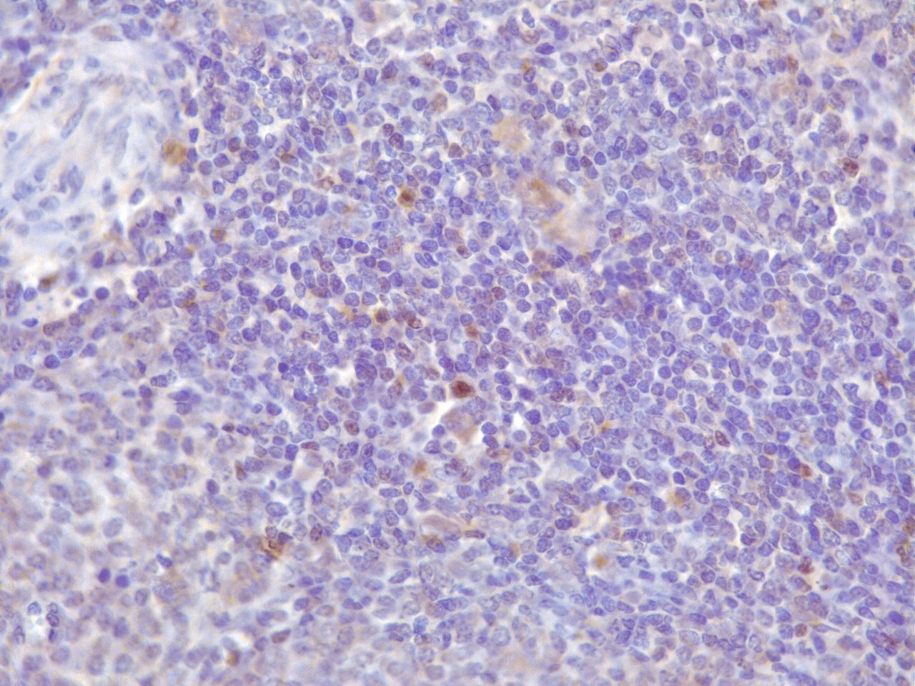
Immunohistochemistry: FoxP3 Antibody [NB100-39002] - Antibody was tested in mouse spleen using DAB with hematoxylin counterstain.![Immunohistochemistry-Paraffin: FoxP3 Antibody - BSA Free [NB100-39002]](https://resources.rndsystems.com/images/products/FoxP3-Antibody-Immunohistochemistry-Paraffin-NB100-39002-img0024.jpg "Immunohistochemistry-Paraffin: FoxP3 Antibody - BSA Free [NB100-39002]")
Immunohistochemistry-Paraffin: FoxP3 Antibody - BSA Free [NB100-39002]
Immunohistochemistry-Paraffin: FoxP3 Antibody [NB100-39002] - FFPE bovine spleen tissue. Deparaffinization and antigen retrieval was carried out with PTlink from Dako, citrate buffer pH 6. Anti-Foxp3 antibody was used at a 1:100 dilution. Secondary reagent: Envision, from Dako. Staining: DAB and hematoxylin. IHC-P image submitted by a verified customer review.![Western Blot: FoxP3 AntibodyBSA Free [NB100-39002]](https://resources.rndsystems.com/images/products/FoxP3-Antibody-Western-Blot-NB100-39002-img0018.jpg "Western Blot: FoxP3 AntibodyBSA Free [NB100-39002]")
Western Blot: FoxP3 AntibodyBSA Free [NB100-39002]
Western Blot: FoxP3 Antibody [NB100-39002] - Jurkat Tag cells were transfected with the expression vector (vector) or with the Foxp3 expression construct (Foxp3). 24 hours after transfection, cells were lysed and analyzed by Western blot with anti-Foxp3 antiserum (1:2000 dilution).![Immunocytochemistry/ Immunofluorescence: FoxP3 Antibody - BSA Free [NB100-39002]](https://resources.rndsystems.com/images/products/FoxP3-Antibody-Immunocytochemistry-Immunofluorescence-NB100-39002-img0012.jpg "Immunocytochemistry/ Immunofluorescence: FoxP3 Antibody - BSA Free [NB100-39002]")
Immunocytochemistry/ Immunofluorescence: FoxP3 Antibody - BSA Free [NB100-39002]
Immunocytochemistry/Immunofluorescence: FoxP3 Antibody [NB100-39002] - Transfectants were stained with anti-Foxp3 antibody and HRPO-conjugated (upper panel) or Cy3-conjugated (lower panels) secondary antibody. Representative images (x100) are shown.![Immunohistochemistry-Paraffin: FoxP3 Antibody - BSA Free [NB100-39002]](https://resources.rndsystems.com/images/products/FoxP3-Antibody-Immunohistochemistry-Paraffin-NB100-39002-img0015.jpg "Immunohistochemistry-Paraffin: FoxP3 Antibody - BSA Free [NB100-39002]")
Immunohistochemistry-Paraffin: FoxP3 Antibody - BSA Free [NB100-39002]
Immunohistochemistry-Paraffin: FoxP3 Antibody [NB100-39002] - Tissue section of human tonsil using 1:200 dilution of rabbit anti-FoxP3 antibody. The staining was developed with HRP-labeled anti-rabbit IgG secondary antibody and DAB reagent, and nuclei of cells were counterstained with hematoxylin. This FoxP3 antibody generated an expected and a specific nuclear staining of FOXP3 in a subset of cells which are potentially the Treg cells.![Flow Cytometry: FoxP3 Antibody - BSA Free [NB100-39002]](https://resources.rndsystems.com/images/products/FoxP3-Antibody-Flow-Cytometry-NB100-39002-img0013.jpg "Flow Cytometry: FoxP3 Antibody - BSA Free [NB100-39002]")
Flow Cytometry: FoxP3 Antibody - BSA Free [NB100-39002]
Flow Cytometry: FoxP3 Antibody [NB100-39002] - Transfectants were permeabilized and stained with anti-Foxp3 antibody and FITC-conjugated secondary antibody. Cells were analyzed by flow cytometry. Percentage of positively stained cells are shown.![Flow Cytometry: FoxP3 Antibody - BSA Free [NB100-39002]](https://resources.rndsystems.com/images/products/FoxP3-Antibody-Flow-Cytometry-NB100-39002-img0021.jpg "Flow Cytometry: FoxP3 Antibody - BSA Free [NB100-39002]")
Flow Cytometry: FoxP3 Antibody - BSA Free [NB100-39002]
Flow Cytometry: FoxP3 Antibody [NB100-39002] - An intracellular stain was performed on MCF7 cells with FoxP3 Antibody NB100-39002PE (blue) and a matched isotype control (orange). Cells were fixed with 4% PFA and then permeablized with 0.1% saponin. Cells were incubated in an antibody dilution of 5 ug/mL for 30 minutes at room temperature. Both antibodies were conjugated to Phycoerythrin.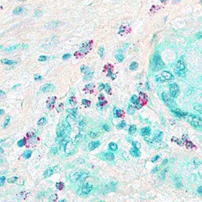

Immunohistochemistry: FoxP3 Antibody - BSA Free [NB100-39002] -
Effects of anaphylatoxins on transcription factor phosphorylation in ARPE-19 cells.(D) Immunofluorescence staining of FoxP3. Upper panel: Lewis rat (albino) eye with experimental uveitis; lower panel: normal rat eye. Asterisks mark immune cells infiltrating the retina from the choroid through the retinal pigment epithelium (RPE) in the eye with uveitis (clinical score of 2, histological score of 1).
FoxP3 in MCF7 Human Cell Line -
FoxP3 was detected in immersion fixed MCF7 human breast cancer cell line using Rabbit anti-FoxP3 Affinity Purified Polyclonal Antibody conjugated to DyLight 488 (Catalog # NB100-39002G) (green) at 10 µg/mL overnight at 4C. Cells were stained counterstained with DAPI (blue). Cells were imaged using a 100X objective and digitally deconvolved.
FoxP3 in MCF7 Human Cell Line.
FoxP3 was detected in immersion fixed MCF7 human breast cancer cell line using Rabbit anti-FoxP3 Affinity Purified Polyclonal Antibody conjugated to DyLight 550 (Catalog # NB100-39002R) (red) at 10 µg/mL overnight at 4C. Cells were stained counterstained with DAPI (blue). Cells were imaged using a 100X objective and digitally deconvolved.
Immunocytochemistry/ Immunofluorescence: FoxP3 Antibody - BSA Free [NB100-39002] -
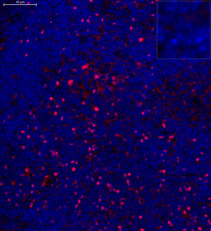
Immunocytochemistry/ Immunofluorescence: FoxP3 Antibody - BSA Free [NB100-39002] - CCR10-expressing CD4+CD25+FOXP3+ Treg cells are enriched in the spinal cord after SCI. (A, B) Representative flow cytometry dot plots (A) & percentage (B) of CD4+CD25+FOXP3+ cells among the CD4+ T cells in the spinal cord after sham or SCI surgery. Cells were gated on 7-AAD negative population & the numbers inside plots refer to % Treg cells (n=5). (C, D) Representative histogram of CCR10 (C) & CCR3 (D) expression in CD4+CD25+FOXP3+ cells as gated in (A). (E) CD4+CD25+FOXP3+ cells in the spinal cord from sham & SCI mice were sorted out & analyzed by Western blotting to detect the expression of CCR10 & CCR3. GAPDH was used as a loading control. (F) Double immunostaining of FOXP3 (red) & CCR10 (green) in the spinal cord sections after sham or SCI surgery. The merged images are also shown. Arrows indicate cells with positive staining & colocalization. Scale bar, 100 μm. Data are mean ± SD. The statistical analysis was performed using Student’s t-test. **, P<0.01. Image collected & cropped by CiteAb from the following publication (https://pubmed.ncbi.nlm.nih.gov/31557129), licensed under a CC-BY license. Not internally tested by Novus Biologicals.
Immunocytochemistry/ Immunofluorescence: FoxP3 Antibody - BSA Free [NB100-39002] -
Immunocytochemistry/ Immunofluorescence: FoxP3 Antibody - BSA Free [NB100-39002] - Effects of anaphylatoxins on transcription factor phosphorylation in ARPE-19 cells. (A–C) Protein phosphorylation of (A) CREB (Ser133); (B) FoxP3 (Ser418); & (C) FOXO1 (Ser256) was examined in ARPE-19 cells treated with C3a, C5a, or the combination of C3a/C5a at 1, 5, 10, & 15 min. Phosphorylation levels were normalized to beta -actin (relative p-protein levels), & slopes of the time course were determined. Anaphylatoxins were applied at concentrations of 300 nM (C3a) & 50 nM (C5a). Data are mean + SEM, n = 3, *p < 0.05, ***p < 0.001 (Student’s t-test). (D) Immunofluorescence staining of FoxP3. Upper panel: Lewis rat (albino) eye with experimental uveitis; lower panel: normal rat eye. Asterisks mark immune cells infiltrating the retina from the choroid through the retinal pigment epithelium (RPE) in the eye with uveitis (clinical score of 2, histological score of 1). Image collected & cropped by CiteAb from the following publication (http://journal.frontiersin.org/article/10.3389/fimmu.2017.00703/full), licensed under a CC-BY license. Not internally tested by Novus Biologicals.Applications for FoxP3 Antibody - BSA Free
Flow Cytometry
Immunoblotting
Immunocytochemistry/ Immunofluorescence
Immunohistochemistry
Immunohistochemistry-Paraffin
Single Cell Western
Western Blot
Reviewed Applications
Read 2 reviews rated 4 using NB100-39002 in the following applications:
Flow Cytometry Panel Builder
Bio-Techne Knows Flow Cytometry
Save time and reduce costly mistakes by quickly finding compatible reagents using the Panel Builder Tool.
Advanced Features
- Spectra Viewer - Custom analysis of spectra from multiple fluorochromes
- Spillover Popups - Visualize the spectra of individual fluorochromes
- Antigen Density Selector - Match fluorochrome brightness with antigen density

Formulation, Preparation, and Storage
Purification
Formulation
Format
Preservative
Concentration
Shipping
Stability & Storage
Background: FoxP3
Long Name
Alternate Names
Entrez Gene IDs
Gene Symbol
UniProt
Additional FoxP3 Products
Product Documents for FoxP3 Antibody - BSA Free
Certificate of Analysis
To download a Certificate of Analysis, please enter a lot or batch number in the search box below.
Product Specific Notices for FoxP3 Antibody - BSA Free
This product is for research use only and is not approved for use in humans or in clinical diagnosis. Primary Antibodies are guaranteed for 1 year from date of receipt.
Related Research Areas
Citations for FoxP3 Antibody - BSA Free
 Powered by Bioz
Customer Reviews for FoxP3 Antibody - BSA Free (2)
Have you used FoxP3 Antibody - BSA Free?
Submit a review and receive an Amazon gift card!
$25/€18/£15/$25CAN/¥2500 Yen for a review with an image
$10/€7/£6/$10CAN/¥1110 Yen for a review without an image
Submit a review
Customer Images
-
Application: Immunohistochemistry-ParaffinSample Tested: FFPE spleen tissueSpecies: Bovine, Sheep and GoatVerified Customer | Posted 10/21/2019FFPE bovine spleen tissueEpitope retrieval: PTlink from Dako with pH6 citrate buffer. Primary antibody dilution: 1:100 DAB staining and hematoxylin contrast.

-
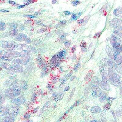
Application: Immunohistochemistry-ParaffinSample Tested: Mouse SpleenSpecies: MouseVerified Customer | Posted 10/06/2015Mouse spleen, cells in white pulp
There are no reviews that match your criteria.
Protocols
Find general support by application which include: protocols, troubleshooting, illustrated assays, videos and webinars.
- 7-Amino Actinomycin D (7-AAD) Cell Viability Flow Cytometry Protocol
- Antigen Retrieval Protocol (PIER)
- Antigen Retrieval for Frozen Sections Protocol
- Appropriate Fixation of IHC/ICC Samples
- Cellular Response to Hypoxia Protocols
- Chromogenic IHC Staining of Formalin-Fixed Paraffin-Embedded (FFPE) Tissue Protocol
- Chromogenic Immunohistochemistry Staining of Frozen Tissue
- ClariTSA™ Fluorophore Kits
- Detection & Visualization of Antibody Binding
- Extracellular Membrane Flow Cytometry Protocol
- Flow Cytometry Protocol for Cell Surface Markers
- Flow Cytometry Protocol for Staining Membrane Associated Proteins
- Flow Cytometry Staining Protocols
- Flow Cytometry Troubleshooting Guide
- Fluorescent IHC Staining of Frozen Tissue Protocol
- Graphic Protocol for Heat-induced Epitope Retrieval
- Graphic Protocol for the Preparation and Fluorescent IHC Staining of Frozen Tissue Sections
- Graphic Protocol for the Preparation and Fluorescent IHC Staining of Paraffin-embedded Tissue Sections
- Graphic Protocol for the Preparation of Gelatin-coated Slides for Histological Tissue Sections
- ICC Cell Smear Protocol for Suspension Cells
- ICC Immunocytochemistry Protocol Videos
- ICC for Adherent Cells
- IHC Sample Preparation (Frozen sections vs Paraffin)
- ISH-IHC Protocol for Chromogenic Detection on Formalin Fixed Paraffin Embedded (FFPE) Tissue
- Immunocytochemistry (ICC) Protocol
- Immunocytochemistry Troubleshooting
- Immunofluorescence of Organoids Embedded in Cultrex Basement Membrane Extract
- Immunofluorescent IHC Staining of Formalin-Fixed Paraffin-Embedded (FFPE) Tissue Protocol
- Immunohistochemistry (IHC) and Immunocytochemistry (ICC) Protocols
- Immunohistochemistry Frozen Troubleshooting
- Immunohistochemistry Paraffin Troubleshooting
- Immunoprecipitation Protocol
- Intracellular Flow Cytometry Protocol Using Alcohol (Methanol)
- Intracellular Flow Cytometry Protocol Using Detergents
- Intracellular Nuclear Staining Flow Cytometry Protocol Using Detergents
- Intracellular Staining Flow Cytometry Protocol Using Alcohol Permeabilization
- Intracellular Staining Flow Cytometry Protocol Using Detergents to Permeabilize Cells
- Preparing Samples for IHC/ICC Experiments
- Preventing Non-Specific Staining (Non-Specific Binding)
- Primary Antibody Selection & Optimization
- Propidium Iodide Cell Viability Flow Cytometry Protocol
- Protocol for Heat-Induced Epitope Retrieval (HIER)
- Protocol for Liperfluo
- Protocol for Making a 4% Formaldehyde Solution in PBS
- Protocol for VisUCyte™ HRP Polymer Detection Reagent
- Protocol for the Characterization of Human Th22 Cells
- Protocol for the Characterization of Human Th9 Cells
- Protocol for the Fluorescent ICC Staining of Cell Smears - Graphic
- Protocol for the Fluorescent ICC Staining of Cultured Cells on Coverslips - Graphic
- Protocol for the Preparation & Fixation of Cells on Coverslips
- Protocol for the Preparation and Chromogenic IHC Staining of Frozen Tissue Sections
- Protocol for the Preparation and Chromogenic IHC Staining of Frozen Tissue Sections - Graphic
- Protocol for the Preparation and Chromogenic IHC Staining of Paraffin-embedded Tissue Sections
- Protocol for the Preparation and Chromogenic IHC Staining of Paraffin-embedded Tissue Sections - Graphic
- Protocol for the Preparation and Fluorescent ICC Staining of Cells on Coverslips
- Protocol for the Preparation and Fluorescent ICC Staining of Non-adherent Cells
- Protocol for the Preparation and Fluorescent ICC Staining of Stem Cells on Coverslips
- Protocol for the Preparation and Fluorescent IHC Staining of Frozen Tissue Sections
- Protocol for the Preparation and Fluorescent IHC Staining of Paraffin-embedded Tissue Sections
- Protocol for the Preparation of Gelatin-coated Slides for Histological Tissue Sections
- Protocol for the Preparation of a Cell Smear for Non-adherent Cell ICC - Graphic
- Protocol: Annexin V and PI Staining by Flow Cytometry
- Protocol: Annexin V and PI Staining for Apoptosis by Flow Cytometry
- R&D Systems Quality Control Western Blot Protocol
- TUNEL and Active Caspase-3 Detection by IHC/ICC Protocol
- The Importance of IHC/ICC Controls
- Troubleshooting Guide: Fluorokine Flow Cytometry Kits
- Troubleshooting Guide: Immunohistochemistry
- Troubleshooting Guide: Western Blot Figures
- Western Blot Conditions
- Western Blot Protocol
- Western Blot Protocol for Cell Lysates
- Western Blot Troubleshooting
- Western Blot Troubleshooting Guide
- View all Protocols, Troubleshooting, Illustrated assays and Webinars
FAQs for FoxP3 Antibody - BSA Free
-
Q: Does FoxP3 antibodies comes in lyophilized form?
A: FoxP3 antibodies, aka JM2 IPEX, and Scurfin antibodies, have the following products in lyophilized form: NBP1-18319, AF3240.
-
Q: What can I use as positive controls for FOXP3 (NB100-39002) other than the mouse spleen tissue?
A: For FOXP3, the best tissues for detection of this protein would be appendix, lymph node, tonsil, spleen, or lung. You can see other tissue expression levels for this protein here: protein atlas site
-
Q: What is the dilution if it will be used in IHC? In other words, how many tests could obtained it used in paraffin based tissues?
A: The recommended dilution is 1:500 for IHC. It is difficult to predict how many tests can be made as the amount of antibody required will vary.
-
Q: What is the immunogen sequence of this FoxP3 antibody?
A: A sequence corresponding to amino acids 43 - 100 of Mouse FoxP3.
-
Q: What the theoretical molecular weight for FoxP3 antibodies?
A: The TMW of FoxP3 antibodies is approximately 47 - 52 kDa.
-
Q: Does FoxP3 antibodies comes in lyophilized form?
A: FoxP3 antibodies, aka JM2 IPEX, and Scurfin antibodies, have the following products in lyophilized form: NBP1-18319, AF3240.
-
Q: What can I use as positive controls for FOXP3 (NB100-39002) other than the mouse spleen tissue?
A: For FOXP3, the best tissues for detection of this protein would be appendix, lymph node, tonsil, spleen, or lung. You can see other tissue expression levels for this protein here: protein atlas site
-
Q: What is the dilution if it will be used in IHC? In other words, how many tests could obtained it used in paraffin based tissues?
A: The recommended dilution is 1:500 for IHC. It is difficult to predict how many tests can be made as the amount of antibody required will vary.
-
Q: What is the immunogen sequence of this FoxP3 antibody?
A: A sequence corresponding to amino acids 43 - 100 of Mouse FoxP3.
-
Q: What the theoretical molecular weight for FoxP3 antibodies?
A: The TMW of FoxP3 antibodies is approximately 47 - 52 kDa.
Associated Pathways

