FoxP3 Antibody - BSA Free
Novus Biologicals | Catalog # NB600-245
![Immunohistochemistry-Frozen: FoxP3 Antibody - BSA Free [NB600-245]](https://resources.rndsystems.com/images/products/FoxP3-Antibody-Immunocytochemistry-Immunofluorescence-NB600-245-img0013.jpg "Immunohistochemistry-Frozen: FoxP3 Antibody - BSA Free [NB600-245]")
Key Product Details
Species Reactivity
Validated:
Cited:
Applications
Validated:
Cited:
Label
Antibody Source
Format
Product Specifications
Immunogen
Reactivity Notes
Clonality
Host
Isotype
Theoretical MW
Disclaimer note: The observed molecular weight of the protein may vary from the listed predicted molecular weight due to post translational modifications, post translation cleavages, relative charges, and other experimental factors.
Scientific Data Images for FoxP3 Antibody - BSA Free
Immunohistochemistry-Frozen: FoxP3 Antibody - BSA Free [NB600-245]
Immunohistochemistry-Frozen: FoxP3 Antibody - BSA Free [NB600-245] - FoxP3 Antibody [NB600-245] - Healthy thymus from a C57BL/6 mouse stained with FoxP3 and CD4.![Immunohistochemistry-Frozen: FoxP3 Antibody - BSA Free [NB600-245]](https://resources.rndsystems.com/images/products/FoxP3-Antibody-Immunocytochemistry-Immunofluorescence-NB600-245-img0012.jpg "Immunohistochemistry-Frozen: FoxP3 Antibody - BSA Free [NB600-245]")
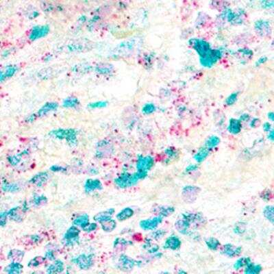
Immunohistochemistry-Frozen: FoxP3 Antibody - BSA Free [NB600-245]
Immunohistochemistry-Frozen: FoxP3 Antibody - BSA Free [NB600-245] - FoxP3 Antibody [NB600-245] - Healthy thymus from a C57BL/6 mouse incubated only with CD4 and no Foxp3.![Immunohistochemistry-Paraffin: FoxP3 Antibody - BSA Free [NB600-245]](https://resources.rndsystems.com/images/products/FoxP3-Antibody-Immunohistochemistry-Paraffin-NB600-245-img0011.jpg "Immunohistochemistry-Paraffin: FoxP3 Antibody - BSA Free [NB600-245]")
Immunohistochemistry-Paraffin: FoxP3 Antibody - BSA Free [NB600-245]
Immunohistochemistry-Paraffin: FoxP3 Antibody [NB600-245] - Fox P3 was detected in immersion fixed paraffin-embedded sections of human tonsil using rabbit anti-human antibody (Catalog # NB600-245) at 1:100 dilution overnight at 4C. Tissue was stained using the VisuCyte anti-rabbit HRP polymer detection reagent (Catalog # VC003) with DAB chromogen (brown) and counterstained with hematoxylin (blue).Images may not be copied, printed or otherwise disseminated without express written permission of Novus Biologicals a bio-techne brand.
![Western Blot: FoxP3 AntibodyBSA Free [NB600-245]](https://resources.rndsystems.com/images/products/FOXP3-Antibody-Western-Blot-NB600-245-img0009.jpg "Western Blot: FoxP3 AntibodyBSA Free [NB600-245]")
Western Blot: FoxP3 AntibodyBSA Free [NB600-245]
Western Blot: FOXP3 Antibody [NB600-245] - Detection of human FOXP3 using NB 600-245. Lane 1: Human CD4+CD25+ PBL, lane 2: HEK293T transfected with human Foxp3 cDNA, lane 3: 293/mouse foxp3, lane 4: 293/empty vector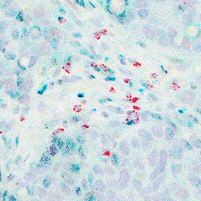

FoxP3 in MCF7 Human Cell Line.
FoxP3 was detected in immersion fixed MCF7 human breast cancer cell line using Rabbit anti-FoxP3 Affinity Purified Polyclonal Antibody conjugated to Alexa Fluor® 647 (Catalog # NB600-245AF647) (light blue) at 10 µg/mL overnight at 4C. Cells were stained counterstained with DAPI (blue). Cells were imaged using a 100X objective and digitally deconvolved.Applications for FoxP3 Antibody - BSA Free
Immunocytochemistry/ Immunofluorescence
Immunohistochemistry
Immunohistochemistry-Frozen
Immunohistochemistry-Paraffin
Western Blot
Formulation, Preparation, and Storage
Purification
Formulation
Format
Preservative
Concentration
Shipping
Stability & Storage
Background: FoxP3
Long Name
Alternate Names
Gene Symbol
UniProt
Additional FoxP3 Products
Product Documents for FoxP3 Antibody - BSA Free
Certificate of Analysis
To download a Certificate of Analysis, please enter a lot or batch number in the search box below.
Product Specific Notices for FoxP3 Antibody - BSA Free
This product is for research use only and is not approved for use in humans or in clinical diagnosis. Primary Antibodies are guaranteed for 1 year from date of receipt.
Related Research Areas
Citations for FoxP3 Antibody - BSA Free
 Powered by Bioz
Customer Reviews for FoxP3 Antibody - BSA Free
There are currently no reviews for this product. Be the first to review FoxP3 Antibody - BSA Free and earn rewards!
Have you used FoxP3 Antibody - BSA Free?
Submit a review and receive an Amazon gift card!
$25/€18/£15/$25CAN/¥2500 Yen for a review with an image
$10/€7/£6/$10CAN/¥1110 Yen for a review without an image
Submit a review
Protocols
View specific protocols for FoxP3 Antibody - BSA Free (NB600-245):
Protocol for Foxp3 staining.
- Cut 5 mm sections in a cryostat and fix them in cold acetone for 10 minutes
- Dry the slides completely at room temperature (30 minutes)
- Permeabilize tissues by immersing them in 0.1% saponin, 3 times (5 minutes each time)
-Block unspecific binding to the slides using 5% normal donkey serum, 5% normal rat serum and Fc block (1:500). Everything has to be dissolved in 0.1% saponin
-Incubate slides in a wet chamber at RT for 30 minutes
-Dip off the blocking solution from slides and add streptavidin (0.1 mg/ml) for 15 minutes at RT
-Wash one time with 0.1% saponin
-Add a similar volume of biotin (0.1 mg/ml) and incubate 15 min at RT
- Wash again with 0.1% saponin
- Add rabbit anti-Foxp3 (1:150) and biotin-rat anti-mouse CD4 (1:100) and incubate ON at RT
- Next day: Wash slides with PBS and add SA-594 or SA-488 (dissolved in PBS) to detect CD4 T cells (Dilution 1:500 from molecular probes). Incubate 1 h at RT
-Wash one time with 0.1% saponin and add biotin-donkey anti rabbit (Fab, Jackson Immunoresearch). Secondary antibody is diluted in PBS 1:500 and incubated by 3 hrs.
- Wash with saponin again and add SA-488 or SA-594 (1:500 in PBS). Incubate 1 h at RT
- Wash with PBS 3 times (10 minutes each wash)
- Mount slides with prolong gold antifade (Molecular probes)
Control: Incubated only with CD4 and no Foxp3
Tissue: thymus from an Influenza-infected mouse or thymus from healthy mice (C57BL/6)
Antigen Unmasking:
Bring slides to a boil in 10 mM sodium citrate buffer (pH 6.0) then maintain at a sub-boiling temperature for 10 minutes. Cool slides on bench-top for 30 minutes (keep slides in the sodium citrate buffer at all times).
Staining:
1. Wash sections in deionized water three times for 5 minutes each.
2. Wash sections in PBS for 5 minutes.
3. Block each section with 100-400 ul blocking solution (1% BSA in PBS) for 1 hour at room temperature.
4. Remove blocking solution and add 100-400 ul diluted primary antibody. Incubate overnight at 4 C.
5. Remove antibody solution and wash sections in wash buffer three times for 5 minutes each.
6. Add 100-400 ul HRP polymer conjugated secondary antibody. Incubate 30 minutes at room temperature.
7. Wash sections three times in wash buffer for 5 minutes each.
8. Add 100-400 ul DAB substrate to each section and monitor staining closely.
9. As soon as the sections develop, immerse slides in deionized water.
10. Counterstain sections in hematoxylin.
11. Wash sections in deionized water two times for 5 minutes each.
12. Dehydrate sections.
13. Mount coverslips.
1. Perform SDS-PAGE on samples to be analyzed, loading 10-25 ug of total protein per lane.
2. Transfer proteins to PVDF membrane according to the instructions provided by the manufacturer of the membrane and transfer apparatus.
3. Stain the membrane with Ponceau S (or similar product) to assess transfer success, and mark molecular weight standards where appropriate.
4. Rinse the blot TBS -0.05% Tween 20 (TBST).
5. Block the membrane in 5% Non-fat milk in TBST (blocking buffer) for at least 1 hour.
6. Wash the membrane in TBST three times for 10 minutes each.
7. Dilute primary antibody in blocking buffer and incubate overnight at 4C with gentle rocking.
8. Wash the membrane in TBST three times for 10 minutes each.
9. Incubate the membrane in diluted HRP conjugated secondary antibody in blocking buffer (as per manufacturer's instructions) for 1 hour at room temperature.
10. Wash the blot in TBST three times for 10 minutes each (this step can be repeated as required to reduce background).
11. Apply the detection reagent of choice in accordance with the manufacturer's instructions.
Find general support by application which include: protocols, troubleshooting, illustrated assays, videos and webinars.
- Antigen Retrieval Protocol (PIER)
- Antigen Retrieval for Frozen Sections Protocol
- Appropriate Fixation of IHC/ICC Samples
- Cellular Response to Hypoxia Protocols
- Chromogenic IHC Staining of Formalin-Fixed Paraffin-Embedded (FFPE) Tissue Protocol
- Chromogenic Immunohistochemistry Staining of Frozen Tissue
- ClariTSA™ Fluorophore Kits
- Detection & Visualization of Antibody Binding
- Fluorescent IHC Staining of Frozen Tissue Protocol
- Graphic Protocol for Heat-induced Epitope Retrieval
- Graphic Protocol for the Preparation and Fluorescent IHC Staining of Frozen Tissue Sections
- Graphic Protocol for the Preparation and Fluorescent IHC Staining of Paraffin-embedded Tissue Sections
- Graphic Protocol for the Preparation of Gelatin-coated Slides for Histological Tissue Sections
- ICC Cell Smear Protocol for Suspension Cells
- ICC Immunocytochemistry Protocol Videos
- ICC for Adherent Cells
- IHC Sample Preparation (Frozen sections vs Paraffin)
- ISH-IHC Protocol for Chromogenic Detection on Formalin Fixed Paraffin Embedded (FFPE) Tissue
- Immunocytochemistry (ICC) Protocol
- Immunocytochemistry Troubleshooting
- Immunofluorescence of Organoids Embedded in Cultrex Basement Membrane Extract
- Immunofluorescent IHC Staining of Formalin-Fixed Paraffin-Embedded (FFPE) Tissue Protocol
- Immunohistochemistry (IHC) and Immunocytochemistry (ICC) Protocols
- Immunohistochemistry Frozen Troubleshooting
- Immunohistochemistry Paraffin Troubleshooting
- Preparing Samples for IHC/ICC Experiments
- Preventing Non-Specific Staining (Non-Specific Binding)
- Primary Antibody Selection & Optimization
- Protocol for Heat-Induced Epitope Retrieval (HIER)
- Protocol for Making a 4% Formaldehyde Solution in PBS
- Protocol for VisUCyte™ HRP Polymer Detection Reagent
- Protocol for the Fluorescent ICC Staining of Cell Smears - Graphic
- Protocol for the Fluorescent ICC Staining of Cultured Cells on Coverslips - Graphic
- Protocol for the Preparation & Fixation of Cells on Coverslips
- Protocol for the Preparation and Chromogenic IHC Staining of Frozen Tissue Sections
- Protocol for the Preparation and Chromogenic IHC Staining of Frozen Tissue Sections - Graphic
- Protocol for the Preparation and Chromogenic IHC Staining of Paraffin-embedded Tissue Sections
- Protocol for the Preparation and Chromogenic IHC Staining of Paraffin-embedded Tissue Sections - Graphic
- Protocol for the Preparation and Fluorescent ICC Staining of Cells on Coverslips
- Protocol for the Preparation and Fluorescent ICC Staining of Non-adherent Cells
- Protocol for the Preparation and Fluorescent ICC Staining of Stem Cells on Coverslips
- Protocol for the Preparation and Fluorescent IHC Staining of Frozen Tissue Sections
- Protocol for the Preparation and Fluorescent IHC Staining of Paraffin-embedded Tissue Sections
- Protocol for the Preparation of Gelatin-coated Slides for Histological Tissue Sections
- Protocol for the Preparation of a Cell Smear for Non-adherent Cell ICC - Graphic
- R&D Systems Quality Control Western Blot Protocol
- TUNEL and Active Caspase-3 Detection by IHC/ICC Protocol
- The Importance of IHC/ICC Controls
- Troubleshooting Guide: Immunohistochemistry
- Troubleshooting Guide: Western Blot Figures
- Western Blot Conditions
- Western Blot Protocol
- Western Blot Protocol for Cell Lysates
- Western Blot Troubleshooting
- Western Blot Troubleshooting Guide
- View all Protocols, Troubleshooting, Illustrated assays and Webinars
FAQs for FoxP3 Antibody - BSA Free
-
Q: Does FoxP3 antibodies comes in lyophilized form?
A: FoxP3 antibodies, aka JM2 IPEX, and Scurfin antibodies, have the following products in lyophilized form: NBP1-18319, AF3240.
-
Q: What is the immunogen sequence of this FoxP3 antibody?
A: A synthetic sequence of amino acids 400 - 431 within the C-terminal region of Human FoxP3.
-
Q: What the theoretical molecular weight for FoxP3 antibodies?
A: The TMW of FoxP3 antibodies is approximately 47 - 52 kDa.
-
Q: Does FoxP3 antibodies comes in lyophilized form?
A: FoxP3 antibodies, aka JM2 IPEX, and Scurfin antibodies, have the following products in lyophilized form: NBP1-18319, AF3240.
-
Q: What is the immunogen sequence of this FoxP3 antibody?
A: A synthetic sequence of amino acids 400 - 431 within the C-terminal region of Human FoxP3.
-
Q: What the theoretical molecular weight for FoxP3 antibodies?
A: The TMW of FoxP3 antibodies is approximately 47 - 52 kDa.
Associated Pathways

