![Western Blot: Furin Antibody [NB100-1903]](https://resources.rndsystems.com/images/products/Furin-Antibody-Western-Blot-NB100-1903-img0008.jpg "Western Blot: Furin Antibody [NB100-1903]")
Loading...
Key Product Details
Species Reactivity
Validated:
Human, Mouse, Rat, Porcine, Canine, Hamster, Primate
Cited:
Human, Primate
Applications
Validated:
Immunohistochemistry, Immunohistochemistry-Paraffin, Western Blot, Block/Neutralize, Flow Cytometry, Immunocytochemistry/ Immunofluorescence, Immunoprecipitation
Cited:
Western Blot
Label
Unconjugated
Antibody Source
Polyclonal Rabbit IgG
Loading...
Product Specifications
Immunogen
Synthetic peptide corresponding to residues R(780) G E R T A F I K D Q S A L(793) of human Furin.
Epitope
Amino acids 781-794.
Reactivity Notes
Hamster reactivity reported in scientific literature (PMID: 16030016). Porcine reactivity reported in scientific literature (PMID: 14581457). Rat reactivity reported in scientific literature (PMID: 11696560). Primate reactivity reported in scientific literature (PMID: 11799113).
Marker
TGN Marker
Specificity
Detects Furin convertase from canine and mouse cells as well as transfected human Furin. This does not detect endogenous Furin from BSC-40, HeLa, J774A.1 BPAEC, or CHO cells nor from rat skeletal muscle, spleen, kidney, ovary, testes, heart, or brain tissues.
Clonality
Polyclonal
Host
Rabbit
Isotype
IgG
Scientific Data Images for Furin Antibody
Western Blot: Furin Antibody [NB100-1903]
Western Blot: Furin Antibody [NB100-1903] - Analysis was performed on membrane enriched extracts (30 ug lysate) of HeLa (Lane 1), HEK 293 (Lane 2), K-562 (Lane 3), SH-SY5Y (Lane 4) and 10uL conditioned media from HeLa cell line (Lane 5).![Immunocytochemistry/ Immunofluorescence: Furin Antibody [NB100-1903]](https://resources.rndsystems.com/images/products/Furin-Antibody-Immunocytochemistry-Immunofluorescence-NB100-1903-img0005.jpg "Immunocytochemistry/ Immunofluorescence: Furin Antibody [NB100-1903]")
Immunocytochemistry/ Immunofluorescence: Furin Antibody [NB100-1903]
Immunocytochemistry/Immunofluorescence: Furin Antibody [NB100-1903] - Immunolocalization of endogenous furin in mouse 3T3 cells.![Flow Cytometry: Furin Antibody [NB100-1903]](https://resources.rndsystems.com/images/products/Furin-Antibody-Flow-Cytometry-NB100-1903-img0007.jpg "Flow Cytometry: Furin Antibody [NB100-1903]")
Flow Cytometry: Furin Antibody [NB100-1903]
Flow Cytometry: Furin Antibody [NB100-1903] - Flow cytometry analysis of Furin Convertase was done on HeLa cells. Cells were fixed with 70% ethanol for 10 minutes, permeabilized with 0.25% Triton (R) X-100 for 20 minutes, and blocked with 5% BSA for 30 minutes at room temperature. Cells were labeled with Furin Convertase Rabbit Polyclonal Antibody or with rabbit isotype control (pink histogram) at 3-5 ug/million cells in 2.5% BSA. After incubation at room temperature for 2 hours, the cells were labeled with Alexa Fluor (R) 488 Goat Anti-Rabbit Secondary Antibody (A11008) at a dilution of 1:400 for 30 minutes at room temperature. The representative 10, 000 cells were acquired and analyzed for each sample using an Attune (R) Acoustic Focusing Cytometer. The purple histogram represents unstained control cells and the green histogram represents no-primary-antibody control.
Western Blot: Furin Antibody [NB100-1903] -
Induction of furin expression by TGF-beta 1. (A) HEK293T, (B) HeLa, (C) Vero E6, (D) Caco-2, (E) Huh-7, and (F) Calu-3 were incubated for 18 h with indicated concentrations of TGF-beta 1 or the solvent control 4 mM HCl 0.1% bovine serum albumin (0 ng/mL TGF-beta 1). Western blot analyses were performed to detect furin and beta -actin. Protein levels of furin and beta -actin were quantified via densitometric analyses utilizing the Image Lab Software. Protein levels without the addition of TGF-beta 1 (0 ng/mL) were set to 1. Image collected and cropped by CiteAb from the following open publication (https://pubmed.ncbi.nlm.nih.gov/35746781), licensed under a CC-BY license. Not internally tested by Novus Biologicals.
Western Blot: Furin Antibody [NB100-1903] -
Decrease of furin expression by the ALK5 inhibitor SB431542. (A) HEK293T, (B) HeLa, (C) Vero E6, (D) CaCo-2, (E) Huh-7, and (F) Calu-3 were incubated for 24 h with the indicated concentrations of SB431542 (SB) or the solvent control DMSO (0 uM). Western Blot analyses were performed to detect furin and beta -actin. Protein levels of furin and beta -actin were quantified via densitometric analyses utilizing the Image Lab Software. Protein levels without SB (0 uM) were set to 1. Image collected and cropped by CiteAb from the following open publication (https://pubmed.ncbi.nlm.nih.gov/35746781), licensed under a CC-BY license. Not internally tested by Novus Biologicals.
Western Blot: Furin Antibody [NB100-1903] -
Induction of furin expression by TGF-beta 1. (A) HEK293T, (B) HeLa, (C) Vero E6, (D) Caco-2, (E) Huh-7, and (F) Calu-3 were incubated for 18 h with indicated concentrations of TGF-beta 1 or the solvent control 4 mM HCl 0.1% bovine serum albumin (0 ng/mL TGF-beta 1). Western blot analyses were performed to detect furin and beta -actin. Protein levels of furin and beta -actin were quantified via densitometric analyses utilizing the Image Lab Software. Protein levels without the addition of TGF-beta 1 (0 ng/mL) were set to 1. Image collected and cropped by CiteAb from the following open publication (https://pubmed.ncbi.nlm.nih.gov/35746781), licensed under a CC-BY license. Not internally tested by Novus Biologicals.
Western Blot: Furin Antibody [NB100-1903] -
Decrease of furin expression by the ALK5 inhibitor SB431542. (A) HEK293T, (B) HeLa, (C) Vero E6, (D) CaCo-2, (E) Huh-7, and (F) Calu-3 were incubated for 24 h with the indicated concentrations of SB431542 (SB) or the solvent control DMSO (0 uM). Western Blot analyses were performed to detect furin and beta -actin. Protein levels of furin and beta -actin were quantified via densitometric analyses utilizing the Image Lab Software. Protein levels without SB (0 uM) were set to 1. Image collected and cropped by CiteAb from the following open publication (https://pubmed.ncbi.nlm.nih.gov/35746781), licensed under a CC-BY license. Not internally tested by Novus Biologicals.
Western Blot: Furin Antibody [NB100-1903] -
Prevention of TGF-beta 1-induced furin expression by the ALK5 inhibitor SB431542. (A) Huh-7 and (B) Calu-3 were incubated for 24 h with 10 uM SB431542 or the solvent control DMSO and subsequently treated for 18 h with the indicated concentrations of TGF-beta 1, the solvent control 4 mM HCl 0.1% bovine serum albumin (0 ng/mL), or were left untreated (−). Western Blot analyses were performed to detect furin and beta -actin. Protein levels of furin and beta -actin were quantified via densitometric analyses utilizing the Image Lab Software. Protein levels without the addition of SB431542 and TGF-beta 1 were set to 1. Image collected and cropped by CiteAb from the following open publication (https://pubmed.ncbi.nlm.nih.gov/35746781), licensed under a CC-BY license. Not internally tested by Novus Biologicals.
Western Blot: Furin Antibody [NB100-1903] -
Induction of furin expression by TGF-beta 1. (A) HEK293T, (B) HeLa, (C) Vero E6, (D) Caco-2, (E) Huh-7, and (F) Calu-3 were incubated for 18 h with indicated concentrations of TGF-beta 1 or the solvent control 4 mM HCl 0.1% bovine serum albumin (0 ng/mL TGF-beta 1). Western blot analyses were performed to detect furin and beta -actin. Protein levels of furin and beta -actin were quantified via densitometric analyses utilizing the Image Lab Software. Protein levels without the addition of TGF-beta 1 (0 ng/mL) were set to 1. Image collected and cropped by CiteAb from the following open publication (https://pubmed.ncbi.nlm.nih.gov/35746781), licensed under a CC-BY license. Not internally tested by Novus Biologicals.
Western Blot: Furin Antibody [NB100-1903] -
Decrease of furin expression by the ALK5 inhibitor SB431542. (A) HEK293T, (B) HeLa, (C) Vero E6, (D) CaCo-2, (E) Huh-7, and (F) Calu-3 were incubated for 24 h with the indicated concentrations of SB431542 (SB) or the solvent control DMSO (0 uM). Western Blot analyses were performed to detect furin and beta -actin. Protein levels of furin and beta -actin were quantified via densitometric analyses utilizing the Image Lab Software. Protein levels without SB (0 uM) were set to 1. Image collected and cropped by CiteAb from the following open publication (https://pubmed.ncbi.nlm.nih.gov/35746781), licensed under a CC-BY license. Not internally tested by Novus Biologicals.
Western Blot: Furin Antibody [NB100-1903] -
Decrease of furin expression by the ALK5 inhibitor SB431542. (A) HEK293T, (B) HeLa, (C) Vero E6, (D) CaCo-2, (E) Huh-7, and (F) Calu-3 were incubated for 24 h with the indicated concentrations of SB431542 (SB) or the solvent control DMSO (0 uM). Western Blot analyses were performed to detect furin and beta -actin. Protein levels of furin and beta -actin were quantified via densitometric analyses utilizing the Image Lab Software. Protein levels without SB (0 uM) were set to 1. Image collected and cropped by CiteAb from the following open publication (https://pubmed.ncbi.nlm.nih.gov/35746781), licensed under a CC-BY license. Not internally tested by Novus Biologicals.Applications for Furin Antibody
Application
Recommended Usage
Flow Cytometry
3 - 5 ug
Immunocytochemistry/ Immunofluorescence
1:10 - 1:500
Immunohistochemistry-Paraffin
1:10 - 1:500
Immunoprecipitation
1:100
Western Blot
1:1000
Application Notes
Blocking usage was reported in scientific literature (PMID: 11741999).
Reviewed Applications
Read 1 review rated 5 using NB100-1903 in the following applications:
Flow Cytometry Panel Builder
Bio-Techne Knows Flow Cytometry
Save time and reduce costly mistakes by quickly finding compatible reagents using the Panel Builder Tool.
Advanced Features
- Spectra Viewer - Custom analysis of spectra from multiple fluorochromes
- Spillover Popups - Visualize the spectra of individual fluorochromes
- Antigen Density Selector - Match fluorochrome brightness with antigen density

Formulation, Preparation, and Storage
Purification
Immunogen affinity purified
Formulation
PBS with 1 mg/ml BSA
Preservative
0.05% Sodium Azide
Concentration
2 mg/ml
Shipping
The product is shipped with polar packs. Upon receipt, store it immediately at the temperature recommended below.
Stability & Storage
Store at -20C. Avoid freeze-thaw cycles.
Background: Furin
Proteolytic cleavage regulates several physiological processes in both health and disease (3). Abnormal activity or mutations in proteases, including furin, is associated with pathologies and diseases including cancer, cardiovascular disorders, diabetes, inflammation, neurological diseases, and autoimmune diseases (3). As mentioned above, furin also acts upon bacterial substrates, including anthrax and Shiga toxin, and many virus families such as Herpes-, Flavi-, and Corona-, leading to host infections. Furthermore, the novel coronavirus disease 2019 (COVID-19) is caused by severe acute respiratory syndrome coronavirus-2 (SARS-CoV-2) present with a S-spike protein that is cleaved by PCs, including furin, at the S1/S2 cleavage site (5, 6). The cleavage allows the SARS-CoV-2 to then attach to the angiotensin-converting enzyme 2 (ACE2) receptor via the S1 domain and the cellular membrane via the S2 domain (5, 6). Although COVID-19 patients mostly present with respiratory symptoms, a variety of other systems are affected including cardiovascular, gastrointestinal (GI), and the liver (5-7). It is suggested that the S1/furin/ACE2 interaction promotes SARS-CoV-2 infection leading to the harmful symptoms and reactions in patients (5, 6). Cardiovascular disease is a common comorbidity in patients, along with hypertension, myocardial damage, and heart palpitations (Ming). Further evidence of furin being a risk factor for infection is the high levels of furin present in the blood of heart failure patients (5). Similarly, the small bowel may be another interaction site for infection as it is rich in furin and the intestinal enterocytes have many ACE2 receptors (6). Furin is also highly expressed in the liver and hepatocytes and cholangiocytes of the liver present ACE2 receptors (3, 7). Studies have shown that up one-third of COVID-19 patients experience GI symptoms which range from diarrhea and loss of appetite to abdominal cramping and bloody stool (6, 7). Additionally, some patients displayed abnormal liver enzyme levels (7). It has been suggested that a possible therapeutic strategy for treating those infected with SARS-CoV-2 is pharmacologically or immunologically modulating furin or ACE2 binding sites to combat COVID-19 infection (3, 5).
References
1. Thomas, G. (2002). Furin at the cutting edge: from protein traffic to emryogenesis and disease. Nature Rev. Mol. Cell Biol. https://doi.org/10.1038/nrm934
2. Zhou A., Paquet, L., & Mains, R.E. (1995). Structural elements that direct specific processing of different mammalian subtilisin-like prohormone convertases. J Biol Chem. https://doi.org/10.1074/jbc.270.37.21509
3. Braun E., & Sauter, D. (2019). Furin-mediated protein processing in infectious diseases and cancer. Clin Transl Immunology. https://doi:10.1002/cti2.1073
4. Atlas of Genetics and Cytogenetics in Oncology and Haematology, FURIN
5. Ming, Y. & Qiang, L. (2020). Involvement of Spike Protein, Furin, and ACE2 in SARS-CoV-2-Related Cardiovascular Complications. SN Compr. Clin. Med. https://doi.org/10.1007/s42399-020-00400-2
6. Monkemuller, K., Fry, L., & Rickes, S. (2020). COVID-19, coronavirus, SARS-CoV-2 and the small bowel. Rev Esp Enferm Dig. https://doi:10.17235/reed.2020.7137/2020
7. Agarwal, A., Chen, A., Ravindran, N., To, C., & Thuluvath, P.J. (2020). Gastrointestinal and Liver Manifestations of COVID-19. J Clin Exp Hepatol. https://doi:10.1016/j.jceh.2020.03.001
Additional Furin Products
Product Documents for Furin Antibody
Certificate of Analysis
To download a Certificate of Analysis, please enter a lot or batch number in the search box below.
Product Specific Notices for Furin Antibody
This product is for research use only and is not approved for use in humans or in clinical diagnosis. Primary Antibodies are guaranteed for 1 year from date of receipt.
Related Research Areas
Citations for Furin Antibody
 Powered by Bioz
Customer Reviews for Furin Antibody (1)
5 out of 5
1 Customer Rating
Have you used Furin Antibody?
Submit a review and receive an Amazon gift card!
$25/€18/£15/$25CAN/¥2500 Yen for a review with an image
$10/€7/£6/$10CAN/¥1110 Yen for a review without an image
Submit a review
Customer Images
Showing
1
-
1 of
1 review
Showing All
Filter By:
-
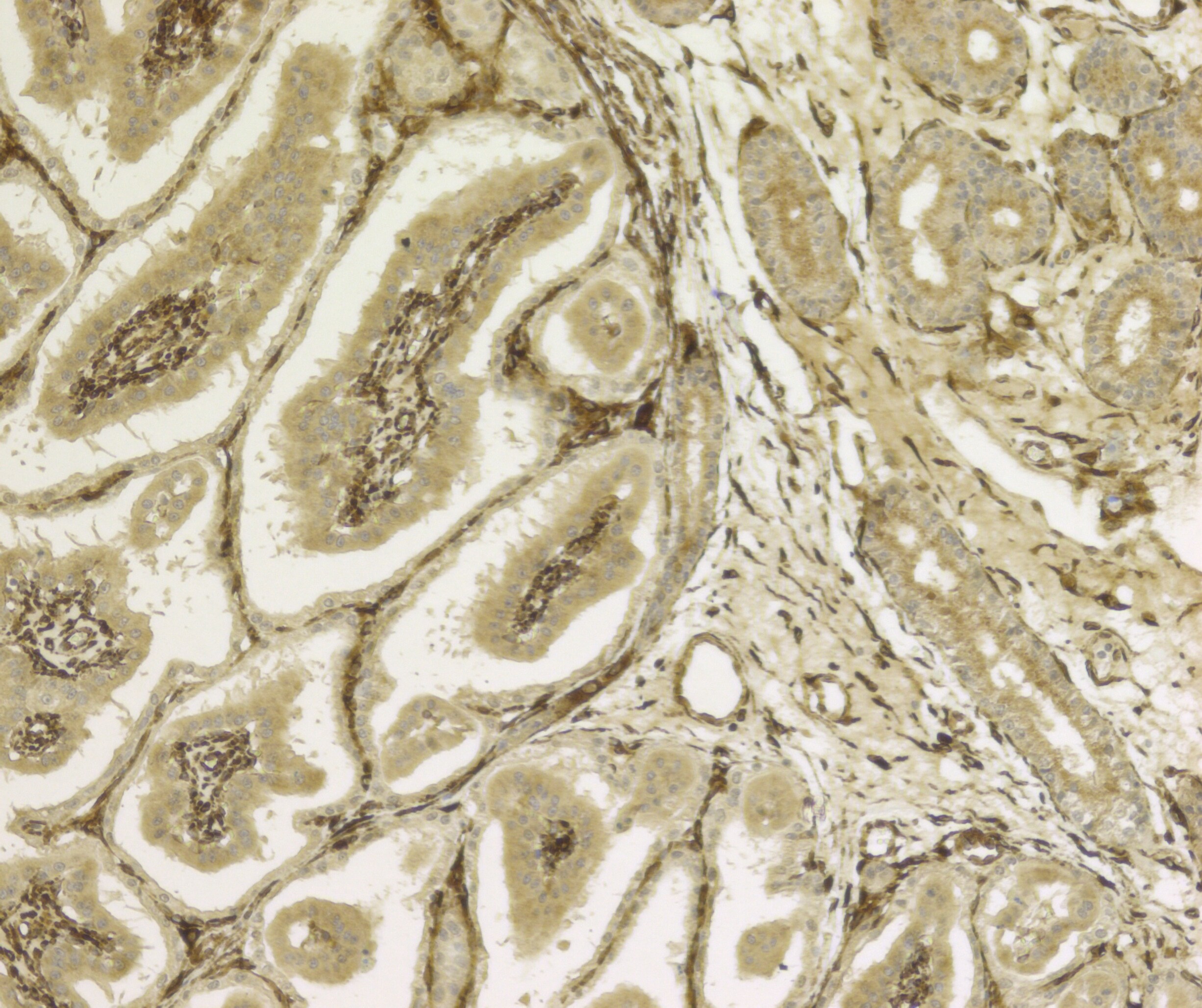
Application: ImmunocytochemistrySample Tested: Placental tissueSpecies: EquineVerified Customer | Posted 05/13/2017Equine placental tissue. Strong staining in the fetal microcotyledons. Moderate staining in endometrium.1:200 dilution. Heat based antigen retrieval using EDTA based solution. No staining on negative when the primary antibody was omitted.

There are no reviews that match your criteria.
Protocols
Find general support by application which include: protocols, troubleshooting, illustrated assays, videos and webinars.
- 7-Amino Actinomycin D (7-AAD) Cell Viability Flow Cytometry Protocol
- Antigen Retrieval Protocol (PIER)
- Antigen Retrieval for Frozen Sections Protocol
- Appropriate Fixation of IHC/ICC Samples
- Cellular Response to Hypoxia Protocols
- Chromogenic IHC Staining of Formalin-Fixed Paraffin-Embedded (FFPE) Tissue Protocol
- Chromogenic Immunohistochemistry Staining of Frozen Tissue
- ClariTSA™ Fluorophore Kits
- Detection & Visualization of Antibody Binding
- Extracellular Membrane Flow Cytometry Protocol
- Flow Cytometry Protocol for Cell Surface Markers
- Flow Cytometry Protocol for Staining Membrane Associated Proteins
- Flow Cytometry Staining Protocols
- Flow Cytometry Troubleshooting Guide
- Fluorescent IHC Staining of Frozen Tissue Protocol
- Graphic Protocol for Heat-induced Epitope Retrieval
- Graphic Protocol for the Preparation and Fluorescent IHC Staining of Frozen Tissue Sections
- Graphic Protocol for the Preparation and Fluorescent IHC Staining of Paraffin-embedded Tissue Sections
- Graphic Protocol for the Preparation of Gelatin-coated Slides for Histological Tissue Sections
- ICC Cell Smear Protocol for Suspension Cells
- ICC Immunocytochemistry Protocol Videos
- ICC for Adherent Cells
- IHC Sample Preparation (Frozen sections vs Paraffin)
- Immunocytochemistry (ICC) Protocol
- Immunocytochemistry Troubleshooting
- Immunofluorescence of Organoids Embedded in Cultrex Basement Membrane Extract
- Immunofluorescent IHC Staining of Formalin-Fixed Paraffin-Embedded (FFPE) Tissue Protocol
- Immunohistochemistry (IHC) and Immunocytochemistry (ICC) Protocols
- Immunohistochemistry Frozen Troubleshooting
- Immunohistochemistry Paraffin Troubleshooting
- Immunoprecipitation Protocol
- Intracellular Flow Cytometry Protocol Using Alcohol (Methanol)
- Intracellular Flow Cytometry Protocol Using Detergents
- Intracellular Nuclear Staining Flow Cytometry Protocol Using Detergents
- Intracellular Staining Flow Cytometry Protocol Using Alcohol Permeabilization
- Intracellular Staining Flow Cytometry Protocol Using Detergents to Permeabilize Cells
- Preparing Samples for IHC/ICC Experiments
- Preventing Non-Specific Staining (Non-Specific Binding)
- Primary Antibody Selection & Optimization
- Propidium Iodide Cell Viability Flow Cytometry Protocol
- Protocol for Heat-Induced Epitope Retrieval (HIER)
- Protocol for Liperfluo
- Protocol for Making a 4% Formaldehyde Solution in PBS
- Protocol for VisUCyte™ HRP Polymer Detection Reagent
- Protocol for the Characterization of Human Th22 Cells
- Protocol for the Characterization of Human Th9 Cells
- Protocol for the Fluorescent ICC Staining of Cell Smears - Graphic
- Protocol for the Fluorescent ICC Staining of Cultured Cells on Coverslips - Graphic
- Protocol for the Preparation & Fixation of Cells on Coverslips
- Protocol for the Preparation and Chromogenic IHC Staining of Frozen Tissue Sections
- Protocol for the Preparation and Chromogenic IHC Staining of Frozen Tissue Sections - Graphic
- Protocol for the Preparation and Chromogenic IHC Staining of Paraffin-embedded Tissue Sections
- Protocol for the Preparation and Chromogenic IHC Staining of Paraffin-embedded Tissue Sections - Graphic
- Protocol for the Preparation and Fluorescent ICC Staining of Cells on Coverslips
- Protocol for the Preparation and Fluorescent ICC Staining of Non-adherent Cells
- Protocol for the Preparation and Fluorescent ICC Staining of Stem Cells on Coverslips
- Protocol for the Preparation and Fluorescent IHC Staining of Frozen Tissue Sections
- Protocol for the Preparation and Fluorescent IHC Staining of Paraffin-embedded Tissue Sections
- Protocol for the Preparation of Gelatin-coated Slides for Histological Tissue Sections
- Protocol for the Preparation of a Cell Smear for Non-adherent Cell ICC - Graphic
- Protocol: Annexin V and PI Staining by Flow Cytometry
- Protocol: Annexin V and PI Staining for Apoptosis by Flow Cytometry
- R&D Systems Quality Control Western Blot Protocol
- TUNEL and Active Caspase-3 Detection by IHC/ICC Protocol
- The Importance of IHC/ICC Controls
- Troubleshooting Guide: Fluorokine Flow Cytometry Kits
- Troubleshooting Guide: Immunohistochemistry
- Troubleshooting Guide: Western Blot Figures
- Western Blot Conditions
- Western Blot Protocol
- Western Blot Protocol for Cell Lysates
- Western Blot Troubleshooting
- Western Blot Troubleshooting Guide
- View all Protocols, Troubleshooting, Illustrated assays and Webinars
Loading...
Associated Pathways
