FUS Antibody - BSA Free
Novus Biologicals | Catalog # NB100-565
![Knockout Validated: FUS Antibody [NB100-565]](https://resources.rndsystems.com/images/products/FUS-Antibody-Knockout-Validated-NB100-565-img0020.jpg "Western Blot: FUS Antibody [NB100-565]")
Key Product Details
Validated by
Knockout/Knockdown, Independent Antibodies
Species Reactivity
Validated:
Human, Mouse, Rat
Cited:
Human, Mouse
Predicted:
Bovine (100%). Backed by our 100% Guarantee.
Applications
Validated:
Knockout Validated, Immunohistochemistry, Immunohistochemistry-Paraffin, Western Blot, Immunocytochemistry/ Immunofluorescence, Immunoprecipitation
Cited:
Immunohistochemistry-Paraffin, Western Blot, Immunocytochemistry/ Immunofluorescence, Immunoprecipitation, IF/IHC
Label
Unconjugated
Antibody Source
Polyclonal Rabbit IgG
Format
BSA Free
Loading...
Product Specifications
Immunogen
The immunogen recognized by this antibody maps to a region between residues 1 and 50 of human fusion (involved in t(12;16) in malignant liposarcoma) using the numbering given in SwissProt entry P35637 (GeneID 2521).
Reactivity Notes
Rat reactivity reported in scientific literature (PMID: 26403203).
Clonality
Polyclonal
Host
Rabbit
Isotype
IgG
Theoretical MW
53 kDa.
Disclaimer note: The observed molecular weight of the protein may vary from the listed predicted molecular weight due to post translational modifications, post translation cleavages, relative charges, and other experimental factors.
Disclaimer note: The observed molecular weight of the protein may vary from the listed predicted molecular weight due to post translational modifications, post translation cleavages, relative charges, and other experimental factors.
Scientific Data Images for FUS Antibody - BSA Free
Western Blot: FUS Antibody [NB100-565]
Western Blot: FUS Antibody [NB100-565] - Western blot shows lysates of HEK293 human embryonic kidney parental cell line and FUS knockout (KO) HEK293 human embryonic kidney cell line. PVDF membrane was probed with 1:1000 of Rabbit Anti-Human FUS Polyclonal Antibody (Catalog # NB100-565) followed by HRP-conjugated Anti-Rabbit IgG Secondary Antibody (Catalog #HAF008). Specific band was detected for FUS at approximately 75 kDa (as indicated) in the parental HEK293 celll line, but is not detectable in the knockout HEK293 cell line. This experiment was conducted under reducing conditions.![Western Blot: FUS Antibody [NB100-565]](https://resources.rndsystems.com/images/products/FUS-Antibody-Western-Blot-NB100-565-img0023.jpg "Western Blot: FUS Antibody [NB100-565]")
Western Blot: FUS Antibody [NB100-565]
FUS-Antibody-Western-Blot-NB100-565-img0023.jpg![Immunocytochemistry/ Immunofluorescence: FUS Antibody [NB100-565]](https://resources.rndsystems.com/images/products/FUS-Antibody-Immunocytochemistry-Immunofluorescence-NB100-565-img0022.jpg "Immunocytochemistry/ Immunofluorescence: FUS Antibody [NB100-565]")
Immunocytochemistry/ Immunofluorescence: FUS Antibody [NB100-565]
Immunocytochemistry/Immunofluorescence: FUS Antibody [NB100-565] - FUS (NB100-565) (green), b-III Tubulin (white), DAPI (blue). FUS antibody dilution: 1:300 in PBST (0.1% Triton X-100) + 10% GS O/N 4C. ICC/IF image submitted by a verified customer review.![Immunohistochemistry-Paraffin: FUS Antibody [NB100-565]](https://resources.rndsystems.com/images/products/FUS-Antibody-Immunohistochemistry-Paraffin-NB100-565-img0017.jpg "Immunohistochemistry-Paraffin: FUS Antibody [NB100-565]")
Immunohistochemistry-Paraffin: FUS Antibody [NB100-565]
Immunohistochemistry-Paraffin: FUS Antibody [NB100-565] - FFPE section of mouse renal cells carcinoma. Affinity purified rabbit anti-FUS used at a dilution of 1:1,000 (1 ug/mL). Detection: DA![Western Blot: FUS Antibody [NB100-565]](https://resources.rndsystems.com/images/products/FUS-Antibody-Western-Blot-NB100-565-img0019.jpg "Western Blot: FUS Antibody [NB100-565]")
Western Blot: FUS Antibody [NB100-565]
Western Blot: FUS Antibody [NB100-565] - Whole cell lysate (50 ug) from HepG2, HeLa, 293T, and mouse NIH3T3 cells prepared using NETN lysis buffer. Affinity purified rabbit anti-FUS antibody used for WB at 0.04 ug/mL. Detection: chemiluminescence with an exposure time of 10 seconds.![Immunohistochemistry-Paraffin: FUS Antibody [NB100-565]](https://resources.rndsystems.com/images/products/FUS-Antibody-Immunohistochemistry-Paraffin-NB100-565-img0015.jpg "Immunohistochemistry-Paraffin: FUS Antibody [NB100-565]")
Immunohistochemistry-Paraffin: FUS Antibody [NB100-565]
Immunohistochemistry-Paraffin: FUS Antibody [NB100-565] - FFPE section of human ovarian carcinoma. Affinity purified rabbit anti-FUS used at a dilution of 1:1,000 (1 ug/mL). Detection: DAB![Immunoprecipitation: FUS Antibody [NB100-565]](https://resources.rndsystems.com/images/products/FUS-Antibody-Immunoprecipitation-NB100-565-img0021.jpg "Immunoprecipitation: FUS Antibody [NB100-565]")
Immunoprecipitation: FUS Antibody [NB100-565]
Immunoprecipitation: FUS Antibody [NB100-565] - Detection of human FUS by Western blot of immunoprecipitates. Samples: Whole cell lysate (1.0 mg per IP reaction; 20% of IP loaded) from HEK293T cells prepared using NETN lysis buffer. Antibodies: Affinity purified rabbit anti-FUS antibody NB100-565 used for IP at 3 ug per reaction. FUS was also immunoprecipitated by rabbit anti-FUS recombinant monoclonal antibody [BLR023E] (NBP2-76415) and rabbit anti-FUS antibody NB100-561. For blotting immunoprecipitated FUS, NB100-565 was used at 1:1000. Detection: Chemiluminescence with an exposure time time of 3 minutes.
Immunocytochemistry/ Immunofluorescence: FUS Antibody [NB100-565] -
Immunocytochemistry/ Immunofluorescence: FUS Antibody [NB100-565] - hnRNP R & hnRNP Q form frequent inclusions in FTLD-FUS. Representative images of FUS, TRN1, hnRNP R & hnRNP Q immunohistochemical staining in the granule cell layer of the dentate fascia of the hippocampus in NIFID & aFTLD-U subtypes of FTLD-FUS. Single arrows indicate neuronal cytoplasmic inclusions & double arrows highlight intranuclear inclusions. Scale bars represent 50 μm in all images Image collected & cropped by CiteAb from the following publication (https://pubmed.ncbi.nlm.nih.gov/30755280), licensed under a CC-BY license. Not internally tested by Novus Biologicals.
Immunocytochemistry/ Immunofluorescence: FUS Antibody [NB100-565] -
Immunocytochemistry/ Immunofluorescence: FUS Antibody [NB100-565] - hnRNP R co-localises with FUS inclusions in NIFID & aFTLD-U cases. Representative images of double-label immunofluorescence in the cortex (a & b) & granular cell layer of the dentate gyrus (c & d) of a NIFID & aFTLD-U case demonstrating colocalisation of FUS (green) & hnRNP R (red) in neuronal cytoplasmic inclusions (white arrows) & intranuclear neuronal inclusions (white arrow heads). Neuronal nuclei are counterstained with DAPI. Scale bars represent 20 μm in all images Image collected & cropped by CiteAb from the following publication (https://pubmed.ncbi.nlm.nih.gov/30755280), licensed under a CC-BY license. Not internally tested by Novus Biologicals.
Western Blot: FUS Antibody [NB100-565] -
Western Blot: FUS Antibody [NB100-565] - Expression of EGFP‐FUS reduces the expression of endogenous FUSHEK293 cells were transfected with control EGFP, EGFP‐FUS, EGFP‐FUSR521C or EGFP‐FUSR518K & 72 h post‐transfection, the samples were probed on immunoblots for FUS (using FUS antibody) & tubulin as a loading control. Image collected & cropped by CiteAb from the following publication (https://pubmed.ncbi.nlm.nih.gov/27418313), licensed under a CC-BY license. Not internally tested by Novus Biologicals.
Western Blot: FUS Antibody [NB100-565] -
Western Blot: FUS Antibody [NB100-565] - FUS does not bind VAPB or PTPIP51 in immunoprecipitation assays from transfected HEK293 cellsCells were transfected as indicated with control vector (CTRL), HA‐FUS + CTRL, myc‐VAPB + CTRL, or myc‐VAPB + either HA‐FUS, HA‐FUSR521C or HA‐FUSR518K. VAPB was immunoprecipitated via the myc‐tag & the samples probed on immunoblots for VAPB using rabbit VAPB antibody & for co‐immunoprecipitating FUS via the HA tag. Input VAPB & FUS were detected using myc & HA antibodies.Cells were transfected as indicated with control vector (CTRL), HA‐FUS + CTRL, HA‐PTPIP51 + CTRL or HA‐PTPIP51 + either HA‐FUS, HA‐FUSR521C or HA‐FUSR518K. PTPIP51 was immunoprecipitated using rat anti‐PTPIP51 & the samples probed for PTPIP51 using rabbit anti‐HA antibody & for co‐immunoprecipitating FUS using rabbit FUS antibody. Input PTPIP51 & FUS were detected using PTPIP51 & EGFP antibodies, respectively. Image collected & cropped by CiteAb from the following publication (https://pubmed.ncbi.nlm.nih.gov/27418313), licensed under a CC-BY license. Not internally tested by Novus Biologicals.
Western Blot: FUS Antibody [NB100-565] -
Western Blot: FUS Antibody [NB100-565] - Expression of wild‐type & ALS/FTD‐mutant FUS reduces ER–mitochondria associations in NSC34 cellsAExpression of FUS does not alter expression of VAPB, PTPIP51 or mitofusin‐2 (MFN2) in transfected NSC34 cells. Immunoblots of NSC34 cells transfected with EGFP as a control (CTRL), or wild‐type or mutant EGFP‐FUS. Transfected cells were purified via EGFP using a cell sorter & the samples probed on immunoblots as indicated. On the FUS immunoblot, samples were probed with FUS antibody to show endogenous & transfected proteins; tubulin is shown as a loading control.BRepresentative electron micrographs of ER–mitochondria associations in NSC34 cells transfected with control EGFP vector (CTRL), EGFP‐FUS, EGFP‐FUSR521C or EGFP‐FUSR518K as indicated; arrowheads with loops show regions of association. Scale bar = 200 nm. Bar chart shows % of the mitochondrial surface closely apposed to ER in the different samples. Data were analysed by one‐way analysis of variance (ANOVA) followed by Tukey's multiple comparison test. N = 27–30 cells & 247–424 mitochondria, error bars are s.e.m.; ***P < 0.001.C, DsiRNA loss of FUS does not affect ER–mitochondria associations or alter expression of VAPB, PTPIP51 or mitofusin‐2 (MFN2) in NSC34 cells. (C) Immunoblots of cells either mock transfected or treated with control (CTRL) or FUS siRNAs; GAPDH is shown as a loading control. (D) Representative electron micrographs of ER–mitochondria associations in control (CTRL) & FUS siRNA‐treated cells. Arrowheads with loops show regions of association. Scale bar = 200 nm. Data analysed by unpaired t‐test. N = 27–28 cells & 193–202 mitochondria, error bars are s.e.m. Image collected & cropped by CiteAb from the following publication (https://pubmed.ncbi.nlm.nih.gov/27418313), licensed under a CC-BY license. Not internally tested by Novus Biologicals.Applications for FUS Antibody - BSA Free
Application
Recommended Usage
Immunocytochemistry/ Immunofluorescence
1:500 - 1:2000
Immunohistochemistry
1:500 - 1:2000
Immunohistochemistry-Paraffin
1:500 - 1:2000
Immunoprecipitation
2-10 ug/mg lysate
Western Blot
1:2000 - 1:10000
Application Notes
Epitope retrieval with citrate buffer pH6.0 is recommended for FFPE tissue sections. ICC/IF reactivity reported in scientific literature (PMID: 28444573). FUS antibody validated for ICC/IF from a verified customer review.
Reviewed Applications
Read 4 reviews rated 5 using NB100-565 in the following applications:
Formulation, Preparation, and Storage
Purification
Immunogen affinity purified
Formulation
Tris-Citrate/Phosphate (pH 7.0 - 8.0)
Format
BSA Free
Preservative
0.09% Sodium Azide
Concentration
1.0 mg/ml
Shipping
The product is shipped with polar packs. Upon receipt, store it immediately at the temperature recommended below.
Stability & Storage
Store at 4C. Do not freeze.
Background: FUS
Long Name
FUS RNA binding protein
Alternate Names
ALS6, altFUS, ETM4, FUS1, HNRNPP2, POMP75, TLS
Gene Symbol
FUS
UniProt
Additional FUS Products
Product Documents for FUS Antibody - BSA Free
Certificate of Analysis
To download a Certificate of Analysis, please enter a lot or batch number in the search box below.
Product Specific Notices for FUS Antibody - BSA Free
This product is for research use only and is not approved for use in humans or in clinical diagnosis. Primary Antibodies are guaranteed for 1 year from date of receipt.
Citations for FUS Antibody - BSA Free
 Powered by Bioz
Customer Reviews for FUS Antibody - BSA Free (4)
5 out of 5
4 Customer Ratings
Have you used FUS Antibody - BSA Free?
Submit a review and receive an Amazon gift card!
$25/€18/£15/$25CAN/¥2500 Yen for a review with an image
$10/€7/£6/$10CAN/¥1110 Yen for a review without an image
Submit a review
Customer Images


Showing
1
-
4 of
4 reviews
Showing All
Filter By:
-
Application: Western BlotSample Tested: Human primary fibroblastSpecies: HumanVerified Customer | Posted 10/31/2020FUS in human primary fibroblasts. Primary antibody incubation: 1:1000 in 5% Milk in PBST (0.1% Tween) O/N 4ºC.Primary antibody incubation: 1:1000 in 5% Milk in PBST (0.1% Tween) O/N 4ºC. Secondary antibody incubation: 1:5000 1h RT Goat anti-rabbit HRP. ECL+. 1s exposure.

-
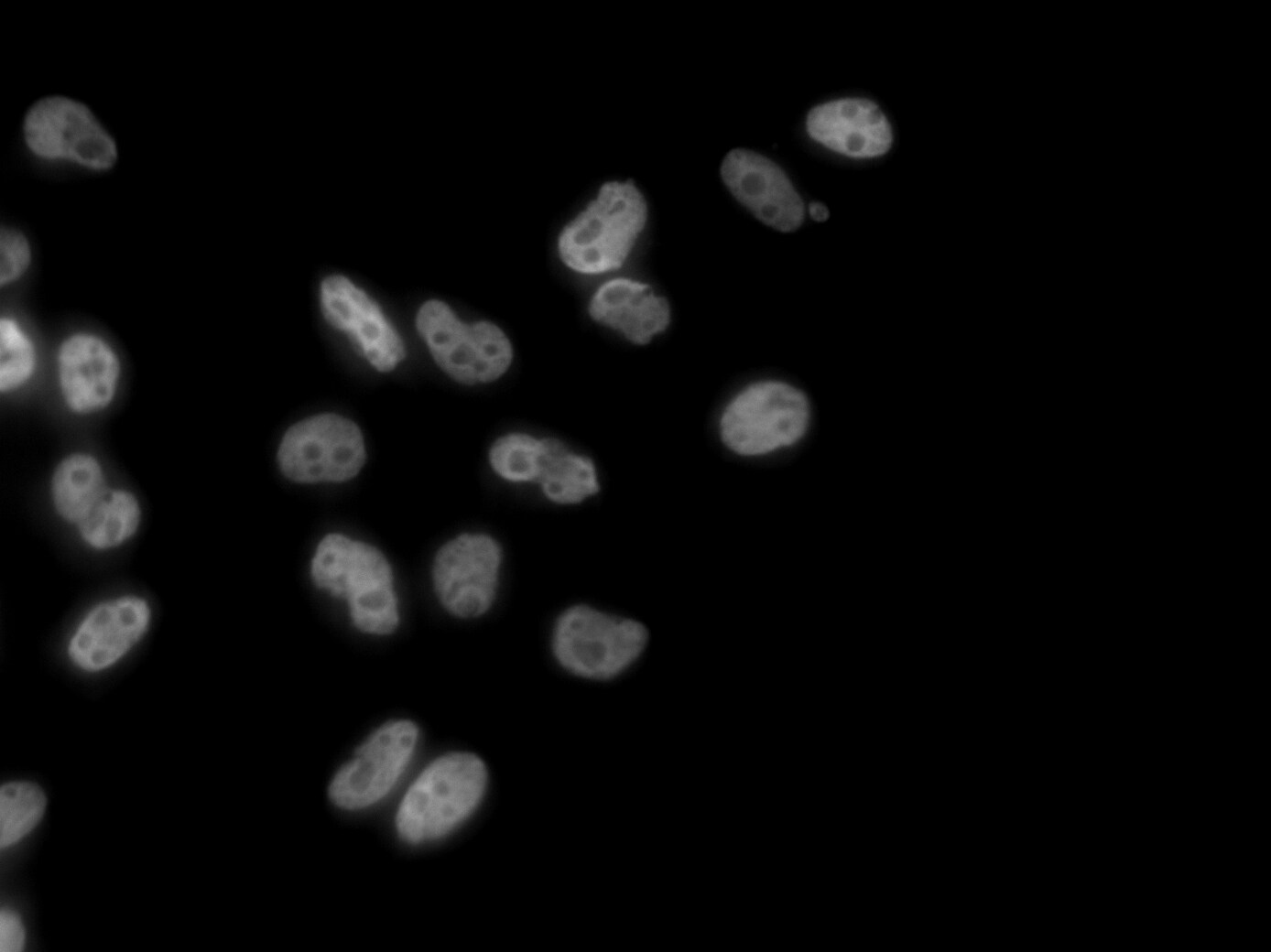
Application: ImmunocytochemistrySample Tested: Cortical neuronsSpecies: MouseVerified Customer | Posted 02/19/2020FUS (NB100-565) (green), b-III Tubulin (white), DAPI (blue). FUS ab dilution: 1:300 in PBST (0.1% triton) + 10% GS O/N 4ºC.FUS ab dilution: 1:300 in PBST (0.1% triton) + 10% GS O/N 4ºC.
-
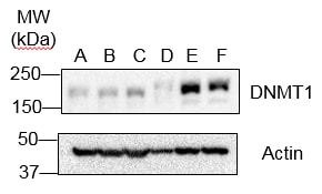
Application: Western BlotSample Tested: cellSpecies: HumanVerified Customer | Posted 01/30/2019DNMT1 WB of human diploid cellsAt least more than 20 μg sample is optimal.
-
Application: ImmunofluorescenceSample Tested: HeLa cellsSpecies: HumanVerified Customer | Posted 10/15/2013
There are no reviews that match your criteria.
Protocols
Find general support by application which include: protocols, troubleshooting, illustrated assays, videos and webinars.
- Antigen Retrieval Protocol (PIER)
- Antigen Retrieval for Frozen Sections Protocol
- Appropriate Fixation of IHC/ICC Samples
- Cellular Response to Hypoxia Protocols
- Chromogenic IHC Staining of Formalin-Fixed Paraffin-Embedded (FFPE) Tissue Protocol
- Chromogenic Immunohistochemistry Staining of Frozen Tissue
- ClariTSA™ Fluorophore Kits
- Detection & Visualization of Antibody Binding
- Fluorescent IHC Staining of Frozen Tissue Protocol
- Graphic Protocol for Heat-induced Epitope Retrieval
- Graphic Protocol for the Preparation and Fluorescent IHC Staining of Frozen Tissue Sections
- Graphic Protocol for the Preparation and Fluorescent IHC Staining of Paraffin-embedded Tissue Sections
- Graphic Protocol for the Preparation of Gelatin-coated Slides for Histological Tissue Sections
- ICC Cell Smear Protocol for Suspension Cells
- ICC Immunocytochemistry Protocol Videos
- ICC for Adherent Cells
- IHC Sample Preparation (Frozen sections vs Paraffin)
- Immunocytochemistry (ICC) Protocol
- Immunocytochemistry Troubleshooting
- Immunofluorescence of Organoids Embedded in Cultrex Basement Membrane Extract
- Immunofluorescent IHC Staining of Formalin-Fixed Paraffin-Embedded (FFPE) Tissue Protocol
- Immunohistochemistry (IHC) and Immunocytochemistry (ICC) Protocols
- Immunohistochemistry Frozen Troubleshooting
- Immunohistochemistry Paraffin Troubleshooting
- Immunoprecipitation Protocol
- Preparing Samples for IHC/ICC Experiments
- Preventing Non-Specific Staining (Non-Specific Binding)
- Primary Antibody Selection & Optimization
- Protocol for Heat-Induced Epitope Retrieval (HIER)
- Protocol for Making a 4% Formaldehyde Solution in PBS
- Protocol for VisUCyte™ HRP Polymer Detection Reagent
- Protocol for the Fluorescent ICC Staining of Cell Smears - Graphic
- Protocol for the Fluorescent ICC Staining of Cultured Cells on Coverslips - Graphic
- Protocol for the Preparation & Fixation of Cells on Coverslips
- Protocol for the Preparation and Chromogenic IHC Staining of Frozen Tissue Sections
- Protocol for the Preparation and Chromogenic IHC Staining of Frozen Tissue Sections - Graphic
- Protocol for the Preparation and Chromogenic IHC Staining of Paraffin-embedded Tissue Sections
- Protocol for the Preparation and Chromogenic IHC Staining of Paraffin-embedded Tissue Sections - Graphic
- Protocol for the Preparation and Fluorescent ICC Staining of Cells on Coverslips
- Protocol for the Preparation and Fluorescent ICC Staining of Non-adherent Cells
- Protocol for the Preparation and Fluorescent ICC Staining of Stem Cells on Coverslips
- Protocol for the Preparation and Fluorescent IHC Staining of Frozen Tissue Sections
- Protocol for the Preparation and Fluorescent IHC Staining of Paraffin-embedded Tissue Sections
- Protocol for the Preparation of Gelatin-coated Slides for Histological Tissue Sections
- Protocol for the Preparation of a Cell Smear for Non-adherent Cell ICC - Graphic
- R&D Systems Quality Control Western Blot Protocol
- TUNEL and Active Caspase-3 Detection by IHC/ICC Protocol
- The Importance of IHC/ICC Controls
- Troubleshooting Guide: Immunohistochemistry
- Troubleshooting Guide: Western Blot Figures
- Western Blot Conditions
- Western Blot Protocol
- Western Blot Protocol for Cell Lysates
- Western Blot Troubleshooting
- Western Blot Troubleshooting Guide
- View all Protocols, Troubleshooting, Illustrated assays and Webinars
Loading...