GABA-A R beta 3 Antibody - Azide Free
Novus Biologicals | Catalog # NB300-199
![Immunohistochemistry: GABA-A R beta 3 Antibody [NB300-199]](https://resources.rndsystems.com/images/products/GABA-A-R-beta-3-Antibody-Immunofluorescence-NB300-199-img0006.jpg "Immunohistochemistry: GABA-A R beta 3 Antibody [NB300-199]")
Key Product Details
Species Reactivity
Validated:
Cited:
Applications
Validated:
Cited:
Label
Antibody Source
Format
Product Specifications
Immunogen
Reactivity Notes
Specificity
Clonality
Host
Isotype
Theoretical MW
Disclaimer note: The observed molecular weight of the protein may vary from the listed predicted molecular weight due to post translational modifications, post translation cleavages, relative charges, and other experimental factors.
Scientific Data Images for GABA-A R beta 3 Antibody - Azide Free
Immunohistochemistry: GABA-A R beta 3 Antibody [NB300-199]
Immunohistochemistry: GABA-A R beta 3 Antibody [NB300-199] - Immunostaining of mouse retina showing specific labeling of the GABAA beta3 subunit in green, calbindin in red and DNA in blue.![Western Blot: GABA-A R beta 3 Antibody [NB300-199]](https://resources.rndsystems.com/images/products/GABA-A-R-beta-3-Antibody-Western-Blot-NB300-199-img0010.jpg "Western Blot: GABA-A R beta 3 Antibody [NB300-199]")
Western Blot: GABA-A R beta 3 Antibody [NB300-199]
GABA-A-R-beta-3-Antibody-Western-Blot-NB300-199-img0010.jpg![Immunohistochemistry: GABA-A R beta 3 Antibody [NB300-199]](https://resources.rndsystems.com/images/products/GABA-A-R-beta-3-Antibody-Immunohistochemistry-NB300-199-img0011.jpg "Immunohistochemistry: GABA-A R beta 3 Antibody [NB300-199]")
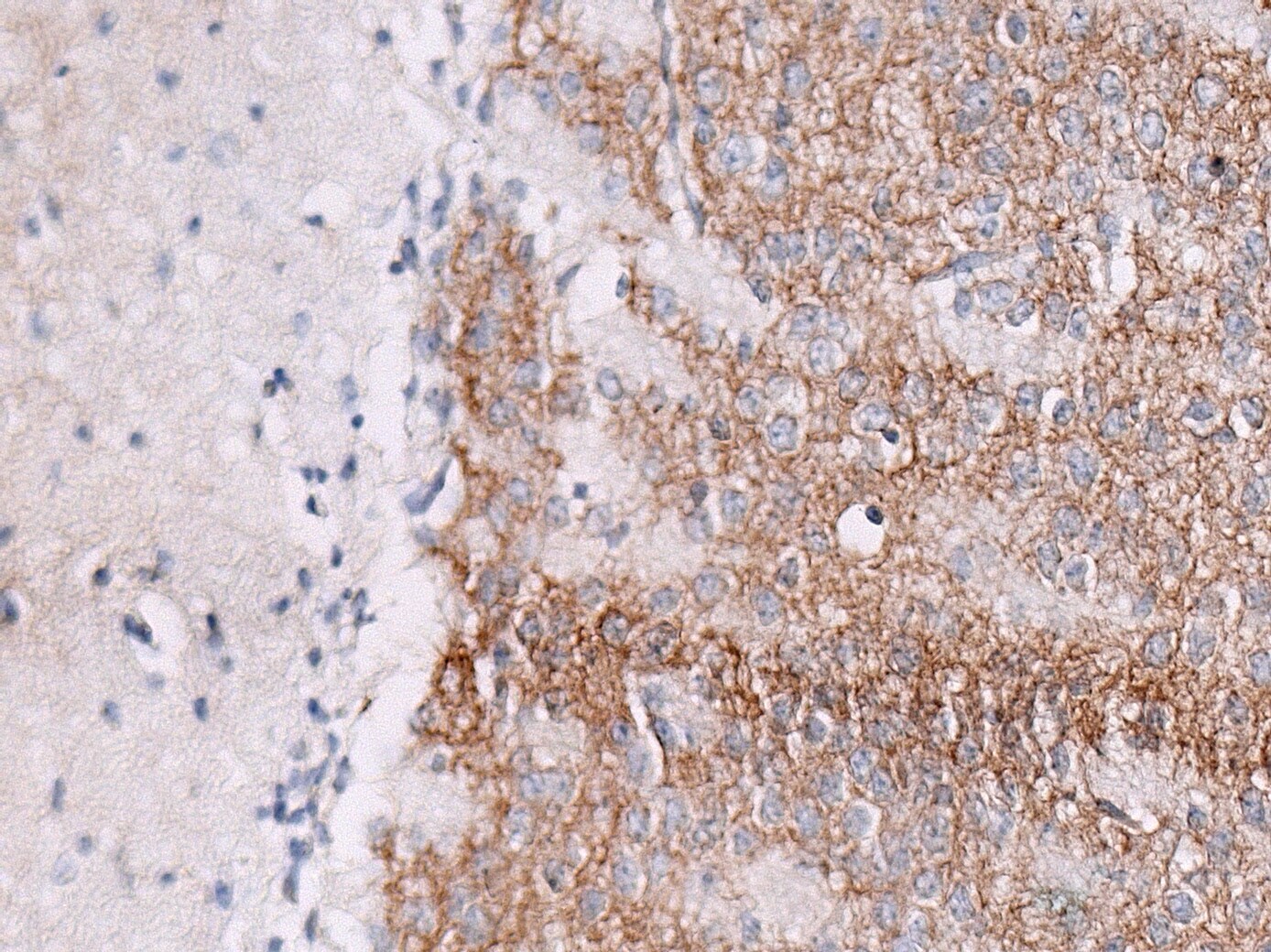
Immunohistochemistry: GABA-A R beta 3 Antibody [NB300-199]
GABA-A-R-beta-3-Antibody-Immunohistochemistry-NB300-199-img0011.jpg![Western Blot: GABA-A R beta 3 Antibody [NB300-199]](https://resources.rndsystems.com/images/products/GABA-A-R-beta-3-Antibody-Western-Blot-NB300-199-img0007.jpg "Western Blot: GABA-A R beta 3 Antibody [NB300-199]")
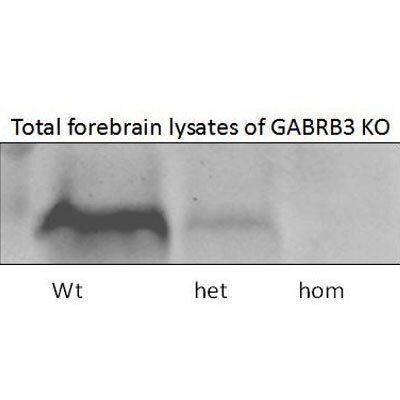
Western Blot: GABA-A R beta 3 Antibody [NB300-199]
Western Blot: GABA-A R beta 3 Antibody [NB300-199] - Rat brain lysate showing specific immunolabeling of the 53 kDa Beta(3)-subunit of the GABA(A)-R![Immunohistochemistry-Paraffin: GABA-A R beta 3 Antibody [NB300-199]](https://resources.rndsystems.com/images/products/GABA-A-R-beta-3-Antibody-Immunohistochemistry-Paraffin-NB300-199-img0008.jpg "Immunohistochemistry-Paraffin: GABA-A R beta 3 Antibody [NB300-199]")
Immunohistochemistry-Paraffin: GABA-A R beta 3 Antibody [NB300-199]
Immunohistochemistry-Paraffin: GABA-A R beta 3 Antibody [NB300-199] - Paraffin-embedded, formalin-fixed mouse head section. WB image submitted by a verified customer review.![Immunohistochemistry-Frozen: GABA-A R beta 3 Antibody [NB300-199]](https://resources.rndsystems.com/images/products/GABA-A-R-beta-3-Antibody-Immunohistochemistry-Frozen-NB300-199-img0009.jpg "Immunohistochemistry-Frozen: GABA-A R beta 3 Antibody [NB300-199]")
Immunohistochemistry-Frozen: GABA-A R beta 3 Antibody [NB300-199]
GABA-A-R-beta-3-Antibody-Immunohistochemistry-Frozen-NB300-199-img0009.jpgApplications for GABA-A R beta 3 Antibody - Azide Free
Immunohistochemistry
Simple Western
Western Blot
Reviewed Applications
Read 2 reviews rated 4.5 using NB300-199 in the following applications:
Formulation, Preparation, and Storage
Purification
Formulation
Format
Preservative
Concentration
Shipping
Stability & Storage
Background: GABA-A R beta 3
Long Name
Alternate Names
Gene Symbol
UniProt
Additional GABA-A R beta 3 Products
Product Documents for GABA-A R beta 3 Antibody - Azide Free
Certificate of Analysis
To download a Certificate of Analysis, please enter a lot or batch number in the search box below.
Product Specific Notices for GABA-A R beta 3 Antibody - Azide Free
This product is for research use only and is not approved for use in humans or in clinical diagnosis. Primary Antibodies are guaranteed for 1 year from date of receipt.
Related Research Areas
Citations for GABA-A R beta 3 Antibody - Azide Free
 Powered by Bioz
Customer Reviews for GABA-A R beta 3 Antibody - Azide Free (2)
Have you used GABA-A R beta 3 Antibody - Azide Free?
Submit a review and receive an Amazon gift card!
$25/€18/£15/$25CAN/¥2500 Yen for a review with an image
$10/€7/£6/$10CAN/¥1110 Yen for a review without an image
Submit a review
Customer Images
-
Application: Immunohistochemistry-ParaffinSample Tested: Mouse brainSpecies: MouseVerified Customer | Posted 08/22/2018IHC was performed on paraffin-embedded, formalin-fixed mouse head sections.

-
Application: Western BlotSample Tested: Mouse forebrain lysateSpecies: MouseVerified Customer | Posted 04/04/2013
There are no reviews that match your criteria.
Protocols
Find general support by application which include: protocols, troubleshooting, illustrated assays, videos and webinars.
- Antigen Retrieval Protocol (PIER)
- Antigen Retrieval for Frozen Sections Protocol
- Appropriate Fixation of IHC/ICC Samples
- Cellular Response to Hypoxia Protocols
- Chromogenic IHC Staining of Formalin-Fixed Paraffin-Embedded (FFPE) Tissue Protocol
- Chromogenic Immunohistochemistry Staining of Frozen Tissue
- ClariTSA™ Fluorophore Kits
- Detection & Visualization of Antibody Binding
- Fluorescent IHC Staining of Frozen Tissue Protocol
- Graphic Protocol for Heat-induced Epitope Retrieval
- Graphic Protocol for the Preparation and Fluorescent IHC Staining of Frozen Tissue Sections
- Graphic Protocol for the Preparation and Fluorescent IHC Staining of Paraffin-embedded Tissue Sections
- Graphic Protocol for the Preparation of Gelatin-coated Slides for Histological Tissue Sections
- ICC Cell Smear Protocol for Suspension Cells
- ICC Immunocytochemistry Protocol Videos
- ICC for Adherent Cells
- IHC Sample Preparation (Frozen sections vs Paraffin)
- Immunocytochemistry (ICC) Protocol
- Immunocytochemistry Troubleshooting
- Immunofluorescence of Organoids Embedded in Cultrex Basement Membrane Extract
- Immunofluorescent IHC Staining of Formalin-Fixed Paraffin-Embedded (FFPE) Tissue Protocol
- Immunohistochemistry (IHC) and Immunocytochemistry (ICC) Protocols
- Immunohistochemistry Frozen Troubleshooting
- Immunohistochemistry Paraffin Troubleshooting
- Preparing Samples for IHC/ICC Experiments
- Preventing Non-Specific Staining (Non-Specific Binding)
- Primary Antibody Selection & Optimization
- Protocol for Heat-Induced Epitope Retrieval (HIER)
- Protocol for Making a 4% Formaldehyde Solution in PBS
- Protocol for VisUCyte™ HRP Polymer Detection Reagent
- Protocol for the Fluorescent ICC Staining of Cell Smears - Graphic
- Protocol for the Fluorescent ICC Staining of Cultured Cells on Coverslips - Graphic
- Protocol for the Preparation & Fixation of Cells on Coverslips
- Protocol for the Preparation and Chromogenic IHC Staining of Frozen Tissue Sections
- Protocol for the Preparation and Chromogenic IHC Staining of Frozen Tissue Sections - Graphic
- Protocol for the Preparation and Chromogenic IHC Staining of Paraffin-embedded Tissue Sections
- Protocol for the Preparation and Chromogenic IHC Staining of Paraffin-embedded Tissue Sections - Graphic
- Protocol for the Preparation and Fluorescent ICC Staining of Cells on Coverslips
- Protocol for the Preparation and Fluorescent ICC Staining of Non-adherent Cells
- Protocol for the Preparation and Fluorescent ICC Staining of Stem Cells on Coverslips
- Protocol for the Preparation and Fluorescent IHC Staining of Frozen Tissue Sections
- Protocol for the Preparation and Fluorescent IHC Staining of Paraffin-embedded Tissue Sections
- Protocol for the Preparation of Gelatin-coated Slides for Histological Tissue Sections
- Protocol for the Preparation of a Cell Smear for Non-adherent Cell ICC - Graphic
- R&D Systems Quality Control Western Blot Protocol
- TUNEL and Active Caspase-3 Detection by IHC/ICC Protocol
- The Importance of IHC/ICC Controls
- Troubleshooting Guide: Immunohistochemistry
- Troubleshooting Guide: Western Blot Figures
- Western Blot Conditions
- Western Blot Protocol
- Western Blot Protocol for Cell Lysates
- Western Blot Troubleshooting
- Western Blot Troubleshooting Guide
- View all Protocols, Troubleshooting, Illustrated assays and Webinars
FAQs for GABA-A R beta 3 Antibody - Azide Free
-
Q: Could you please confirm the concentration for cat# NB300-199? The lot is WS712G.
A: The concentration of that lot # is 0.15 mg/mL.
-
Q: Could you please provide the immunogen sequence for Anti-GABA A Receptor beta 3 antibody and Anti-GABA A Receptor beta 2 antibody, catalog number NB300-199 and NB300-198 at your earliest convenience? If the exact sequence is not available, could you please tell me a broader range of amino acids or the region of the peptide from which it is derived? Also, please let me know if this information can be made public. Thank you for your help in this matter.
A: The immunogen for NB300-199 falls within 340-420 amino acids, while the immunogen for NB300-198 falls within the range of 327-451.
-
Q: Which species is the sequence from?
A: This sequence came from rat.
-
Q: Could you please confirm the concentration for cat# NB300-199? The lot is WS712G.
A: The concentration of that lot # is 0.15 mg/mL.
-
Q: Could you please provide the immunogen sequence for Anti-GABA A Receptor beta 3 antibody and Anti-GABA A Receptor beta 2 antibody, catalog number NB300-199 and NB300-198 at your earliest convenience? If the exact sequence is not available, could you please tell me a broader range of amino acids or the region of the peptide from which it is derived? Also, please let me know if this information can be made public. Thank you for your help in this matter.
A: The immunogen for NB300-199 falls within 340-420 amino acids, while the immunogen for NB300-198 falls within the range of 327-451.
-
Q: Which species is the sequence from?
A: This sequence came from rat.
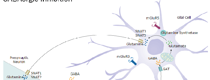
Associated Pathways