GAP-43 Antibody - BSA Free
Novus Biologicals | Catalog # NBP1-41123
![Western Blot: GAP-43 Antibody [NBP1-41123]](https://resources.rndsystems.com/images/products/GAP-43-Antibody-Western-Blot-NBP1-41123-img0008.jpg "Western Blot: GAP-43 Antibody [NBP1-41123]")
![Immunohistochemistry: GAP-43 Antibody [NBP1-41123]](https://resources.rndsystems.com/images/products/GAP-43-Antibody-Immunohistochemistry-NBP1-41123-img0009.jpg "Immunohistochemistry: GAP-43 Antibody [NBP1-41123]")
Key Product Details
Validated by
Species Reactivity
Validated:
Cited:
Applications
Validated:
Cited:
Label
Antibody Source
Format
Product Specifications
Immunogen
Localization
Marker
Clonality
Host
Isotype
Theoretical MW
Disclaimer note: The observed molecular weight of the protein may vary from the listed predicted molecular weight due to post translational modifications, post translation cleavages, relative charges, and other experimental factors.
Scientific Data Images for GAP-43 Antibody - BSA Free
Immunohistochemistry: GAP-43 Antibody [NBP1-41123]
Immunohistochemistry: GAP-43 Antibody [NBP1-41123] - Coronal sections of the developing (A-H) eye at (A-D) E14.5, (E-H) E17.5. were immunostained for (A-H) EVA1C and (A-H) GAP43. All sections were counterstained with DAPI. EVA1C was strongly co-expressed with GAP43 by retinal cell ganglionic axons (A-H) as they exit the retina and form the optic nerve (arrowheads). Scale bars represent 100 uM. PLoS One. 2013 Sep 9;8(9):e74115. doi: 10.1371/journal.pone.0074115.![Immunocytochemistry/ Immunofluorescence: GAP-43 Antibody [NBP1-41123]](https://resources.rndsystems.com/images/products/GAP-43-Antibody-Immunocytochemistry-Immunofluorescence-NBP1-41123-img0005.jpg "Immunocytochemistry/ Immunofluorescence: GAP-43 Antibody [NBP1-41123]")
Immunocytochemistry/ Immunofluorescence: GAP-43 Antibody [NBP1-41123]
Immunocytochemistry/Immunofluorescence: GAP-43 Antibody [NBP1-41123] - Staining of a mature retinal ganglion cell demonstrating axon outgrowth.![Western Blot: GAP-43 Antibody [NBP1-41123]](https://resources.rndsystems.com/images/products/GAP-43-Antibody-Western-Blot-NBP1-41123-img0006.jpg "Western Blot: GAP-43 Antibody [NBP1-41123]")
Western Blot: GAP-43 Antibody [NBP1-41123]
Western Blot: GAP-43 Antibody [NBP1-41123] - Analysis of GAP43 in human brain protein![Western Blot: GAP-43 Antibody [NBP1-41123]](https://resources.rndsystems.com/images/products/GAP-43-Antibody-Western-Blot-NBP1-41123-img0007.jpg "Western Blot: GAP-43 Antibody [NBP1-41123]")
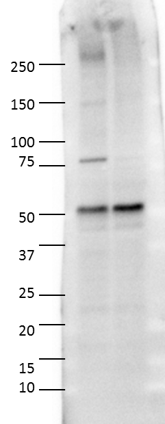
Western Blot: GAP-43 Antibody [NBP1-41123]
Western Blot: GAP-43 Antibody [NBP1-41123] - Total protein from Human, Mouse and Rat brain, and SHSY-5Y cells was separated on a 12% gel by SDS-PAGE, transferred to PVDF membrane and blocked in 5% non-fat milk in TBST. The membrane was probed with 2.0 ug/ml anti-GAP43 in 1% non-fat milk in TBST and detected with an anti-sheep HRP secondary antibody using chemiluminescence.Applications for GAP-43 Antibody - BSA Free
Immunoblotting
Immunocytochemistry/ Immunofluorescence
Immunohistochemistry
Immunohistochemistry-Frozen
Immunohistochemistry-Paraffin
Western Blot
Reviewed Applications
Read 1 review rated 4 using NBP1-41123 in the following applications:
Formulation, Preparation, and Storage
Purification
Formulation
Format
Preservative
Concentration
Shipping
Stability & Storage
Background: GAP-43
Long Name
Alternate Names
Entrez Gene IDs
Gene Symbol
Additional GAP-43 Products
Product Documents for GAP-43 Antibody - BSA Free
Certificate of Analysis
To download a Certificate of Analysis, please enter a lot or batch number in the search box below.
Product Specific Notices for GAP-43 Antibody - BSA Free
This product is for research use only and is not approved for use in humans or in clinical diagnosis. Primary Antibodies are guaranteed for 1 year from date of receipt.
Citations for GAP-43 Antibody - BSA Free
 Powered by Bioz
Customer Reviews for GAP-43 Antibody - BSA Free (1)
Have you used GAP-43 Antibody - BSA Free?
Submit a review and receive an Amazon gift card!
$25/€18/£15/$25CAN/¥2500 Yen for a review with an image
$10/€7/£6/$10CAN/¥1110 Yen for a review without an image
Submit a review
Customer Images
-
Application: Western BlotSample Tested: Rat olfactory epithelium tissue lysateSpecies: RatVerified Customer | Posted 05/28/2015Rat Olfactory epthelium tissue, 10ug/lane (lane1: saline, lane2: ZnSO4)

There are no reviews that match your criteria.
Protocols
View specific protocols for GAP-43 Antibody - BSA Free (NBP1-41123):
Western Blot Protocol
1. Perform SDS-PAGE (4-12% MOPS) on samples to be analyzed, loading 25 ug of total protein per lane.
2. Transfer proteins to Nitrocellulose according to the instructions provided by the manufacturer of the transfer apparatus.
3. Rinse membrane with dH2O and then stain the blot using Ponceau S for 1-2 minutes to access the transfer of proteins onto the nitrocellulose membrane. Rinse the blot in water to remove excess stain and mark the lane locations and locations of molecular weight markers using a pencil.
4. Rinse the blot in TBS for approximately 5 minutes.
5. Block the membrane using 5% NFDM + 1% BSA in TBS + Tween, 1 hour at RT.
6. Rinse the membrane in dH2O and then wash the membrane in wash buffer [TBS + 0.1% Tween] 3 times for 10 minutes each.
7. Dilute the anti-GAP43 primary antibody (NBP1-41123) in blocking buffer and incubate 1 hour at room temperature.
8. Rinse the membrane in dH2O and then wash the membrane in wash buffer [TBS + 0.1% Tween] 3 times for 10 minutes each.
9. Apply the diluted sheep-IgG HRP-conjugated secondary antibody in blocking buffer (as per manufacturers instructions) and incubate 1 hour at room temperature.
10. Wash the blot in wash buffer [TBS + 0.1% Tween] 3 times for 10 minutes each (this step can be repeated as required to reduce background).
11. Apply the detection reagent of choice in accordance with the manufacturers instructions (Pierce ECL).
Note: Tween-20 can be added to the blocking or antibody dilution buffer at a final concentration of 0.05-0.2%, provided it does not interfere with antibody-antigen binding.
Find general support by application which include: protocols, troubleshooting, illustrated assays, videos and webinars.
- Antigen Retrieval Protocol (PIER)
- Antigen Retrieval for Frozen Sections Protocol
- Appropriate Fixation of IHC/ICC Samples
- Cellular Response to Hypoxia Protocols
- Chromogenic IHC Staining of Formalin-Fixed Paraffin-Embedded (FFPE) Tissue Protocol
- Chromogenic Immunohistochemistry Staining of Frozen Tissue
- ClariTSA™ Fluorophore Kits
- Detection & Visualization of Antibody Binding
- Fluorescent IHC Staining of Frozen Tissue Protocol
- Graphic Protocol for Heat-induced Epitope Retrieval
- Graphic Protocol for the Preparation and Fluorescent IHC Staining of Frozen Tissue Sections
- Graphic Protocol for the Preparation and Fluorescent IHC Staining of Paraffin-embedded Tissue Sections
- Graphic Protocol for the Preparation of Gelatin-coated Slides for Histological Tissue Sections
- ICC Cell Smear Protocol for Suspension Cells
- ICC Immunocytochemistry Protocol Videos
- ICC for Adherent Cells
- IHC Sample Preparation (Frozen sections vs Paraffin)
- Immunocytochemistry (ICC) Protocol
- Immunocytochemistry Troubleshooting
- Immunofluorescence of Organoids Embedded in Cultrex Basement Membrane Extract
- Immunofluorescent IHC Staining of Formalin-Fixed Paraffin-Embedded (FFPE) Tissue Protocol
- Immunohistochemistry (IHC) and Immunocytochemistry (ICC) Protocols
- Immunohistochemistry Frozen Troubleshooting
- Immunohistochemistry Paraffin Troubleshooting
- Preparing Samples for IHC/ICC Experiments
- Preventing Non-Specific Staining (Non-Specific Binding)
- Primary Antibody Selection & Optimization
- Protocol for Heat-Induced Epitope Retrieval (HIER)
- Protocol for Making a 4% Formaldehyde Solution in PBS
- Protocol for VisUCyte™ HRP Polymer Detection Reagent
- Protocol for the Fluorescent ICC Staining of Cell Smears - Graphic
- Protocol for the Fluorescent ICC Staining of Cultured Cells on Coverslips - Graphic
- Protocol for the Preparation & Fixation of Cells on Coverslips
- Protocol for the Preparation and Chromogenic IHC Staining of Frozen Tissue Sections
- Protocol for the Preparation and Chromogenic IHC Staining of Frozen Tissue Sections - Graphic
- Protocol for the Preparation and Chromogenic IHC Staining of Paraffin-embedded Tissue Sections
- Protocol for the Preparation and Chromogenic IHC Staining of Paraffin-embedded Tissue Sections - Graphic
- Protocol for the Preparation and Fluorescent ICC Staining of Cells on Coverslips
- Protocol for the Preparation and Fluorescent ICC Staining of Non-adherent Cells
- Protocol for the Preparation and Fluorescent ICC Staining of Stem Cells on Coverslips
- Protocol for the Preparation and Fluorescent IHC Staining of Frozen Tissue Sections
- Protocol for the Preparation and Fluorescent IHC Staining of Paraffin-embedded Tissue Sections
- Protocol for the Preparation of Gelatin-coated Slides for Histological Tissue Sections
- Protocol for the Preparation of a Cell Smear for Non-adherent Cell ICC - Graphic
- R&D Systems Quality Control Western Blot Protocol
- TUNEL and Active Caspase-3 Detection by IHC/ICC Protocol
- The Importance of IHC/ICC Controls
- Troubleshooting Guide: Immunohistochemistry
- Troubleshooting Guide: Western Blot Figures
- Western Blot Conditions
- Western Blot Protocol
- Western Blot Protocol for Cell Lysates
- Western Blot Troubleshooting
- Western Blot Troubleshooting Guide
- View all Protocols, Troubleshooting, Illustrated assays and Webinars
FAQs for GAP-43 Antibody - BSA Free
-
Q: Can you confirm the tested application of NBP1-41123 includes Immunohistochemistry on Formalin-Fixed Paraffin Embedded sections?
A: NBP1-41123 has only been validated in IHC on frozen sections at this time. However, NB300-143 has been validated in IHC on both frozen and paraffin-embedded tissues.
-
Q: Could you tell us the sequence homology between the immunogen of this antibody and the GAP43 protein of canine?
A: The immunogen for NB300-143 shares 82% similarity with canine. NBP1-41123 may be a better choice. It is known to work in dog and in IHC. It has also been published with 10 times in IHC and some of these publications almost certainly used paraffin-embedded tissues.
-
Q: Can you confirm the tested application of NBP1-41123 includes Immunohistochemistry on Formalin-Fixed Paraffin Embedded sections?
A: NBP1-41123 has only been validated in IHC on frozen sections at this time. However, NB300-143 has been validated in IHC on both frozen and paraffin-embedded tissues.
-
Q: Could you tell us the sequence homology between the immunogen of this antibody and the GAP43 protein of canine?
A: The immunogen for NB300-143 shares 82% similarity with canine. NBP1-41123 may be a better choice. It is known to work in dog and in IHC. It has also been published with 10 times in IHC and some of these publications almost certainly used paraffin-embedded tissues.