GFAP Antibody (5C10) - BSA Free
Novus Biologicals | Catalog # NBP1-05197
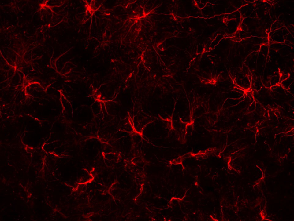
![Immunohistochemistry Free-Floating: GFAP Antibody (5C10) [NBP1-05197]](https://resources.rndsystems.com/images/products/GFAP-Antibody-5C10-Immunohistochemistry-Free-Floating-NBP1-05197-img0007.jpg "Immunohistochemistry Free-Floating: GFAP Antibody (5C10) [NBP1-05197]")
Key Product Details
Species Reactivity
Validated:
Human, Mouse, Rat, Porcine, Bovine, Equine
Cited:
Human, Mouse, Rat, Porcine
Applications
Validated:
Immunohistochemistry, Immunohistochemistry-Paraffin, Immunohistochemistry-Frozen, Immunohistochemistry Free-Floating, Western Blot, Immunocytochemistry/ Immunofluorescence, Simple Western
Cited:
Immunohistochemistry, Immunohistochemistry-Paraffin, Immunohistochemistry-Frozen, Western Blot, Immunocytochemistry/ Immunofluorescence, IF/IHC, IHC/IF
Label
Unconjugated
Antibody Source
Monoclonal Mouse IgG1 Clone # 5C10
Format
BSA Free
Loading...
Product Specifications
Immunogen
This GFAP Antibody (5C10) was developed against a preparation of purified pig spinal cord GFAP
Localization
Cytoplasm. Note: Associated with intermediate filaments.
Marker
Astrocyte Marker
Clonality
Monoclonal
Host
Mouse
Isotype
IgG1
Theoretical MW
50 kDa.
Disclaimer note: The observed molecular weight of the protein may vary from the listed predicted molecular weight due to post translational modifications, post translation cleavages, relative charges, and other experimental factors.
Disclaimer note: The observed molecular weight of the protein may vary from the listed predicted molecular weight due to post translational modifications, post translation cleavages, relative charges, and other experimental factors.
Scientific Data Images for GFAP Antibody (5C10) - BSA Free
Immunohistochemistry Free-Floating: GFAP Antibody (5C10) [NBP1-05197]
Immunohistochemistry Free-Floating: GFAP Antibody (5C10) [NBP1-05197] - Analysis of rat cerebellum section stained with mouse GFAP mAb, dilution 1:1,000 (Green), costained with rabbit neurofilament NF-L pAb, dilution 1:2,000 (Red). Following transcardial perfusion with 4% paraformaldehyde, brain was post fixed for 24hrs, cut to 45uM, and free-floating sections were stained with antibodies. The GFAP antibody stains a network of astroglial cells, while the NF-L antibody labels neuronal cells and their processes.![Simple Western: GFAP Antibody (5C10) [NBP1-05197]](https://resources.rndsystems.com/images/products/GFAP-Antibody-5C10-Simple-Western-NBP1-05197-img0004.jpg "Simple Western: GFAP Antibody (5C10) [NBP1-05197]")
Simple Western: GFAP Antibody (5C10) [NBP1-05197]
Simple Western: GFAP Antibody (5C10) [NBP1-05197] - Simple Western lane view shows a specific band for GFAP in 0.05 mg/ml of Human Brain lysate. This experiment was performed under reducing conditions using the 12-230 kDa separation system.![Immunohistochemistry: GFAP Antibody (5C10) [NBP1-05197]](https://resources.rndsystems.com/images/products/GFAP-Antibody-5C10-Immunohistochemistry-NBP1-05197-img0008.jpg "Immunohistochemistry: GFAP Antibody (5C10) [NBP1-05197]")
Immunohistochemistry: GFAP Antibody (5C10) [NBP1-05197]
GFAP-Antibody-5C10-Immunohistochemistry-NBP1-05197-img0008.jpg![Immunohistochemistry: GFAP Antibody (5C10) [NBP1-05197]](https://resources.rndsystems.com/images/products/GFAP-Antibody-5C10-Immunocytochemistry-Immunofluorescence-NBP1-05197-img0005.jpg "Immunohistochemistry: GFAP Antibody (5C10) [NBP1-05197]")
Immunohistochemistry: GFAP Antibody (5C10) [NBP1-05197]
Immunohistochemistry: GFAP Antibody (5C10) [NBP1-05197] - Analysis of rat cerebellum section stained with mouse mAb to GFAP, NBP1-05197, dilution 1:1,000, in green, costained with rabbit pAb to neurofilament NF-L, dilution 1:2,000, in red. Following transcardial perfusion of rat with 4% paraformaldehyde, brain was post fixed for 24 hours, cut to 45uM, and free-floating sections were stained with above antibodies. The NBP1-05197 antibody stains a network of astroglial cells, while the NF-L antibody labels neuronal cells and their processes.![Western Blot: GFAP Antibody (5C10) [NBP1-05197]](https://resources.rndsystems.com/images/products/GFAP-Antibody-5C10-Western-Blot-NBP1-05197-img0006.jpg "Western Blot: GFAP Antibody (5C10) [NBP1-05197]")
Western Blot: GFAP Antibody (5C10) [NBP1-05197]
Western Blot: GFAP Antibody (5C10) [NBP1-05197] - Analysis of whole tissue lysates using mouse mAb to GFAP, NBP1-05197, dilution 1:2,000, in green: [1] protein standard (red), [2] rat brain, [3] rat spinal cord, [4] mouse brain, [5] mouse spinal cord. The strong band at about 50kDa corresponds to the GFAP protein.Applications for GFAP Antibody (5C10) - BSA Free
Application
Recommended Usage
Immunocytochemistry/ Immunofluorescence
1:1000
Immunohistochemistry
1:1000
Immunohistochemistry Free-Floating
1:1000
Immunohistochemistry-Frozen
1:1000
Immunohistochemistry-Paraffin
1:1000
Simple Western
1:3000
Western Blot
1:5000
Application Notes
This GFAP (5C10) antibody is useful for Immunocytochemistry/Immunofluorescence, Immunohistochemistry on paraffin-embedded and frozen sections, and Western blot. In WB, a band can be seen at approx. 50 kDa.
In Simple Western only 10 - 15 uL of the recommended dilution is used per data point.
See Simple Western Antibody Database for Simple Western validation: Tested in Human Brain lysate 0.05 mg/mL, separated by Size, antibody dilution of 1:3000, apparent MW was 51 kDa. Separated by Size-Wes, Sally Sue/Peggy Sue.
Reviewed Applications
Read 1 review rated 3 using NBP1-05197 in the following applications:
Formulation, Preparation, and Storage
Purification
Immunogen affinity purified
Formulation
PBS, 50% glycerol
Format
BSA Free
Preservative
5mM Sodium Azide
Concentration
1.0 mg/ml
Shipping
The product is shipped with polar packs. Upon receipt, store it immediately at the temperature recommended below.
Stability & Storage
Store at 4C short term. Aliquot and store at -20C long term. Avoid freeze-thaw cycles.
Background: GFAP
An increase in GFAP levels is often associated with neuroinflammation which results in the activation and proliferation of astroglia cell population (1,2). GFAP expression is also observed in brains of patients with neurodegenerative diseases including Alzheimer's and Parkinson's, epilepsy disorders, and brain injuries (1-4). Lesion sites associated with neurodegeneration can exhibit an array of gliosis characteristics from glial scarring with reduced astrocyte proliferation to activated, GFAP-positive astrocytes surrounding amyloid plaques (2). Furthermore, the GFAP gene is a target of single nucleotide polymorphisms in the coding region, considered a gain-of-function mutation, characterized by astrocytic inclusions, termed Rosenthal fibers, resulting in Alexander Disease (1-4). GFAP is also a center of many post-translational modifications, such as phosphorylation, which can alter various aspects of filament assembly (1,4).
References
1. Yang, Z., & Wang, K. K. (2015). Glial fibrillary acidic protein: from intermediate filament assembly and gliosis to neurobiomarker. Trends in Neurosciences. https://doi.org/10.1016/j.tins.2015.04.003
2. Hol, E. M., & Capetanaki, Y. (2017). Type III Intermediate Filaments Desmin, Glial Fibrillary Acidic Protein (GFAP), Vimentin, and Peripherin. Cold Spring Harbor Perspectives in Biology. https://doi.org/10.1101/cshperspect.a021642
3. Potokar, M., Morita, M., Wiche, G., & Jorgacevski, J. (2020). The Diversity of Intermediate Filaments in Astrocytes. Cells. https://doi.org/10.3390/cells9071604
4. Viedma-Poyatos, a., Pajares, M. A., & Perez-Sala, D. (2020). Type III intermediate filaments as targets and effectors of electrophiles and oxidants. Redox Biology. https://doi.org/10.1016/j.redox.2020.101582
Long Name
Glial Fibrillary Acidic Protein
Alternate Names
ALXDRD, FLJ45472, GFAP, GFAP astrocytes, glial fibrillary acidic protein
Gene Symbol
GFAP
Additional GFAP Products
Product Documents for GFAP Antibody (5C10) - BSA Free
Certificate of Analysis
To download a Certificate of Analysis, please enter a lot or batch number in the search box below.
Product Specific Notices for GFAP Antibody (5C10) - BSA Free
This product is for research use only and is not approved for use in humans or in clinical diagnosis. Primary Antibodies are guaranteed for 1 year from date of receipt.
Related Research Areas
Citations for GFAP Antibody (5C10) - BSA Free
 Powered by Bioz
Customer Reviews for GFAP Antibody (5C10) - BSA Free (1)
3 out of 5
1 Customer Rating
Have you used GFAP Antibody (5C10) - BSA Free?
Submit a review and receive an Amazon gift card!
$25/€18/£15/$25CAN/¥2500 Yen for a review with an image
$10/€7/£6/$10CAN/¥1110 Yen for a review without an image
Submit a review
Customer Images
Showing
1
-
1 of
1 review
Showing All
Filter By:
-
Application: ImmunofluorescenceSample Tested: mouse spinal cordSpecies: MouseVerified Customer | Posted 05/11/2016

There are no reviews that match your criteria.
Protocols
Find general support by application which include: protocols, troubleshooting, illustrated assays, videos and webinars.
- Antigen Retrieval Protocol (PIER)
- Antigen Retrieval for Frozen Sections Protocol
- Appropriate Fixation of IHC/ICC Samples
- Cellular Response to Hypoxia Protocols
- Chromogenic IHC Staining of Formalin-Fixed Paraffin-Embedded (FFPE) Tissue Protocol
- Chromogenic Immunohistochemistry Staining of Frozen Tissue
- ClariTSA™ Fluorophore Kits
- Detection & Visualization of Antibody Binding
- Fluorescent IHC Staining of Frozen Tissue Protocol
- Graphic Protocol for Heat-induced Epitope Retrieval
- Graphic Protocol for the Preparation and Fluorescent IHC Staining of Frozen Tissue Sections
- Graphic Protocol for the Preparation and Fluorescent IHC Staining of Paraffin-embedded Tissue Sections
- Graphic Protocol for the Preparation of Gelatin-coated Slides for Histological Tissue Sections
- ICC Cell Smear Protocol for Suspension Cells
- ICC Immunocytochemistry Protocol Videos
- ICC for Adherent Cells
- IHC Sample Preparation (Frozen sections vs Paraffin)
- Immunocytochemistry (ICC) Protocol
- Immunocytochemistry Troubleshooting
- Immunofluorescence of Organoids Embedded in Cultrex Basement Membrane Extract
- Immunofluorescent IHC Staining of Formalin-Fixed Paraffin-Embedded (FFPE) Tissue Protocol
- Immunohistochemistry (IHC) and Immunocytochemistry (ICC) Protocols
- Immunohistochemistry Frozen Troubleshooting
- Immunohistochemistry Paraffin Troubleshooting
- Preparing Samples for IHC/ICC Experiments
- Preventing Non-Specific Staining (Non-Specific Binding)
- Primary Antibody Selection & Optimization
- Protocol for Heat-Induced Epitope Retrieval (HIER)
- Protocol for Making a 4% Formaldehyde Solution in PBS
- Protocol for VisUCyte™ HRP Polymer Detection Reagent
- Protocol for the Fluorescent ICC Staining of Cell Smears - Graphic
- Protocol for the Fluorescent ICC Staining of Cultured Cells on Coverslips - Graphic
- Protocol for the Preparation & Fixation of Cells on Coverslips
- Protocol for the Preparation and Chromogenic IHC Staining of Frozen Tissue Sections
- Protocol for the Preparation and Chromogenic IHC Staining of Frozen Tissue Sections - Graphic
- Protocol for the Preparation and Chromogenic IHC Staining of Paraffin-embedded Tissue Sections
- Protocol for the Preparation and Chromogenic IHC Staining of Paraffin-embedded Tissue Sections - Graphic
- Protocol for the Preparation and Fluorescent ICC Staining of Cells on Coverslips
- Protocol for the Preparation and Fluorescent ICC Staining of Non-adherent Cells
- Protocol for the Preparation and Fluorescent ICC Staining of Stem Cells on Coverslips
- Protocol for the Preparation and Fluorescent IHC Staining of Frozen Tissue Sections
- Protocol for the Preparation and Fluorescent IHC Staining of Paraffin-embedded Tissue Sections
- Protocol for the Preparation of Gelatin-coated Slides for Histological Tissue Sections
- Protocol for the Preparation of a Cell Smear for Non-adherent Cell ICC - Graphic
- R&D Systems Quality Control Western Blot Protocol
- TUNEL and Active Caspase-3 Detection by IHC/ICC Protocol
- The Importance of IHC/ICC Controls
- Troubleshooting Guide: Immunohistochemistry
- Troubleshooting Guide: Western Blot Figures
- Western Blot Conditions
- Western Blot Protocol
- Western Blot Protocol for Cell Lysates
- Western Blot Troubleshooting
- Western Blot Troubleshooting Guide
- View all Protocols, Troubleshooting, Illustrated assays and Webinars
Loading...
Associated Pathways
