Best Seller
GFP Antibody - BSA Free
Novus Biologicals | Catalog # NB100-1770

Key Product Details
Species Reactivity
Validated:
Non-species specific
Cited:
Human, Mouse, Non-species specific, Rabbit
Applications
Validated:
Knockout Validated, Immunohistochemistry, Immunohistochemistry-Paraffin, Immunohistochemistry-Frozen, Immunohistochemistry Free-Floating, Western Blot, ELISA, Flow Cytometry, Immunocytochemistry/ Immunofluorescence, Immunoprecipitation, Proximity Ligation Assay, Electron Microscopy, Knockdown Validated
Cited:
Knockout Validated, Immunohistochemistry, Immunohistochemistry-Paraffin, Immunohistochemistry-Frozen, Immunohistochemistry Free-Floating, Western Blot, ELISA, Block/Neutralize, Flow Cytometry, Immunofluorescence, Immunocytochemistry/ Immunofluorescence, Proximity Ligation Assay, IF/IHC, Electron Microscopy, Knockdown Validated
Label
Unconjugated
Antibody Source
Polyclonal Goat IgG
Format
BSA Free
Loading...
Product Specifications
Immunogen
The immunogen is a Green Fluorescent Protein (GFP) fusion protein corresponding to the full length amino acid sequence (246aa) derived from the jellyfish Aequorea victoria. (Uniprot: P42212)
Reactivity Notes
No reaction was observed against Human, or Rat serum proteins. Known Cross Reactivity: rGFP. YFP differs from GFP due to a mutation at Thr203Tyr; antibodies raised against full-length GFP should also detect YFP and other variants. Reactivity in transgenic mice with GFP. Reactivity in human cell lines transfected will a GFP construct.
Specificity
No reaction was observed against Human, Mouse or Rat serum proteins.
Clonality
Polyclonal
Host
Goat
Isotype
IgG
Description
GFP antibody was prepared from monospecific antiserum by immunoaffinity chromatography using Green Fluorescent Protein (Aequorea victoria) coupled to agarose beads followed by solid phase adsorption(s) to remove any unwanted reactivities. Assay by immunoelectrophoresis resulted in a single precipitin arc against anti-Goat Serum and purified and partially purified Green Fluorescent Protein (Aequorea victoria)
Store this antibody at -20C prior to opening. Aliquot contents and freeze at -20C or below for extended storage. Avoid cycles of freezing and thawing. Centrifuge product if not completely clear after standing at room temperature. This product is stable for several weeks at 4C as an undiluted liquid. Dilute only prior to immediate use.
Store this antibody at -20C prior to opening. Aliquot contents and freeze at -20C or below for extended storage. Avoid cycles of freezing and thawing. Centrifuge product if not completely clear after standing at room temperature. This product is stable for several weeks at 4C as an undiluted liquid. Dilute only prior to immediate use.
Scientific Data Images for GFP Antibody - BSA Free
Immunofluorescent Staining of Mouse Pancreas Using Biotin Conjugated GFP Antibody
Analysis using the Biotin conjugate of NB100-1770. Staining of transgenic mouse pancreas, expressing GFP in beta cells. Image courtesy of product review by Dr. Yves Heremans of Vrije Universiteit Brussel.
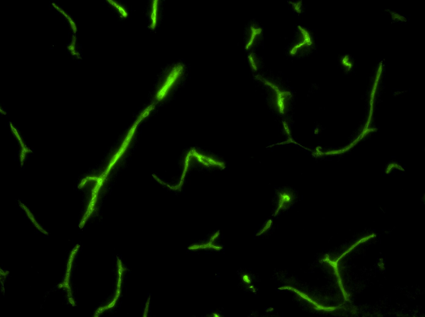
Immunohistochemical Staining of Drosophila Central Nervous System Tissue Using Biotin Conjugated GFP Antibody
Analysis of Biotin conjugate of NB100-1770. Tissue: Drosophila melanogaster late stage embryonic central nervous system. Fixation: 0.5% PFA. Antigen retrieval: not required. Primary antibody: Anti-GFP antibody at a 1:1000 for 1 h at RT.
Western Blot Detection of GFP Using FITC Conjugated Antibody
Analysis using the FITC conjugate of NB100-1770. Lane 1: GFP (50 ug). Primary antibody: None. Secondary antibody: Fluorescein goat secondary antibody at 1:1000 for 60 min at RT. Block for 30 minutes.
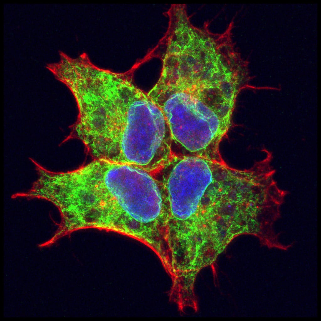
Immunocytochemistry/Immunofluorescence Staining of GFP in Transfected HEK293 Cells
HEK293 cells stable transfected with integrin alpha8-GFP, were immunostained for GFP (green) and counterstained with phalloidin (red) and DAPI (BLUE). ICC/IF image submitted by a verified customer review.
Western Blot Detection of GFP Using Alkaline Phosphatase Conjugated Antibody
Analysis using the Alkaline Phosphatase conjugate of NB100-1770. Lane 1: GFP (50 ng). Alkaline Phosphatase GFP secondary antibody at 1:1000 for 60 min at RT. Predicted/Observed size: 28 kDa for GFP.
Western Blot Detection of GFP in Transfected HeLa Cells
GFP-Antibody-Western-Blot-NB100-1770-img0049.jpg
Immunohistochemical Detection of GFP in Transgenic Mouse Embryo
E5.5 Hex-GFP transgenic mouse embryo tissue. Primary antibody: Goat anti-GFP at 1:500 dilution. Secondary antibody: Fluorochrome conjugated Anti-goat IgG antibody at 1:10,000 for 45 min at RT. Staining: GFP as green fluorescent signal with DAPI blue counterstain.
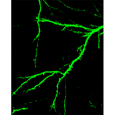
Immunofluorescent Staining of GFP in Mouse Brain
IF analysis of GFP in mouse brain. Image courtesy of product review by Tatyana Pivneva.
Immunohistochemical Detection of GFP in Free Floating Mouse Brain
Tissue: Sf-1:Cre mice crossed to the Z/EG reporter line. Mouse brain (coronal view, 20X magnification). Fixation: 4%PFA/PBS o/n, and subsequently transferred to a 30% sucrose solution. Antigen retrieval: frozen in OCT freezing medium (Sakura) and cryostat sectioned at 40 microns. Primary antibody: Goat anti-GFP was used at 1:500 dilution in free floating immunohistochemistry to detect GFP. Secondary antibody: Fluorochrome conjugated Anti-goat IgG antibody at 1:500 for 45 min at RT. Localization: Sf-1+ neurons and their processes of the ventromedial nucleus of the hypothalamus. Staining: eGFP as green fluorescent signal and sections were counterstained with DAPI.
Immunofluorescent Staining of Human Breast Carcinoma Tissue Using DyLight 488 Conjugated GFP Antibody
Analysis of DyLight 488 conjugate of NB100-1770. Human breast carcinoma tissue. Fixation: 0.5% PFA. Antigen retrieval: not required. Primary antibody: Anti-Histone and Anti-Tubulin antibody at 10 ug/mL for 1 h at RT. Secondary antibody: DyLight 488.
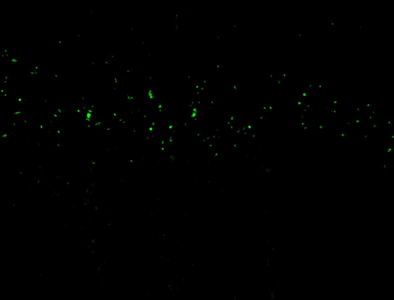
Immunohistochemical Detection of GFP in Transplanted NPCs
GFP-positive transplanted NPCs have morphological features of hippocampal pyramidal neurons at day 90 after grafting. IHC image submitted by a verified customer review.
Western Blot Detection of GFP in HeLa Cells
Lane 1: HeLa cells. Lane 2: mock transfected HeLa cell lysate. Load: 35 ug per lane. Primary antibody: GFP antibody at 1 ug/ml for 1 h at room temperature. Secondary antibody: IRDye(R) 800 conjugated Donkey-a-Goat IgG [H&L] MX7 () secondary antibody at 1:2,500 for 45 min at RT. Block: 5% BLOTTO overnight at 4C. Predicted/Observed size: 27 kDa, 33 kDa for GFP. Other band(s): none.
Use of Anti-GFP Antibody in Western Blot
Western Blot of GFP Antibody. Lane 1: Opal Prestained Molecular Weight Marker
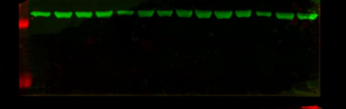
Use of Anti-GFP Antibody in Multi-Lysate Western Blot
Multi-lysate Western Blot of GFP Antibody. Marker: Opal Pre-stained ladder
Immunocytochemistry/ Immunofluorescence: GFP Antibody [NB100-1770] -
Immunocytochemistry/ Immunofluorescence: GFP Antibody [NB100-1770] - Viral recombination of Oxtr in anterior DG hilar neurons impairs discrimination of social, but not non-social, stimuli. a Schematic illustrating viral injection & behavioral testing timeline. b Representative images of Cre & GFP virus infection in aDG. Scale bar, 200 μm. c Representative images & quantifications of cFos immunoreactivity in granule cell layer of aDG (GFP: n = 7, Cre: n = 4). Scale bar, 75 μm. d Behavioral schematic (top) & quantification (bottom) of single object exploration (GFP: n = 11, Cre: n = 6). e Behavioral schematic (top) & quantification (bottom) of novel objection recognition (GFP: n = 11, Cre: n = 6). Quantifications are displayed as Habituation (trials 1–3), Test (trial 4), & discrimination ratio (trial 4). f Behavioral schematic (top) & quantification (bottom) of social exploration test (GFP: n = 11, Cre: n = 6). g Behavioral schematic (top) & quantification (bottom) of social discrimination task (GFP: n = 11, Cre: n = 6). Quantifications are displayed as Habituation (trials 1–3), Test (trial 4), & discrimination ratio (trial 4). All data are displayed as mean ± SEM Image collected & cropped by CiteAb from the following publication (https://pubmed.ncbi.nlm.nih.gov/29222469), licensed under a CC-BY license. Not internally tested by Novus Biologicals.
Immunohistochemistry: GFP Antibody [NB100-1770] -
Immunohistochemistry: GFP Antibody [NB100-1770] - Macrophages regulate dormancy in tumor cells.a Representative image of triple immunofluorescently stained in E0771-GFP primary tumor tissue for tumor cells, macrophages, & NR2F1. Green = GFP; Red = NR2F1; White = IBA-1; Blue = DAPI. White arrow shows a macrophage. The yellow arrow contact between an NR2F1-positive tumor cell & a macrophage. Mϕ=Macrophage. Scale bar=20 μm. b Quantification showing frequency of distances between NR2F1+ tumor cells to nearest macrophage in primary tumor. Data is normalized to frequency of distances between all DAPI+ nuclei to nearest TMEM. Bar = mean. Error bars = ±SEM. n = 34 fields of view (551 × 316 µm2) in 4 animals. For comparison between 0 & 200 µm bins a two-tailed Mann-Whitney test used (p < 0.0001). ****p < 0.0001. c Representative immunofluorescence images of NR2F1 expression in E0771-GFP tumor cells cultured alone, in direct contact w/ BAC1.2F5 macrophages, or in direct contact w/ HUVEC endothelial cells. White arrows show macrophages or endothelial cells in direct contact w/ a tumor cell. Green = GFP; Red = NR2F1; Blue = DAPI. TC = Tumor Cell. Mϕ = Macrophage. EC = Endothelial Cell. Scale bar = 15 μm. d Percentage of NR2F1-positive tumor cells from each group in C. TC alone: n = 777 cells in 9 independent experiments; TC+Mϕ; n = 226 cells in 6 independent experiments, TC+EC = n = 359 cells in 4 independent experiments. Bar = mean. Error bars = ±SEM. For TC vs. TC+Mϕ (p = 0.0039), & for TC vs. TC+EC (p = 1), a two-tailed Kruskal-Wallis test w/ Dunn’s multiple comparisons adjustment used. For TC+Mϕ vs. TC+EC (0.012), a two-tailed one-way ANOVA w/ Sidak’s multiple comparison adjustment used. *p < 0.05. **p < 0.01; ns = not significant. Source data are provided as a Source Data file. Image collected & cropped by CiteAb from following publication (https://pubmed.ncbi.nlm.nih.gov/35110548), licensed under a CC-BY license. Not internally tested by Novus Biologicals.
Immunocytochemistry/ Immunofluorescence: GFP Antibody [NB100-1770] -
Immunocytochemistry/ Immunofluorescence: GFP Antibody [NB100-1770] - Viral recombination of Oxtr in aCA2/CA3distal impairs discrimination of social, but not non-social, stimuli. a Schematic illustrating viral injection & behavioral testing timeline. b Representative images of Cre & GFP virus infection in aCA2/CA3. Scale bar, 200 μm. c Behavioral schematic (top) & quantification (bottom) of single object exploration (GFP: n = 7, Cre: n = 10). d Behavioral schematic (top) & quantification (bottom) of novel objection recognition (GFP: n = 7, Cre: n = 10). Quantifications are displayed as Habituation (trials 1–3), Test (trial 4), & discrimination ratio (trial 4). e Behavioral schematic (top) & quantification (bottom) of social exploration test (GFP: n = 7, Cre: n = 10). f Behavioral schematic (top) & quantification (bottom) of social discrimination task (GFP: n = 7, Cre: n = 10). Quantifications are displayed as Habituation (trials 1–3), Test (trial 4), & discrimination ratio (trial 4). All data are displayed as mean ± SEM Image collected & cropped by CiteAb from the following publication (https://pubmed.ncbi.nlm.nih.gov/29222469), licensed under a CC-BY license. Not internally tested by Novus Biologicals.
Immunocytochemistry/ Immunofluorescence: GFP Antibody [NB100-1770] -
Immunocytochemistry/ Immunofluorescence: GFP Antibody [NB100-1770] - Transduction of mature retinal ganglion cells.Dissociated retinal ganglion cells (RGCs) isolated from adult rats (a–c), mice (d–f) & zebrafish (g–i) were transduced with baculovirus (bv) encoding fGFP (rat & mice) or DsRed (zebrafish). Representative pictures of vehicle- (-) & bv -treated cultures (a,d,g) visualize transduced RGCs (GFP & DsRed, respectively) that were either co-stained with the neuronal marker beta III-tubulin (Tub, red) at 2 days after transduction (d.a.t) for rat & mice RGCs (a,d) or identified by EGFP expression for zebrafish RGCs at 6 d.a.t. (g). The percentage of transduced RGCs was determined at 1, 2 & 3 days after transduction (d.a.t.) for rat & mice (b,e) & at 4 & 6 d.a.t. for zebrafish (h). Treatment effects compared to vehicle-treated controls: ***p < 0.001, **p < 0.01. Scale bars: 50 μm. RGC survival (c,f,i) was not affected by baculovirus application (bv) compared to vehicle-treated controls (-). (j) Delayed transduction of adult zebrafish RGCs with DsRed-bv after 4 days in culture. Scale bar: 50 μm. (k,l) Co-transduction of adult rat RGCs with fGFP-bv & DsRed-bv. The two viruses were added to retinal cultures simultaneously at half the concentration of single transductions. Representative pictures show co-transduced, fGFP- & DsRed-expressing RGCs (green & red, respectively) that were co-stained against the neuronal marker beta III-tubulin (white) at 2 d.a.t. Scale bar: 50 μm. (k). The percentage of transduced RGCs was determined at 2 & 3 d.a.t. (l). Non-significant difference in co-tranduction efficiency compared to single transductions. Image collected & cropped by CiteAb from the following publication (https://www.nature.com/articles/srep38928), licensed under a CC-BY license. Not internally tested by Novus Biologicals.
Immunocytochemistry/ Immunofluorescence: GFP Antibody [NB100-1770] -
Immunocytochemistry/ Immunofluorescence: GFP Antibody [NB100-1770] - Transduction of dorsal root ganglion neurons.Dissociated dorsal root ganglion neurons (DRG) isolated from postnatal (a–c) or adult (d–f) rats & adult mice (g–i) were transduced with fGFP-baculovirus (bv). Representative pictures of vehicle (-) & bv -treated cultures (a,d,g) show transduced, GFP-expressing neurons (green) that were co-stained with the neuronal marker beta III-tubulin (Tub, red) at 2 days after transduction (d.a.t.). The percentage of transduced, GFP-expressing DRG neurons was determined at 1, 2 & 3 d.a.t. (b,e,h), revealing similar transduction efficiencies of 80–90% in postnatal & adult DRG neurons. Cell survival in postnatal (c) & adult (f, i) DRG cultures was not affected by bv-application compared to vehicle-treated controls (-). Scale bars: 100 μm. Image collected & cropped by CiteAb from the following publication (https://www.nature.com/articles/srep38928), licensed under a CC-BY license. Not internally tested by Novus Biologicals.
Immunocytochemistry/ Immunofluorescence: GFP Antibody [NB100-1770] -
Immunocytochemistry/ Immunofluorescence: GFP Antibody [NB100-1770] - hnRNPA3 knockout enhances DNA damage in DPR expressing HeLa cells. a Immunocytochemical detection of gamma H2AX foci in GFP-tagged DPR-expressing HeLa WT & A3KO HeLa cells. b Fold change of gamma H2AX foci in HeLa WT & A3KO cells expressing the indicated DPRs relative to HeLa WT cells with GFP expression. N = 47–127 cells from 2 biological replicates. c Immunocytochemical detection of pATM foci in GFP-tagged DPR-expressing HeLa WT & A3KO HeLa cells. d Fold change of pATM foci in HeLa WT & A3KO cells expressing the indicated DPRs relative to HeLa WT cells with GFP expression. N = 71–263 cells from 2 biological replicates. GFP EGFP transfected, GA EGFP-tagged poly-GA 175 repeats transfected, GR EGFP-tagged poly-GR 177 repeats transfected, PR: EGFP-tagged poly-PR 176 repeats transfected. All graphs are shown as mean ± SEM. *p < 0.05, **p < 0.01; one-way ANOVA & Tukey’s post-hoc test. Scale bar 10 μm Image collected & cropped by CiteAb from the following publication (https://pubmed.ncbi.nlm.nih.gov/31642962), licensed under a CC-BY license. Not internally tested by Novus Biologicals.Applications for GFP Antibody - BSA Free
Application
Recommended Usage
ELISA
1:10000-1:30000
Electron Microscopy
1:10 - 1:500
Flow Cytometry
1:10 - 1:1000
Immunocytochemistry/ Immunofluorescence
1:500
Immunohistochemistry
1:200-1:1000
Immunohistochemistry Free-Floating
1:10 - 1:500
Immunohistochemistry-Frozen
1:10 - 1:500
Immunohistochemistry-Paraffin
1:10 - 1:500
Immunoprecipitation
1:10 - 1:500
Western Blot
1:1000-1:10000
Application Notes
This product is designed to detect GFP and its variants. Goat This product has been tested by ELISA, SDS-PAGE, Western blot, and Immunofluorescence. This product is ideal for western blotting, ELISA, immunofluorescence, IHC, and IP. This antibody can be used to detect GFP by ELISA (sandwich or capture) for the direct binding of antigen and recognizes wild type, recombinant and enhanced forms of GFP. Biotin conjugated polyclonal anti-GFP used in a sandwich ELISA is well suited to titrate GFP in solution using this antibody in combination with monoclonal anti-GFP using either form of the antibody as the capture or detection antibody. However, use the monoclonal form only for the detection of wild type or recombinant GFP as this form does not sufficiently detect 'enhanced' GFP. The detection antibody is typically conjugated to biotin and subsequently reacted with streptavidin-HRP. Fluorochrome conjugated polyclonal anti-GFP can be used to detect GFP by immunofluorescence microscopy in prokaryotic (E.coli) and eukaryotic (CHO cells) expression systems and detects GFP containing inserts. Significant amplification of signal is achieved using fluorochrome conjugated polyclonal anti-GFP relative to the fluorescence of GFP alone. For immunoblotting use either alkaline phosphatase or peroxidase conjugated polyclonal anti-GFP to detect GFP or GFP-containing proteins on western blots. Researchers should determine optimal titers for applications.
Reviewed Applications
Read 7 reviews rated 4.7 using NB100-1770 in the following applications:
Flow Cytometry Panel Builder
Bio-Techne Knows Flow Cytometry
Save time and reduce costly mistakes by quickly finding compatible reagents using the Panel Builder Tool.
Advanced Features
- Spectra Viewer - Custom analysis of spectra from multiple fluorochromes
- Spillover Popups - Visualize the spectra of individual fluorochromes
- Antigen Density Selector - Match fluorochrome brightness with antigen density

Formulation, Preparation, and Storage
Purification
Immunogen affinity purified
Formulation
0.02 M Potassium Phosphate, 0.15 M Sodium Chloride, pH 7.2
Format
BSA Free
Preservative
0.01% Sodium Azide
Concentration
Please see the vial label for concentration. If unlisted please contact technical services.
Shipping
The product is shipped with polar packs. Upon receipt, store it immediately at the temperature recommended below.
Stability & Storage
Store at -20C. Avoid freeze-thaw cycles.
Background: GFP
References
1. Shi, C., Pan, F. C., Kim, J. N., Washington, M. K., Padmanabhan, C., Meyer, C. T.,... Means, A. L. (2019). Differential Cell Susceptibilities to Kras(G12D) in the Setting of Obstructive Chronic Pancreatitis. Cell Mol Gastroenterol Hepatol. doi:10.1016/j.jcmgh.2019.07.001
2. Zhao, S., Fortier, T. M., & Baehrecke, E. H. (2018). Autophagy Promotes Tumor-like Stem Cell Niche Occupancy. Curr Biol, 28(19), 3056-3064.e3053. doi:10.1016/j.cub.2018.07.075
3. Zusso, M., Lunardi, V., Franceschini, D., Pagetta, A., Lo, R., Stifani, S.,... Moro, S. (2019). Ciprofloxacin and levofloxacin attenuate microglia inflammatory response via TLR4/NF-kB pathway. J Neuroinflammation, 16(1), 148. doi:10.1186/s12974-019-1538-9
Long Name
Green Fluorescent Protein
Alternate Names
eGFP, GFPuv
Additional GFP Products
Product Documents for GFP Antibody - BSA Free
Certificate of Analysis
To download a Certificate of Analysis, please enter a lot or batch number in the search box below.
Product Specific Notices for GFP Antibody - BSA Free
This product is for research use only and is not approved for use in humans or in clinical diagnosis. Primary Antibodies are guaranteed for 1 year from date of receipt.
Citations for GFP Antibody - BSA Free
 Powered by Bioz
Customer Reviews for GFP Antibody - BSA Free (7)
4.7 out of 5
7 Customer Ratings
Have you used GFP Antibody - BSA Free?
Submit a review and receive an Amazon gift card!
$25/€18/£15/$25CAN/¥2500 Yen for a review with an image
$10/€7/£6/$10CAN/¥1110 Yen for a review without an image
Submit a review
Customer Images
-(01-mg)_NB100-1770_8391.jpg)
Showing
1
-
5 of
7 reviews
Showing All
Filter By:
-
Application: Immunohistochemistry-FrozenSample Tested: Mouse brainSpecies: MouseVerified Customer | Posted 05/04/2021Endogenous Laminin y11:200 dilution

-
Application: Western BlotSample Tested: 4T1 mouse breast cancer cell lineSpecies: MouseVerified Customer | Posted 03/08/2020GFP was used in WB as 1:2000 dilution
-
Application: ImmunofluorescenceSample Tested: Tumor sectionsSpecies: MouseVerified Customer | Posted 10/21/2019GFP expressing epitopes in murine breast cancer tissue
-
Application: ImmunocytochemistrySample Tested: Itga8-GFP transfected HEK293 cellsSpecies: cellsVerified Customer | Posted 05/08/2018HEK293 cells stable transfected with integrin alpha8-GFP, were immunostained for GFP (green) and counterstained with phalloidin (red) and DAPI (BLUE)
-
Application: ImmunofluorescenceSample Tested: See PMID 24753232Species: MouseVerified Customer | Posted 12/12/2014
-
Application: ImmunohistochemistrySample Tested:Species: MouseVerified Customer | Posted 06/19/2014GFP-positive transplanted NPCs have morphological features of hippocampal pyramidal neurons at day 90 after grafting.
-
Application: ImmunohistochemistrySample Tested: Mouse brainSpecies: MouseVerified Customer | Posted 05/15/2012GFP-positive dendrites with spines
There are no reviews that match your criteria.
Protocols
Find general support by application which include: protocols, troubleshooting, illustrated assays, videos and webinars.
- 7-Amino Actinomycin D (7-AAD) Cell Viability Flow Cytometry Protocol
- Antigen Retrieval Protocol (PIER)
- Antigen Retrieval for Frozen Sections Protocol
- Appropriate Fixation of IHC/ICC Samples
- Cellular Response to Hypoxia Protocols
- Chromogenic IHC Staining of Formalin-Fixed Paraffin-Embedded (FFPE) Tissue Protocol
- Chromogenic Immunohistochemistry Staining of Frozen Tissue
- ClariTSA™ Fluorophore Kits
- Detection & Visualization of Antibody Binding
- ELISA Sample Preparation & Collection Guide
- ELISA Troubleshooting Guide
- Extracellular Membrane Flow Cytometry Protocol
- Flow Cytometry Protocol for Cell Surface Markers
- Flow Cytometry Protocol for Staining Membrane Associated Proteins
- Flow Cytometry Staining Protocols
- Flow Cytometry Troubleshooting Guide
- Fluorescent IHC Staining of Frozen Tissue Protocol
- Graphic Protocol for Heat-induced Epitope Retrieval
- Graphic Protocol for the Preparation and Fluorescent IHC Staining of Frozen Tissue Sections
- Graphic Protocol for the Preparation and Fluorescent IHC Staining of Paraffin-embedded Tissue Sections
- Graphic Protocol for the Preparation of Gelatin-coated Slides for Histological Tissue Sections
- How to Run an R&D Systems DuoSet ELISA
- How to Run an R&D Systems Quantikine ELISA
- How to Run an R&D Systems Quantikine™ QuicKit™ ELISA
- ICC Cell Smear Protocol for Suspension Cells
- ICC Immunocytochemistry Protocol Videos
- ICC for Adherent Cells
- IHC Sample Preparation (Frozen sections vs Paraffin)
- Immunocytochemistry (ICC) Protocol
- Immunocytochemistry Troubleshooting
- Immunofluorescence of Organoids Embedded in Cultrex Basement Membrane Extract
- Immunofluorescent IHC Staining of Formalin-Fixed Paraffin-Embedded (FFPE) Tissue Protocol
- Immunohistochemistry (IHC) and Immunocytochemistry (ICC) Protocols
- Immunohistochemistry Frozen Troubleshooting
- Immunohistochemistry Paraffin Troubleshooting
- Immunoprecipitation Protocol
- Intracellular Flow Cytometry Protocol Using Alcohol (Methanol)
- Intracellular Flow Cytometry Protocol Using Detergents
- Intracellular Nuclear Staining Flow Cytometry Protocol Using Detergents
- Intracellular Staining Flow Cytometry Protocol Using Alcohol Permeabilization
- Intracellular Staining Flow Cytometry Protocol Using Detergents to Permeabilize Cells
- Preparing Samples for IHC/ICC Experiments
- Preventing Non-Specific Staining (Non-Specific Binding)
- Primary Antibody Selection & Optimization
- Propidium Iodide Cell Viability Flow Cytometry Protocol
- Protocol for Heat-Induced Epitope Retrieval (HIER)
- Protocol for Liperfluo
- Protocol for Making a 4% Formaldehyde Solution in PBS
- Protocol for VisUCyte™ HRP Polymer Detection Reagent
- Protocol for the Characterization of Human Th22 Cells
- Protocol for the Characterization of Human Th9 Cells
- Protocol for the Fluorescent ICC Staining of Cell Smears - Graphic
- Protocol for the Fluorescent ICC Staining of Cultured Cells on Coverslips - Graphic
- Protocol for the Preparation & Fixation of Cells on Coverslips
- Protocol for the Preparation and Chromogenic IHC Staining of Frozen Tissue Sections
- Protocol for the Preparation and Chromogenic IHC Staining of Frozen Tissue Sections - Graphic
- Protocol for the Preparation and Chromogenic IHC Staining of Paraffin-embedded Tissue Sections
- Protocol for the Preparation and Chromogenic IHC Staining of Paraffin-embedded Tissue Sections - Graphic
- Protocol for the Preparation and Fluorescent ICC Staining of Cells on Coverslips
- Protocol for the Preparation and Fluorescent ICC Staining of Non-adherent Cells
- Protocol for the Preparation and Fluorescent ICC Staining of Stem Cells on Coverslips
- Protocol for the Preparation and Fluorescent IHC Staining of Frozen Tissue Sections
- Protocol for the Preparation and Fluorescent IHC Staining of Paraffin-embedded Tissue Sections
- Protocol for the Preparation of Gelatin-coated Slides for Histological Tissue Sections
- Protocol for the Preparation of a Cell Smear for Non-adherent Cell ICC - Graphic
- Protocol: Annexin V and PI Staining by Flow Cytometry
- Protocol: Annexin V and PI Staining for Apoptosis by Flow Cytometry
- Quantikine HS ELISA Kit Assay Principle, Alkaline Phosphatase
- Quantikine HS ELISA Kit Principle, Streptavidin-HRP Polymer
- R&D Systems Quality Control Western Blot Protocol
- Sandwich ELISA (Colorimetric) – Biotin/Streptavidin Detection Protocol
- Sandwich ELISA (Colorimetric) – Direct Detection Protocol
- TUNEL and Active Caspase-3 Detection by IHC/ICC Protocol
- The Importance of IHC/ICC Controls
- Troubleshooting Guide: ELISA
- Troubleshooting Guide: Fluorokine Flow Cytometry Kits
- Troubleshooting Guide: Immunohistochemistry
- Troubleshooting Guide: Western Blot Figures
- Western Blot Conditions
- Western Blot Protocol
- Western Blot Protocol for Cell Lysates
- Western Blot Troubleshooting
- Western Blot Troubleshooting Guide
- View all Protocols, Troubleshooting, Illustrated assays and Webinars
FAQs for GFP Antibody - BSA Free
Showing
1
-
2 of
2 FAQs
Showing All
-
Q: Does this antibody NB100-1770 detect eGFP?
A: NB100-1770 has not yet been tested against eGFP by our lab. Theoretically, you should be able to detect the eGFP with this antibody, as the two are very homologous; however, we cannot guarantee it. If you would like to test an antibody in an untested species and/or application and share your results with us, I can recommend our Innovator's Reward program. Our Innovator's Reward program was created to allow researchers the opportunity to try our primary antibodies in an untested species or application, without the financial risk of failure. To participate you simply submit an online review detailing your positive or negative results. In return, you receive a discount voucher for 100% of the purchase price of the reviewed product. Reviews may also be submitted by emailing Innovators Reward.
-
Q: We are looking for a rabbit anti-GFP antibody as a primary antibody for immunofluorescence and WB. I've seen that you carry several anti-GFP antibodies and I would like to ask which one you would recommend. The one with Cat.No NB600-308 has been used in a lot of publications and I reckon it is a solid antibody. Is this our best choice, or is there a better alternative?
A: NB600-308 is an excellent GFP antibody and I think it would work very well for you. It has been reviewed and published with which usually makes our customer feel much more confident, seeing as it worked in other people's hand.
-
Q: Does this antibody NB100-1770 detect eGFP?
A: NB100-1770 has not yet been tested against eGFP by our lab. Theoretically, you should be able to detect the eGFP with this antibody, as the two are very homologous; however, we cannot guarantee it. If you would like to test an antibody in an untested species and/or application and share your results with us, I can recommend our Innovator's Reward program. Our Innovator's Reward program was created to allow researchers the opportunity to try our primary antibodies in an untested species or application, without the financial risk of failure. To participate you simply submit an online review detailing your positive or negative results. In return, you receive a discount voucher for 100% of the purchase price of the reviewed product. Reviews may also be submitted by emailing Innovators Reward.
-
Q: We are looking for a rabbit anti-GFP antibody as a primary antibody for immunofluorescence and WB. I've seen that you carry several anti-GFP antibodies and I would like to ask which one you would recommend. The one with Cat.No NB600-308 has been used in a lot of publications and I reckon it is a solid antibody. Is this our best choice, or is there a better alternative?
A: NB600-308 is an excellent GFP antibody and I think it would work very well for you. It has been reviewed and published with which usually makes our customer feel much more confident, seeing as it worked in other people's hand.
Loading...