GLI-2 Antibody - BSA Free
Novus Biologicals | Catalog # NB600-874
![Western Blot: GLI-2 Antibody [NB600-874]](https://resources.rndsystems.com/images/products/GLI-2-Antibody-Western-Blot-NB600-874-img0012.jpg "Western Blot: GLI-2 Antibody [NB600-874]")
Key Product Details
Validated by
Biological Validation
Species Reactivity
Validated:
Human, Mouse, Rat
Cited:
Human, Mouse
Applications
Validated:
Immunohistochemistry, Immunohistochemistry-Paraffin, Western Blot, ELISA, Flow (Intracellular), Chromatin Immunoprecipitation (ChIP)
Cited:
Immunohistochemistry-Paraffin, Western Blot, Chemotaxis
Label
Unconjugated
Antibody Source
Polyclonal Rabbit IgG
Format
BSA Free
Loading...
Product Specifications
Immunogen
This affinity purified GLI-2 Antibody was prepared from whole rabbit serum produced by repeated immunizations with a synthetic peptide corresponding to an internal region near amino acids 30-65 of human GLI-2 (isoform a). (Uniprot: P10070)
Reactivity Notes
A BLAST analysis was used to suggest cross reactivity with Gli-2 from chimpanzee based on the immunizing sequence
Mouse reactivity reported in scientific literature (PMID: 27760825).
Mouse reactivity reported in scientific literature (PMID: 27760825).
Localization
Nuclear
Clonality
Polyclonal
Host
Rabbit
Isotype
IgG
Description
This affinity-purified antibody is directed against human Gli-2 protein. The product was affinity purified from monospecific antiserum by immunoaffinity purification. A BLAST analysis was used to suggest cross reactivity with GLI-2 from human and chimpanzee based on the immunizing sequence.
Store vial at -20C prior to opening. Aliquot contents and freeze at -20C or below for extended storage. Avoid cycles of freezing and thawing. Centrifuge product if not completely clear after standing at room temperature. This product is stable for several weeks at 4C as an undiluted liquid. Dilute only prior to immediate use.
Store vial at -20C prior to opening. Aliquot contents and freeze at -20C or below for extended storage. Avoid cycles of freezing and thawing. Centrifuge product if not completely clear after standing at room temperature. This product is stable for several weeks at 4C as an undiluted liquid. Dilute only prior to immediate use.
Scientific Data Images for GLI-2 Antibody - BSA Free
Western Blot: GLI-2 Antibody [NB600-874]
Western Blot: GLI-2 Antibody [NB600-874] - Analysis of Gli-2 protein in rat testes (lane 1) and human HEK293 (lane 2) whole cell lysates (arrowhead). See Ruppert et al for testing conditions. Each lane contains approximately 35 ug of lysate. Primary antibody was used at a 1:400 dilution in 5% BLOTTO in PBS overnight at 4C. The membrane was washed and reacted with a 1:10,000 dilution of IRDye 800 conjugated Gt-a-Rabbit IgG [H&L] MX10 for 45 min at room temperature (800 nm channel, green).![Immunohistochemistry-Paraffin: GLI-2 Antibody [NB600-874]](https://resources.rndsystems.com/images/products/GLI-2-Antibody-Immunohistochemistry-Paraffin-NB600-874-img0013.jpg "Immunohistochemistry-Paraffin: GLI-2 Antibody [NB600-874]")
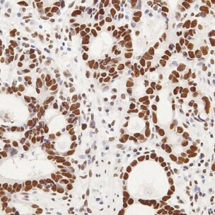
Immunohistochemistry-Paraffin: GLI-2 Antibody [NB600-874]
Immunohistochemistry-Paraffin: GLI-2 Antibody [NB600-874] - Mouse adenocarcinoma section shows high and diffuse nuclear Gli2 expression. IHC-P image submitted by a verified customer review.![Flow (Intracellular): GLI-2 Antibody [NB600-874]](https://resources.rndsystems.com/images/products/GLI-2-Antibody-Flow-Intracellular-NB600-874-img0006.jpg "Flow (Intracellular): GLI-2 Antibody [NB600-874]")
Flow (Intracellular): GLI-2 Antibody [NB600-874]
Flow (Intracellular): GLI-2 Antibody [NB600-874] - intracellular staining for GLI-2 in untreated human B cell lines (BCWM.1, MWCL-1 and RPCI-WM1) using anti-GLI-2 antibody. Rabbit IgG Isotype Control (Cat# NBP2-36463) was used as a negative control. Image courtesy of Sherine Elsawa, Northern Illinois University.![Immunohistochemistry: GLI-2 Antibody [NB600-874]](https://resources.rndsystems.com/images/products/GLI-2-Antibody-Immunohistochemistry-NB600-874-img0010.jpg "Immunohistochemistry: GLI-2 Antibody [NB600-874]")
Immunohistochemistry: GLI-2 Antibody [NB600-874]
Immunohistochemistry: GLI-2 Antibody [NB600-874] - Staining of tumor cells in human breast tissue. Tissue was formalin-fixed and paraffin embedded. Brown color indicates presence of protein, blue color shows cell nuclei.
GLI-2 Antibody
Affinity Purified anti-Gli2 antibody shows strong cytoplasmic and membranous staining of tumor cells in human breast tissue. Tissue was formalin-fixed and paraffin embedded. Brown color indicates presence of protein, blue color shows cell nuclei. Personal Communication, Kenneth Wester, www.proteinatlas.org, Uppsala, Sweden.
GLI-2 Antibody
Western blot using affinity purified antibody shows detection of Gli-2 protein. Lane 1: rat testes
Western Blot: GLI-2 Antibody - BSA Free [NB600-874] -
Analysis of Ptch receptor-dependent inhibition of Hh signaling. (A,C,E) Immunocytochemistry detection of Gli3 (A), Gli1 (C) and Gli2 (E) in the wild-type, Ptch1−/−, and Ptch2−/− MEFs. Scale bar 50 μm. (B,D,F) Quantification of the intensity of the nuclear GLI3 (B), GLI1 (D), and GLI2 (F) in wild-type, Ptch1−/−, and Ptch2−/− MEFs. **p ≤ 0.005, ***p ≤ 0.0005, ****p < 0.0001. (G) Western blot showing nuclear (N) and cellular (C) expression of Gli2 (FL = full length and RF = repressor form) in wild-type, Ptch1−/− and Ptch2−/− MEFs. Image collected and cropped by CiteAb from the following open publication (https://pubmed.ncbi.nlm.nih.gov/35574464), licensed under a CC-BY license. Not internally tested by Novus Biologicals.
Immunohistochemistry: GLI-2 Antibody - BSA Free [NB600-874] -
Immunohistochemical analysis of the expressions of Sonic Hedgehog (SHH), Gli1, and Gli2 in human melanoma samples. (A) Representative images of SHH, Gli1, and Gli2 expressions in normal dermal tissue and melanoma. SHH, Gli1, and Gli2 are expressed not only in melanoma cells but also in tumor vascular endothelial cells in the stroma. Scale bar: 200 um. Arrowhead: tumor vasculature. (B) The numbers of SHH-, Gli1-, and Gli2-positive cells/mm2 are significantly higher in melanoma tissues than in normal skin tissues. The data from a typical experiment (mean +/- SD) are presented. * p < 0.05 between the indicated groups. (C) Staining intensity (SI, A) is evaluated visually: negative (0), weak (1), moderate (2), and strong (3). SI3 (SHH and Gli1, n = 15), SI3 (SHH and Gli2, n = 12), SI2 (SHH and Gli1, n = 7), and SI2 (SHH and Gli2, n = 4). Spearman’s correlations between the intensities of SHH and Gli1 (left) or Gli2 (right) were analyzed using GraphPad Prism 6.0. (D) Immunohistochemical staining for SHH, Gli1, and Gli2 in osteolytic malignant melanoma of the maxilla. Each photo is a magnification of the rectangle-delimited area corresponding to a melanoma bone-destructive area. Scale bars: 200 um (upper) and 100 um (lower). Arrowhead: osteoclasts. Triangular arrowheads: tumor vasculature. Bn: bone. Image collected and cropped by CiteAb from the following open publication (https://pubmed.ncbi.nlm.nih.gov/37240209), licensed under a CC-BY license. Not internally tested by Novus Biologicals.
Immunocytochemistry/ Immunofluorescence: GLI-2 Antibody - BSA Free [NB600-874] -
Analysis of Ptch receptor-dependent inhibition of Hh signaling. (A,C,E) Immunocytochemistry detection of Gli3 (A), Gli1 (C) and Gli2 (E) in the wild-type, Ptch1−/−, and Ptch2−/− MEFs. Scale bar 50 μm. (B,D,F) Quantification of the intensity of the nuclear GLI3 (B), GLI1 (D), and GLI2 (F) in wild-type, Ptch1−/−, and Ptch2−/− MEFs. **p ≤ 0.005, ***p ≤ 0.0005, ****p < 0.0001. (G) Western blot showing nuclear (N) and cellular (C) expression of Gli2 (FL = full length and RF = repressor form) in wild-type, Ptch1−/− and Ptch2−/− MEFs. Image collected and cropped by CiteAb from the following open publication (https://pubmed.ncbi.nlm.nih.gov/35574464), licensed under a CC-BY license. Not internally tested by Novus Biologicals.Applications for GLI-2 Antibody - BSA Free
Application
Recommended Usage
Chromatin Immunoprecipitation (ChIP)
1:10-1:500
ELISA
1:10000-1:30000
Immunohistochemistry
1:500-1:2000
Immunohistochemistry-Paraffin
1:10-1:500
Western Blot
1:500-1:2000
Application Notes
This antibody has been tested for use in ELISA, immunohistochemistry and western blot. Specific conditions for reactivity should be optimized by the end user. See figure legend for expectations by western blot. Multiple splice variants have been reported for this protein a, b, g and d (133.3, 131.6, 88.1 and 86.4 kDa respectively). Detection of Gli-2 by western blot may be enhanced if nuclear extracts are used instead of whole cell lysates as the expression/abundance of Gli-2 is likely to be low. Furthermore, Gli-2 expression is likely to be developmentally regulated and induced, making it difficult to detect in whole tissue homogenates.
Use in chromatin immunoprecipitation reported in scientific literature (PMID: 27760825).
Use in chromatin immunoprecipitation reported in scientific literature (PMID: 27760825).
Reviewed Applications
Read 1 review rated 5 using NB600-874 in the following applications:
Flow Cytometry Panel Builder
Bio-Techne Knows Flow Cytometry
Save time and reduce costly mistakes by quickly finding compatible reagents using the Panel Builder Tool.
Advanced Features
- Spectra Viewer - Custom analysis of spectra from multiple fluorochromes
- Spillover Popups - Visualize the spectra of individual fluorochromes
- Antigen Density Selector - Match fluorochrome brightness with antigen density

Formulation, Preparation, and Storage
Purification
Immunogen affinity purified
Formulation
0.02 M Potassium Phosphate, 0.15 M Sodium Chloride, pH 7.2
Format
BSA Free
Preservative
0.01% Sodium Azide
Concentration
Please see the vial label for concentration. If unlisted please contact technical services.
Shipping
The product is shipped with polar packs. Upon receipt, store it immediately at the temperature recommended below.
Stability & Storage
Store at -20C short term. Aliquot and store at -80C long term. Avoid freeze-thaw cycles.
Background: GLI-2
Long Name
GLI-Kruppel family member GLI2
Alternate Names
GLI2, THP2
Entrez Gene IDs
2736 (Human)
Gene Symbol
GLI2
UniProt
Additional GLI-2 Products
Product Documents for GLI-2 Antibody - BSA Free
Certificate of Analysis
To download a Certificate of Analysis, please enter a lot or batch number in the search box below.
Product Specific Notices for GLI-2 Antibody - BSA Free
This product is for research use only and is not approved for use in humans or in clinical diagnosis. Primary Antibodies are guaranteed for 1 year from date of receipt.
Related Research Areas
Citations for GLI-2 Antibody - BSA Free
 Powered by Bioz
Customer Reviews for GLI-2 Antibody - BSA Free (1)
5 out of 5
1 Customer Rating
Have you used GLI-2 Antibody - BSA Free?
Submit a review and receive an Amazon gift card!
$25/€18/£15/$25CAN/¥2500 Yen for a review with an image
$10/€7/£6/$10CAN/¥1110 Yen for a review without an image
Submit a review
Customer Images
Showing
1
-
1 of
1 review
Showing All
Filter By:
-
Application: Immunohistochemistry-ParaffinSample Tested: adenocarcinomasSpecies: MouseVerified Customer | Posted 02/28/2021Mouse adenocarcinomas section show high and diffuse nuclear Gli2 expression.

There are no reviews that match your criteria.
Protocols
Find general support by application which include: protocols, troubleshooting, illustrated assays, videos and webinars.
- 7-Amino Actinomycin D (7-AAD) Cell Viability Flow Cytometry Protocol
- Antigen Retrieval Protocol (PIER)
- Antigen Retrieval for Frozen Sections Protocol
- Appropriate Fixation of IHC/ICC Samples
- Cellular Response to Hypoxia Protocols
- ChIP Protocol Video
- Chromatin Immunoprecipitation (ChIP) Protocol
- Chromatin Immunoprecipitation Protocol
- Chromogenic IHC Staining of Formalin-Fixed Paraffin-Embedded (FFPE) Tissue Protocol
- Chromogenic Immunohistochemistry Staining of Frozen Tissue
- ClariTSA™ Fluorophore Kits
- Detection & Visualization of Antibody Binding
- ELISA Sample Preparation & Collection Guide
- ELISA Troubleshooting Guide
- Extracellular Membrane Flow Cytometry Protocol
- Flow Cytometry Protocol for Cell Surface Markers
- Flow Cytometry Protocol for Staining Membrane Associated Proteins
- Flow Cytometry Staining Protocols
- Flow Cytometry Troubleshooting Guide
- Fluorescent IHC Staining of Frozen Tissue Protocol
- Graphic Protocol for Heat-induced Epitope Retrieval
- Graphic Protocol for the Preparation and Fluorescent IHC Staining of Frozen Tissue Sections
- Graphic Protocol for the Preparation and Fluorescent IHC Staining of Paraffin-embedded Tissue Sections
- Graphic Protocol for the Preparation of Gelatin-coated Slides for Histological Tissue Sections
- How to Run an R&D Systems DuoSet ELISA
- How to Run an R&D Systems Quantikine ELISA
- How to Run an R&D Systems Quantikine™ QuicKit™ ELISA
- IHC Sample Preparation (Frozen sections vs Paraffin)
- Immunofluorescent IHC Staining of Formalin-Fixed Paraffin-Embedded (FFPE) Tissue Protocol
- Immunohistochemistry (IHC) and Immunocytochemistry (ICC) Protocols
- Immunohistochemistry Frozen Troubleshooting
- Immunohistochemistry Paraffin Troubleshooting
- Intracellular Flow Cytometry Protocol Using Alcohol (Methanol)
- Intracellular Flow Cytometry Protocol Using Detergents
- Intracellular Nuclear Staining Flow Cytometry Protocol Using Detergents
- Intracellular Staining Flow Cytometry Protocol Using Alcohol Permeabilization
- Intracellular Staining Flow Cytometry Protocol Using Detergents to Permeabilize Cells
- Preparing Samples for IHC/ICC Experiments
- Preventing Non-Specific Staining (Non-Specific Binding)
- Primary Antibody Selection & Optimization
- Propidium Iodide Cell Viability Flow Cytometry Protocol
- Protocol for Heat-Induced Epitope Retrieval (HIER)
- Protocol for Liperfluo
- Protocol for Making a 4% Formaldehyde Solution in PBS
- Protocol for VisUCyte™ HRP Polymer Detection Reagent
- Protocol for the Characterization of Human Th22 Cells
- Protocol for the Characterization of Human Th9 Cells
- Protocol for the Preparation & Fixation of Cells on Coverslips
- Protocol for the Preparation and Chromogenic IHC Staining of Frozen Tissue Sections
- Protocol for the Preparation and Chromogenic IHC Staining of Frozen Tissue Sections - Graphic
- Protocol for the Preparation and Chromogenic IHC Staining of Paraffin-embedded Tissue Sections
- Protocol for the Preparation and Chromogenic IHC Staining of Paraffin-embedded Tissue Sections - Graphic
- Protocol for the Preparation and Fluorescent IHC Staining of Frozen Tissue Sections
- Protocol for the Preparation and Fluorescent IHC Staining of Paraffin-embedded Tissue Sections
- Protocol for the Preparation of Gelatin-coated Slides for Histological Tissue Sections
- Protocol: Annexin V and PI Staining by Flow Cytometry
- Protocol: Annexin V and PI Staining for Apoptosis by Flow Cytometry
- Quantikine HS ELISA Kit Assay Principle, Alkaline Phosphatase
- Quantikine HS ELISA Kit Principle, Streptavidin-HRP Polymer
- R&D Systems Quality Control Western Blot Protocol
- Sandwich ELISA (Colorimetric) – Biotin/Streptavidin Detection Protocol
- Sandwich ELISA (Colorimetric) – Direct Detection Protocol
- TUNEL and Active Caspase-3 Detection by IHC/ICC Protocol
- The Importance of IHC/ICC Controls
- Troubleshooting Guide: ELISA
- Troubleshooting Guide: Fluorokine Flow Cytometry Kits
- Troubleshooting Guide: Immunohistochemistry
- Troubleshooting Guide: Western Blot Figures
- Western Blot Conditions
- Western Blot Protocol
- Western Blot Protocol for Cell Lysates
- Western Blot Troubleshooting
- Western Blot Troubleshooting Guide
- View all Protocols, Troubleshooting, Illustrated assays and Webinars
Loading...