GLI-3 Antibody - BSA Free
Novus Biologicals | Catalog # NBP2-29627
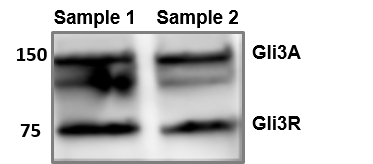
![Western Blot: GLI-3 AntibodyBSA Free [NBP2-29627]](https://resources.rndsystems.com/images/products/GLI-3-Antibody-Western-Blot-NBP2-29627-img0017.jpg "Western Blot: GLI-3 AntibodyBSA Free [NBP2-29627]")
Key Product Details
Validated by
Biological Validation
Species Reactivity
Human, Mouse
Applications
Validated:
Immunohistochemistry, Immunohistochemistry-Paraffin, Western Blot, Flow Cytometry, Flow (Intracellular), Immunocytochemistry/ Immunofluorescence
Cited:
Immunohistochemistry-Paraffin
Label
Unconjugated
Antibody Source
Polyclonal Rabbit IgG
Format
BSA Free
Loading...
Product Specifications
Immunogen
A synthetic peptide made to an internal portion of human Gli3 protein (between residues 50-120) [UniProt P10071]
Reactivity Notes
Immunogen displays the following percentage of sequence identity for non-tested species: Xenopus (84%) and Chicken (85%).
Localization
Nucleus. Cytoplasm.
Clonality
Polyclonal
Host
Rabbit
Isotype
IgG
Theoretical MW
190 kDa.
Disclaimer note: The observed molecular weight of the protein may vary from the listed predicted molecular weight due to post translational modifications, post translation cleavages, relative charges, and other experimental factors.
Disclaimer note: The observed molecular weight of the protein may vary from the listed predicted molecular weight due to post translational modifications, post translation cleavages, relative charges, and other experimental factors.
Scientific Data Images for GLI-3 Antibody - BSA Free
Western Blot: GLI-3 AntibodyBSA Free [NBP2-29627]
Western Blot: GLI-3 Antibody [NBP2-29627] - Adult mouse brain tissue lysates. Image from verified customer review.![Immunocytochemistry/ Immunofluorescence: GLI-3 Antibody - BSA Free [NBP2-29627]](https://resources.rndsystems.com/images/products/GLI-3-Antibody-Immunofluorescence-NBP2-29627-img0012.jpg "Immunocytochemistry/ Immunofluorescence: GLI-3 Antibody - BSA Free [NBP2-29627]")
Immunocytochemistry/ Immunofluorescence: GLI-3 Antibody - BSA Free [NBP2-29627]
Immunocytochemistry/Immunofluorescence: GLI-3 Antibody [NBP2-29627] - HeLa cells with FITC (green). Nuclei and alpha-tubulin were counterstained with DAPI (blue) and DyLight 550 (red).![Immunohistochemistry-Paraffin: GLI-3 Antibody - BSA Free [NBP2-29627]](https://resources.rndsystems.com/images/products/GLI-3-Antibody-Immunohistochemistry-Paraffin-NBP2-29627-img0015.jpg "Immunohistochemistry-Paraffin: GLI-3 Antibody - BSA Free [NBP2-29627]")
Immunohistochemistry-Paraffin: GLI-3 Antibody - BSA Free [NBP2-29627]
Immunohistochemistry-Paraffin: GLI-3 Antibody [NBP2-29627] - Analysis of in mouse liver.![Flow (Intracellular): GLI-3 Antibody - BSA Free [NBP2-29627]](https://resources.rndsystems.com/images/products/GLI-3-Antibody-Flow-Intracellular-NBP2-29627-img0010.jpg "Flow (Intracellular): GLI-3 Antibody - BSA Free [NBP2-29627]")
Flow (Intracellular): GLI-3 Antibody - BSA Free [NBP2-29627]
Flow (Intracellular): GLI-3 Antibody [NBP2-29627] - Intracellular staining for GLI-3 in untreated human B cell lines (BCWM.1, MWCL-1 and RPCI-WM1) using anti-GLI-3 antibody. Rabbit IgG Isotype Control (Cat# NBP2-36463) was used as a negative control. Image courtesy of Sherine Elsawa, Northern Illinois University.![Western Blot: GLI-3 AntibodyBSA Free [NBP2-29627]](https://resources.rndsystems.com/images/products/GLI-3-Antibody-Western-Blot-NBP2-29627-img0016.jpg "Western Blot: GLI-3 AntibodyBSA Free [NBP2-29627]")
Western Blot: GLI-3 AntibodyBSA Free [NBP2-29627]
Western Blot: GLI-3 Antibody [NBP2-29627] - Analysis of GLI-3 in Hela cell lysate and MEF cell lysate.![Immunohistochemistry-Paraffin: GLI-3 Antibody - BSA Free [NBP2-29627]](https://resources.rndsystems.com/images/products/GLI-3-Antibody-Immunohistochemistry-Paraffin-NBP2-29627-img0014.jpg "Immunohistochemistry-Paraffin: GLI-3 Antibody - BSA Free [NBP2-29627]")
Immunohistochemistry-Paraffin: GLI-3 Antibody - BSA Free [NBP2-29627]
Immunohistochemistry-Paraffin: GLI-3 Antibody [NBP2-29627] - Analysis of in mouse testis.Applications for GLI-3 Antibody - BSA Free
Application
Recommended Usage
Flow Cytometry
reported in scientific literature (PMID 26238488)
Immunocytochemistry/ Immunofluorescence
1:50
Immunohistochemistry
1:100
Immunohistochemistry-Paraffin
1:100
Western Blot
1:1000
Application Notes
In Western blot, bands were observed at ~75 kDa (GLI3R) in both HeLa and MEF cell lysates while an additional band was observed ~150 kDa (GLI3A) in MEF cell lysate only. The observed molecular weight of the protein may vary from the listed predicted molecular weight due to post translational modifications, post translation cleavages, relative charges, and other experimental factors.
Reviewed Applications
Read 1 review rated 4 using NBP2-29627 in the following applications:
Flow Cytometry Panel Builder
Bio-Techne Knows Flow Cytometry
Save time and reduce costly mistakes by quickly finding compatible reagents using the Panel Builder Tool.
Advanced Features
- Spectra Viewer - Custom analysis of spectra from multiple fluorochromes
- Spillover Popups - Visualize the spectra of individual fluorochromes
- Antigen Density Selector - Match fluorochrome brightness with antigen density

Formulation, Preparation, and Storage
Purification
Immunogen affinity purified
Formulation
PBS
Format
BSA Free
Preservative
0.05% Sodium Azide
Concentration
1.0 mg/ml
Shipping
The product is shipped with polar packs. Upon receipt, store it immediately at the temperature recommended below.
Stability & Storage
Store at 4C short term. Aliquot and store at -20C long term. Avoid freeze-thaw cycles.
Background: GLI-3
Long Name
GLI-Kruppel family member GLI3
Alternate Names
ACLS, ADD, Bph, GCPS, GLI3, PAPA1, PAPB, Pdn, PHS, PPDIV, Xt
Entrez Gene IDs
2737 (Human)
Gene Symbol
GLI3
UniProt
Additional GLI-3 Products
Product Documents for GLI-3 Antibody - BSA Free
Certificate of Analysis
To download a Certificate of Analysis, please enter a lot or batch number in the search box below.
Product Specific Notices for GLI-3 Antibody - BSA Free
This product is for research use only and is not approved for use in humans or in clinical diagnosis. Primary Antibodies are guaranteed for 1 year from date of receipt.
Related Research Areas
Citations for GLI-3 Antibody - BSA Free
 Powered by Bioz
Customer Reviews for GLI-3 Antibody - BSA Free (1)
4 out of 5
1 Customer Rating
Have you used GLI-3 Antibody - BSA Free?
Submit a review and receive an Amazon gift card!
$25/€18/£15/$25CAN/¥2500 Yen for a review with an image
$10/€7/£6/$10CAN/¥1110 Yen for a review without an image
Submit a review
Customer Images
Showing
1
-
1 of
1 review
Showing All
Filter By:
-
Application: Western BlotSample Tested: mouse brain lysates and Adult mouse brain tissueSpecies: MouseVerified Customer | Posted 07/09/2019Azure c600mice brain samples for western blot.

There are no reviews that match your criteria.
Protocols
View specific protocols for GLI-3 Antibody - BSA Free (NBP2-29627):
GLI-3 Antibody:
Immunocytochemistry Protocol
Culture cells to appropriate density in 35 mm culture dishes or 6-well plates.
1. Remove culture medium and add 10% formalin to the dish. Fix at room temperature for 30 minutes.
2. Remove the formalin and add ice cold methanol. Incubate for 5-10 minutes.
3. Remove methanol and add washing solution (i.e. PBS). Be sure to not let the specimen dry out. Wash three times for 10 minutes.
4. To block nonspecific antibody binding incubate in 10% normal goat serum from 1 hour to overnight at room temperature.
5. Add primary antibody at appropriate dilution and incubate at room temperature from 2 hours to overnight at room temperature.
6. Remove primary antibody and replace with washing solution. Wash three times for 10 minutes.
7. Add secondary antibody at appropriate dilution. Incubate for 1 hour at room temperature.
8. Remove antibody and replace with wash solution, then wash for 10 minutes. Add Hoechst 33258 to wash solution at 1:25,0000 and incubate for 10 minutes. Wash a third time for 10 minutes.
9. Cells can be viewed directly after washing. The plates can also be stored in PBS containing Azide covered in Parafilm (TM). Cells can also be cover-slipped using Fluoromount, with appropriate sealing.
*The above information is only intended as a guide. The researcher should determine what protocol best meets their needs. Please follow safe laboratory procedures.
GLI-3 Antibody:
Immunohistochemistry-Paraffin Embedded Sections
Antigen Unmasking:
Bring slides to a boil in 10 mM sodium citrate buffer (pH 6.0) then maintain at a sub-boiling temperature for 10 minutes. Cool slides on bench-top for 30 minutes.
Staining:
1. Wash sections in deionized water three times for 5 minutes each.
2. Wash sections in wash buffer for 5 minutes.
3. Block each section with 100-400 ul blocking solution for 1 hour at room temperature.
4. Remove blocking solution and add 100-400 ul diluted primary antibody. Incubate overnight at 4 C.
5. Remove antibody solution and wash sections in wash buffer three times for 5 minutes each.
6. Add 100-400 ul biotinylated diluted secondary antibody. Incubate 30 minutes at room temperature.
7. Remove secondary antibody solution and wash sections three times with wash buffer for 5 minutes each.
8. Add 100-400 ul Streptavidin-HRP reagent to each section and incubate for 30 minutes at room temperature.
9. Wash sections three times in wash buffer for 5 minutes each.
10. Add 100-400 ul DAB substrate to each section and monitor staining closely.
11. As soon as the sections develop, immerse slides in deionized water.
12. Counterstain sections in hematoxylin.
13. Wash sections in deionized water two times for 5 minutes each.
14. Dehydrate sections.
15. Mount coverslips.
GLI-3 Antibody:
Western Blot Protocol
1. Perform SDS-PAGE on samples to be analyzed, loading 25 ug of total protein per lane.
2. Transfer proteins to membrane according to the instructions provided by the manufacturer of the membrane and transfer apparatus.
3. Stain according to standard Ponceau S procedure (or similar product) to assess transfer success, and mark molecular weight standards where appropriate.
4. Rinse the blot.
5. Block the membrane using standard blocking buffer for at least 1 hour.
6. Wash the membrane in wash buffer three times for 10 minutes each.
7. Dilute anti-Gli3 primary antibody in blocking buffer and incubate 1 hour at room temperature.
8. Wash the membrane in wash buffer three times for 10 minutes each.
9. Apply the diluted anti-rabbit HRP conjugated secondary antibody in blocking buffer (as per manufacturers instructions) and incubate 1 hour at room temperature.
10. Wash the blot in wash buffer three times for 10 minutes each (this step can be repeated as required to reduce background).
11. Apply the detection reagent of choice in accordance with the manufacturers instructions.
Note: Tween-20 can be added to the blocking or antibody dilution buffer at a final concentration of 0.05-0.2%.
Find general support by application which include: protocols, troubleshooting, illustrated assays, videos and webinars.
- 7-Amino Actinomycin D (7-AAD) Cell Viability Flow Cytometry Protocol
- Antigen Retrieval Protocol (PIER)
- Antigen Retrieval for Frozen Sections Protocol
- Appropriate Fixation of IHC/ICC Samples
- Cellular Response to Hypoxia Protocols
- Chromogenic IHC Staining of Formalin-Fixed Paraffin-Embedded (FFPE) Tissue Protocol
- Chromogenic Immunohistochemistry Staining of Frozen Tissue
- ClariTSA™ Fluorophore Kits
- Detection & Visualization of Antibody Binding
- Extracellular Membrane Flow Cytometry Protocol
- Flow Cytometry Protocol for Cell Surface Markers
- Flow Cytometry Protocol for Staining Membrane Associated Proteins
- Flow Cytometry Staining Protocols
- Flow Cytometry Troubleshooting Guide
- Fluorescent IHC Staining of Frozen Tissue Protocol
- Graphic Protocol for Heat-induced Epitope Retrieval
- Graphic Protocol for the Preparation and Fluorescent IHC Staining of Frozen Tissue Sections
- Graphic Protocol for the Preparation and Fluorescent IHC Staining of Paraffin-embedded Tissue Sections
- Graphic Protocol for the Preparation of Gelatin-coated Slides for Histological Tissue Sections
- ICC Cell Smear Protocol for Suspension Cells
- ICC Immunocytochemistry Protocol Videos
- ICC for Adherent Cells
- IHC Sample Preparation (Frozen sections vs Paraffin)
- Immunocytochemistry (ICC) Protocol
- Immunocytochemistry Troubleshooting
- Immunofluorescence of Organoids Embedded in Cultrex Basement Membrane Extract
- Immunofluorescent IHC Staining of Formalin-Fixed Paraffin-Embedded (FFPE) Tissue Protocol
- Immunohistochemistry (IHC) and Immunocytochemistry (ICC) Protocols
- Immunohistochemistry Frozen Troubleshooting
- Immunohistochemistry Paraffin Troubleshooting
- Intracellular Flow Cytometry Protocol Using Alcohol (Methanol)
- Intracellular Flow Cytometry Protocol Using Detergents
- Intracellular Nuclear Staining Flow Cytometry Protocol Using Detergents
- Intracellular Staining Flow Cytometry Protocol Using Alcohol Permeabilization
- Intracellular Staining Flow Cytometry Protocol Using Detergents to Permeabilize Cells
- Preparing Samples for IHC/ICC Experiments
- Preventing Non-Specific Staining (Non-Specific Binding)
- Primary Antibody Selection & Optimization
- Propidium Iodide Cell Viability Flow Cytometry Protocol
- Protocol for Heat-Induced Epitope Retrieval (HIER)
- Protocol for Liperfluo
- Protocol for Making a 4% Formaldehyde Solution in PBS
- Protocol for VisUCyte™ HRP Polymer Detection Reagent
- Protocol for the Characterization of Human Th22 Cells
- Protocol for the Characterization of Human Th9 Cells
- Protocol for the Fluorescent ICC Staining of Cell Smears - Graphic
- Protocol for the Fluorescent ICC Staining of Cultured Cells on Coverslips - Graphic
- Protocol for the Preparation & Fixation of Cells on Coverslips
- Protocol for the Preparation and Chromogenic IHC Staining of Frozen Tissue Sections
- Protocol for the Preparation and Chromogenic IHC Staining of Frozen Tissue Sections - Graphic
- Protocol for the Preparation and Chromogenic IHC Staining of Paraffin-embedded Tissue Sections
- Protocol for the Preparation and Chromogenic IHC Staining of Paraffin-embedded Tissue Sections - Graphic
- Protocol for the Preparation and Fluorescent ICC Staining of Cells on Coverslips
- Protocol for the Preparation and Fluorescent ICC Staining of Non-adherent Cells
- Protocol for the Preparation and Fluorescent ICC Staining of Stem Cells on Coverslips
- Protocol for the Preparation and Fluorescent IHC Staining of Frozen Tissue Sections
- Protocol for the Preparation and Fluorescent IHC Staining of Paraffin-embedded Tissue Sections
- Protocol for the Preparation of Gelatin-coated Slides for Histological Tissue Sections
- Protocol for the Preparation of a Cell Smear for Non-adherent Cell ICC - Graphic
- Protocol: Annexin V and PI Staining by Flow Cytometry
- Protocol: Annexin V and PI Staining for Apoptosis by Flow Cytometry
- R&D Systems Quality Control Western Blot Protocol
- TUNEL and Active Caspase-3 Detection by IHC/ICC Protocol
- The Importance of IHC/ICC Controls
- Troubleshooting Guide: Fluorokine Flow Cytometry Kits
- Troubleshooting Guide: Immunohistochemistry
- Troubleshooting Guide: Western Blot Figures
- Western Blot Conditions
- Western Blot Protocol
- Western Blot Protocol for Cell Lysates
- Western Blot Troubleshooting
- Western Blot Troubleshooting Guide
- View all Protocols, Troubleshooting, Illustrated assays and Webinars
Loading...
Associated Pathways
