Glu-Glu Epitope Tag Antibody - BSA Free
Novus Biologicals | Catalog # NB600-354
![Western Blot: Glu-Glu Epitope Tag AntibodyBSA Free [NB600-354]](https://resources.rndsystems.com/images/products/Glu-Glu-Epitope-Tag-Antibody-Western-Blot-NB600-354-img0003.jpg "Western Blot: Glu-Glu Epitope Tag AntibodyBSA Free [NB600-354]")
Key Product Details
Validated by
Biological Validation
Species Reactivity
Epitope Tag
Applications
Western Blot, ELISA, Immunocytochemistry/ Immunofluorescence, Immunoprecipitation
Label
Unconjugated
Antibody Source
Polyclonal Rabbit IgG
Format
BSA Free
Loading...
Product Specifications
Immunogen
Rabbits were immunized with glu-glu cleavage site (EYMPME) conjugated to KLH. Antibody was isolated by affinity chromatography using the peptide immobilized on solid support. Rabbit anti- human glu-glu affinity purified antibodies were coupled to peptide immobilized on solid support.
Clonality
Polyclonal
Host
Rabbit
Isotype
IgG
Scientific Data Images for Glu-Glu Epitope Tag Antibody - BSA Free
Western Blot: Glu-Glu Epitope Tag AntibodyBSA Free [NB600-354]
Western Blot: Glu-Glu Epitope Tag Antibody [NB600-354] - Analysis using the HRP conjugate of NB600-354. Detection of 200, 100, or 50 ng of E. coli whole cell lysate expressing a multi-tag fusion protein. Antibody used at 0.2 ug/ml (1:5,000).![Western Blot: Glu-Glu Epitope Tag AntibodyBSA Free [NB600-354]](https://resources.rndsystems.com/images/products/Glu-Glu-Epitope-Tag-Antibody-Western-Blot-NB600-354-img0002.jpg "Western Blot: Glu-Glu Epitope Tag AntibodyBSA Free [NB600-354]")
Western Blot: Glu-Glu Epitope Tag AntibodyBSA Free [NB600-354]
Western Blot: Glu-Glu Epitope Tag Antibody [NB600-354] - 200, 100, or 50 ng of E. coli whole cell lysate expressing a multi-tag fusion protein. Antibody used at 0.04 ug/ml (1:25,000).![Western Blot: Glu-Glu Epitope Tag AntibodyBSA Free [NB600-354]](https://resources.rndsystems.com/images/products/Glu-Glu-Epitope-Tag-Antibody-Western-Blot-NB600-354-img0001.jpg "Western Blot: Glu-Glu Epitope Tag AntibodyBSA Free [NB600-354]")
Western Blot: Glu-Glu Epitope Tag AntibodyBSA Free [NB600-354]
Western Blot: Glu-Glu Epitope Tag Antibody [NB600-354] - Triton X-100(1%0 Whole cell lysate(20mcg) from Cos7 cells transiently transfected with control vector (v) or an expression vector driving expression of Glu-Glu tagged RalBP1 (a to d).Applications for Glu-Glu Epitope Tag Antibody - BSA Free
Application
Recommended Usage
ELISA
Primary-1:1000-1:30000; Coating: 1:100-1:500
Immunocytochemistry/ Immunofluorescence
1:100-1:400
Immunoprecipitation
1 - 4 ug/mg lysate
Western Blot
1:1000-1:30000
Application Notes
Suggested working dilutions: * ELISA Coating 1:100-1:500Primary 1:1000-1:30,000 Western Blots Colorimetric detection 1:1000-1:10,000ECL 1:1000-1:30,000 Cyto 1:100-1:400 *The investigator should determine the optimal working dilution for a specific application. ELISA, Western blot and immunocytochemistry.
Reviewed Applications
Read 1 review rated 1 using NB600-354 in the following applications:
Formulation, Preparation, and Storage
Purification
Immunogen affinity purified
Formulation
PBS
Format
BSA Free
Preservative
0.09% Sodium Azide
Concentration
1.0 mg/ml
Shipping
The product is shipped with polar packs. Upon receipt, store it immediately at the temperature recommended below.
Stability & Storage
Store at 4C. Do not freeze.
Background: Glu-Glu Epitope Tag
Alternate Names
EYMPME epitope tag, EYMPME tag, Glu-Glu epitope tag
Additional Glu-Glu Epitope Tag Products
Product Documents for Glu-Glu Epitope Tag Antibody - BSA Free
Certificate of Analysis
To download a Certificate of Analysis, please enter a lot or batch number in the search box below.
Product Specific Notices for Glu-Glu Epitope Tag Antibody - BSA Free
This product is for research use only and is not approved for use in humans or in clinical diagnosis. Primary Antibodies are guaranteed for 1 year from date of receipt.
Citations for Glu-Glu Epitope Tag Antibody - BSA Free
 Powered by Bioz
Customer Reviews for Glu-Glu Epitope Tag Antibody - BSA Free (1)
1 out of 5
1 Customer Rating
Have you used Glu-Glu Epitope Tag Antibody - BSA Free?
Submit a review and receive an Amazon gift card!
$25/€18/£15/$25CAN/¥2500 Yen for a review with an image
$10/€7/£6/$10CAN/¥1110 Yen for a review without an image
Submit a review
Customer Images
Showing
1
-
1 of
1 review
Showing All
Filter By:
-
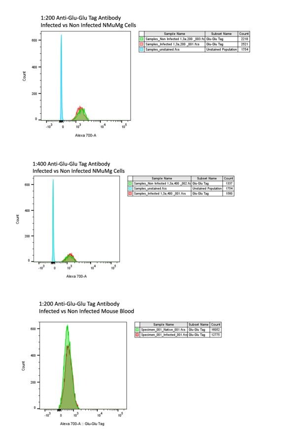
Application: Flow CytometrySample Tested: NMuMG mouse mammary gland epithelial cell line and Mouse bloodSpecies: MouseVerified Customer | Posted 08/24/2023Histogram results from flow cytometry on mouse polyomavirus infected and non-infected NMuMg cells and mouse blood. No difference is seen between the infected and non-infected groups.Based on the principle that the Glu-Glu epitope tag is derived from the sequence for mouse polyomavirus Middle T we hypothesized that the anti-Glu-Glu tag might be able to stain middle T antigen in mouse polyomavirus infected cells. We grew two groups of mice NMuMg cells, infecting one with A2 strain mouse polyomavirus for 24 hours and leaving one culture uninfected. The cells were lifted from the plates and prepped for flow cytometry including permeabilizing and staining with the anti-Glu-Glu Tag. The antibody stained a majority of the cells in both groups as positive, demonstrating that the antibody is not specific. Unstained cells were included for comparison. The same experiment was repeated with mouse blood from both infected and non-infected animals producing the same results. In conclusion the Glu-Glu tag antibody does not specifically recognize middle T in cells infected by mouse polyomavirus by flow cytometry. This was an unvalidated application.
 Bio-Techne ResponseThis review was submitted through the legacy Novus Innovators Program, reflecting a new species or application tested on a primary antibody.
There are no reviews that match your criteria.
Protocols
Find general support by application which include: protocols, troubleshooting, illustrated assays, videos and webinars.
- Appropriate Fixation of IHC/ICC Samples
- Cellular Response to Hypoxia Protocols
- ClariTSA™ Fluorophore Kits
- Detection & Visualization of Antibody Binding
- ELISA Sample Preparation & Collection Guide
- ELISA Troubleshooting Guide
- How to Run an R&D Systems DuoSet ELISA
- How to Run an R&D Systems Quantikine ELISA
- How to Run an R&D Systems Quantikine™ QuicKit™ ELISA
- ICC Cell Smear Protocol for Suspension Cells
- ICC Immunocytochemistry Protocol Videos
- ICC for Adherent Cells
- Immunocytochemistry (ICC) Protocol
- Immunocytochemistry Troubleshooting
- Immunofluorescence of Organoids Embedded in Cultrex Basement Membrane Extract
- Immunohistochemistry (IHC) and Immunocytochemistry (ICC) Protocols
- Immunoprecipitation Protocol
- Preparing Samples for IHC/ICC Experiments
- Preventing Non-Specific Staining (Non-Specific Binding)
- Primary Antibody Selection & Optimization
- Protocol for VisUCyte™ HRP Polymer Detection Reagent
- Protocol for the Fluorescent ICC Staining of Cell Smears - Graphic
- Protocol for the Fluorescent ICC Staining of Cultured Cells on Coverslips - Graphic
- Protocol for the Preparation and Fluorescent ICC Staining of Cells on Coverslips
- Protocol for the Preparation and Fluorescent ICC Staining of Non-adherent Cells
- Protocol for the Preparation and Fluorescent ICC Staining of Stem Cells on Coverslips
- Protocol for the Preparation of a Cell Smear for Non-adherent Cell ICC - Graphic
- Quantikine HS ELISA Kit Assay Principle, Alkaline Phosphatase
- Quantikine HS ELISA Kit Principle, Streptavidin-HRP Polymer
- R&D Systems Quality Control Western Blot Protocol
- Sandwich ELISA (Colorimetric) – Biotin/Streptavidin Detection Protocol
- Sandwich ELISA (Colorimetric) – Direct Detection Protocol
- TUNEL and Active Caspase-3 Detection by IHC/ICC Protocol
- The Importance of IHC/ICC Controls
- Troubleshooting Guide: ELISA
- Troubleshooting Guide: Western Blot Figures
- Western Blot Conditions
- Western Blot Protocol
- Western Blot Protocol for Cell Lysates
- Western Blot Troubleshooting
- Western Blot Troubleshooting Guide
- View all Protocols, Troubleshooting, Illustrated assays and Webinars
Loading...