Glut1 Antibody - BSA Free
Novus Biologicals | Catalog # NB110-39113
![Western Blot: Glut1 AntibodyBSA Free [NB110-39113]](https://resources.rndsystems.com/images/products/Glut1-Antibody-Western-Blot-NB110-39113-img0019.jpg "Western Blot: Glut1 AntibodyBSA Free [NB110-39113]")
Key Product Details
Validated by
Biological Validation
Species Reactivity
Validated:
Human, Mouse, Rat, Rabbit
Cited:
Human, Mouse, Rat, Rabbit
Predicted:
Bovine (93%), Primate (100%). Backed by our 100% Guarantee.
Applications
Validated:
Immunohistochemistry, Immunohistochemistry-Paraffin, Immunohistochemistry-Frozen, Western Blot, Flow Cytometry, Flow (Intracellular), Immunocytochemistry/ Immunofluorescence, Simple Western, Chromatin Immunoprecipitation, Chromatin Immunoprecipitation (ChIP), In vitro assay
Cited:
Immunohistochemistry-Paraffin, Immunohistochemistry-Frozen, Western Blot, Flow Cytometry, Immunocytochemistry/ Immunofluorescence, Chemotaxis, In vitro assay, IF/IHC, PCR
Label
Unconjugated
Antibody Source
Polyclonal Rabbit IgG
Format
BSA Free
Loading...
Product Specifications
Immunogen
This Glut1 antibody is made against a synthetic peptide made to an N-terminal region of the human GLUT1 protein (between residues 1-100). [Swiss-Prot# P11166].
Reactivity Notes
Rabbit reactivity reported in scientific literature (PMID: 29456650). 100% sequence identity with primate, 93% sequence identity with bovine.
Localization
Cell membrane, cytoplasm (near membranes), melanosome
Marker
Plasma Membrane Marker
Clonality
Polyclonal
Host
Rabbit
Isotype
IgG
Theoretical MW
54.1 kDa.
Disclaimer note: The observed molecular weight of the protein may vary from the listed predicted molecular weight due to post translational modifications, post translation cleavages, relative charges, and other experimental factors.
Disclaimer note: The observed molecular weight of the protein may vary from the listed predicted molecular weight due to post translational modifications, post translation cleavages, relative charges, and other experimental factors.
Scientific Data Images for Glut1 Antibody - BSA Free
![Western Blot: Glut1 AntibodyBSA Free [NB110-39113]](https://resources.rndsystems.com/images/products/Glut1-Antibody-Western-Blot-NB110-39113-img0006.jpg "Western Blot: Glut1 AntibodyBSA Free [NB110-39113]")
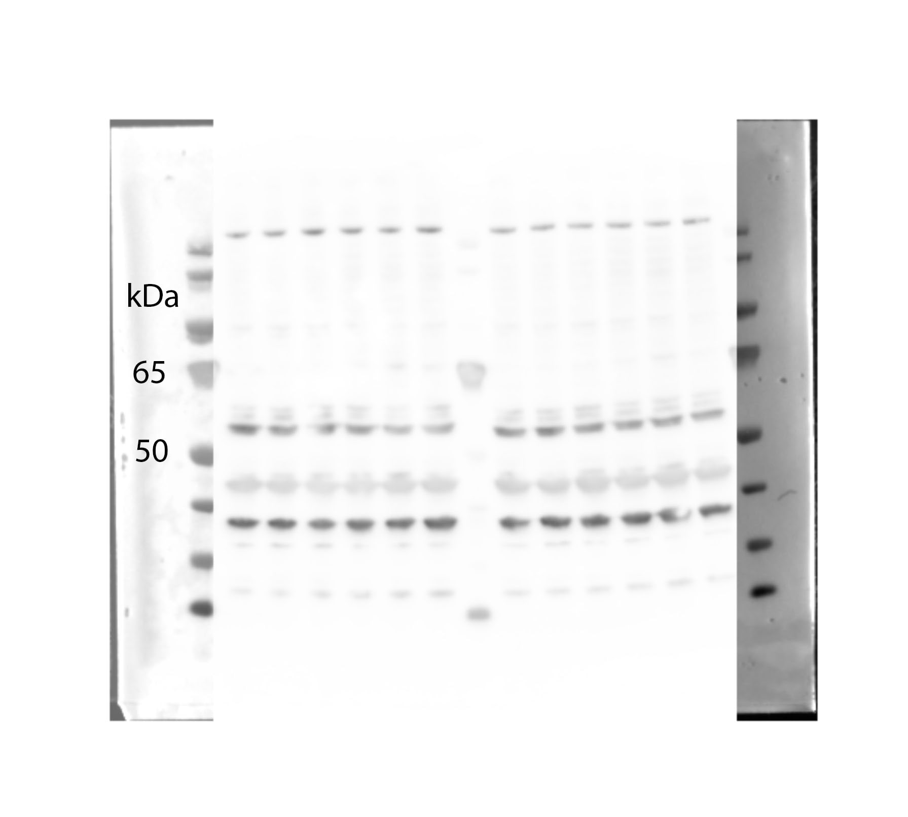
Western Blot: Glut1 AntibodyBSA Free [NB110-39113]
Western Blot: Glut1 Antibody [NB110-39113] - Western blot of GLUT1 on mouse kidney membrane protein (lane A) and rat kidney membrane protein (lane B).![Immunohistochemistry-Paraffin: Glut1 Antibody - BSA Free [NB110-39113]](https://resources.rndsystems.com/images/products/Glut1-Antibody-Immunohistochemistry-Paraffin-NB110-39113-img0011.jpg "Immunohistochemistry-Paraffin: Glut1 Antibody - BSA Free [NB110-39113]")
Immunohistochemistry-Paraffin: Glut1 Antibody - BSA Free [NB110-39113]
Immunohistochemistry-Paraffin: Glut1 Antibody [NB110-39113] - Immunohistochemical analysis of FFPE tissue section of human placenta using 1:200 dilution of Glut1 antibody. The staining was developed using HRP-DAB detection method and the sections were further counterstained with hematoxylin. This antibody generated a specific strong membrane cytoplasmic staining of Glut1 primarily in the syncytiotrophoblast layers of various villi and in the red blood cells (RBCs). Cytotrophoblasts showed a very weak expression of this protein.![Flow Cytometry: Glut1 Antibody - BSA Free [NB110-39113]](https://resources.rndsystems.com/images/products/Glut1-Antibody-Flow-Cytometry-NB110-39113-img0018.jpg "Flow Cytometry: Glut1 Antibody - BSA Free [NB110-39113]")
Flow Cytometry: Glut1 Antibody - BSA Free [NB110-39113]
Flow Cytometry: Glut1 Antibody [NB110-39113] - An intracellular stain was performed on HepG2 with NB110-39113 and a matched isotype control. Cells were fixed with 4% PFA and then permeablized with 0.1% saponin. Cells were incubated in an antibody dilution of 5 ug/mL for 30 minutes at room temperature, followed by Rabbit IgG (H+L) Cross-Adsorbed Secondary Antibody.![Immunocytochemistry/ Immunofluorescence: Glut1 Antibody - BSA Free [NB110-39113]](https://resources.rndsystems.com/images/products/Glut1-Antibody-Immunocytochemistry-Immunofluorescence-NB110-39113-img0013.jpg "Immunocytochemistry/ Immunofluorescence: Glut1 Antibody - BSA Free [NB110-39113]")
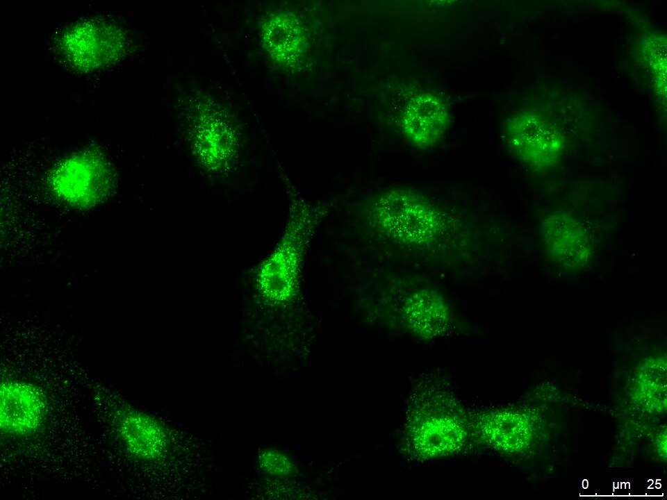
Immunocytochemistry/ Immunofluorescence: Glut1 Antibody - BSA Free [NB110-39113]
Immunocytochemistry/Immunofluorescence: Glut1 Antibody [NB110-39113] - HepG2 cells were fixed for 10 minutes using 10% formalin and then permeabilized for 5 minutes using 1X TBS + 0.5% Triton X-100. The cells were incubated with anti-GLUT1 at a 1:200 dilution overnight at 4C and detected with an anti-rabbit DyLight 488 (Green) at a 1:500 dilution. Alpha tubulin (DM1A) NB100-690 was used as a co-stain at a 1:1000 dilution and detected with an anti-mouse DyLight 550 (Red) at a 1:500 dilution. Nuclei were counterstained with DAPI (Blue). Cells were imaged using a 40X objective![Western Blot: Glut1 AntibodyBSA Free [NB110-39113]](https://resources.rndsystems.com/images/products/Glut1-Antibody-Western-Blot-NB110-39113-img0007.jpg "Western Blot: Glut1 AntibodyBSA Free [NB110-39113]")
Western Blot: Glut1 AntibodyBSA Free [NB110-39113]
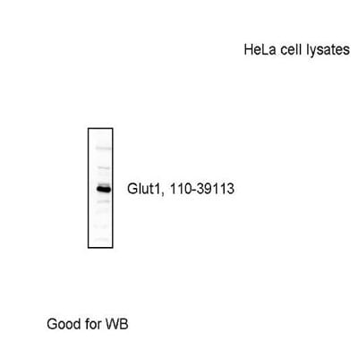
Western Blot: Glut1 Antibody [NB110-39113] - Analysis of HeLa lysates using NB110-39113. Image courtesy of Gregg Semenza (PMID: 21620138).![Western Blot: Glut1 AntibodyBSA Free [NB110-39113]](https://resources.rndsystems.com/images/products/Glut1-Antibody-Western-Blot-NB110-39113-img0016.jpg "Western Blot: Glut1 AntibodyBSA Free [NB110-39113]")
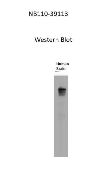
Western Blot: Glut1 AntibodyBSA Free [NB110-39113]
Western Blot: Glut1 Antibody [NB110-39113] - Glut1 in human brain lysate (55 kDa). Antibody at 1:500. Western blot image submitted by a verified customer review.![Flow Cytometry: Glut1 Antibody - BSA Free [NB110-39113]](https://resources.rndsystems.com/images/products/Glut1-Antibody-Flow-Cytometry-NB110-39113-img0014.jpg "Flow Cytometry: Glut1 Antibody - BSA Free [NB110-39113]")
Flow Cytometry: Glut1 Antibody - BSA Free [NB110-39113]
Flow Cytometry: Glut1 Antibody [NB110-39113] - Flow cytometry analysis using the PE conjugate of NB110-39113. Staining of Glut 1 expression on CD4+ T cells stimulated with anti-CD3/CD28 beads and insulin (1ug/mL) for 5 days in culture media with additional glucose provided. FMO control (red) and isotype control (blue, NBP2-24983) were compared to CD4+ T cells (orange), and this PE conjugated Glut 1 antibody positively stained CD4+ lymphocytes isolated from Mouse. Flow cytometry image submitted by a verified customer review.
Immunocytochemistry/ Immunofluorescence: Glut1 Antibody - BSA Free [NB110-39113] -
Immunocytochemistry/ Immunofluorescence: Glut1 Antibody - BSA Free [NB110-39113] - LAIR-1 inhibits Glut1-related glucose uptake in OS cells. a Heatmap showing the levels of differentially expressed mRNAs. b Top 20 KEGG pathway annotation categories for target gene functions of predicted mRNAs. c Selected significantly differentially expressed mRNA-related to EMT in RNA-seq data between two groups, ***P < 0.001. d qPCR validation of differentially expressed EMT-related genes in LV-NC & LV-LAIR-1-overexpressing OS cells, **P < 0.01. e Glut1 expression analyzed by western blotting. f Immunofluorescence staining of Glut1 in the LV-LAIR-1-overexpressing OS cells. Scale bar = 50 μm. Data were obtained from at least two independent experiments. Image collected & cropped by CiteAb from the following publication (https://pubmed.ncbi.nlm.nih.gov/32563267), licensed under a CC-BY license. Not internally tested by Novus Biologicals.
Western Blot: Glut1 Antibody - BSA Free [NB110-39113] -
Western Blot: Glut1 Antibody - BSA Free [NB110-39113] - LAIR-1 inhibits Glut1-related glucose uptake in OS cells. a Heatmap showing the levels of differentially expressed mRNAs. b Top 20 KEGG pathway annotation categories for target gene functions of predicted mRNAs. c Selected significantly differentially expressed mRNA-related to EMT in RNA-seq data between two groups, ***P < 0.001. d qPCR validation of differentially expressed EMT-related genes in LV-NC & LV-LAIR-1-overexpressing OS cells, **P < 0.01. e Glut1 expression analyzed by western blotting. f Immunofluorescence staining of Glut1 in the LV-LAIR-1-overexpressing OS cells. Scale bar = 50 μm. Data were obtained from at least two independent experiments. Image collected & cropped by CiteAb from the following publication (https://pubmed.ncbi.nlm.nih.gov/32563267), licensed under a CC-BY license. Not internally tested by Novus Biologicals.
Simple Western: Glut1 Antibody - BSA Free [NB110-39113] -
Simple Western: Glut1 Antibody - BSA Free [NB110-39113] - Protein changes in ONHAs corroborate bioenergetics data. (A) Glucose transporter-1 protein levels in Stretched ONH astrocytes are significantly higher than Control (*p = 0.0225, n = 7 Control, n = 8 Stretch). Retinal lysate from a 2 month-old mouse was used as a positive control for each protein analyzed, while negative control was the signal obtained when no primary antibody was included in the capillary. (B) Lactate dehydrogenase-A, the astrocyte-specific isoform of the enzyme that catalyzes the interconversion of pyruvate & lactate, has equivalent protein levels in Control & Stretch ONH astrocytes. (C) Glucose-6-phosphate dehydrogenase, the enzyme that shunts glucose into the pentose phosphate pathway, is no different in Control & Stretch ONH astrocytes. (D) Glutamine synthetase, the enzyme that synthesizes glutamine from glutamate, is no different in Control & Stretch ONH astrocytes. (E) The monomeric form of glutamate-aspartate transporter (GLAST) has significantly higher protein levels in the Stretch as compared to the Control ONH astrocytes (p = 0.020; n = 4 Control, n = 5 Stretch). (F) GLAST dimer protein levels are no different in Control & Stretch ONH astrocytes. Image collected & cropped by CiteAb from the following publication (https://pubmed.ncbi.nlm.nih.gov/35992925), licensed under a CC-BY license. Not internally tested by Novus Biologicals.Applications for Glut1 Antibody - BSA Free
Application
Recommended Usage
Chromatin Immunoprecipitation
reported in scientific literature
Flow (Intracellular)
1 ug/ml
Flow Cytometry
1 ug/ml. Use reported by customer review
Immunocytochemistry/ Immunofluorescence
1:1000
Immunohistochemistry
1:200
Immunohistochemistry-Frozen
1:200. Use reported in scientific literature
Immunohistochemistry-Paraffin
1:200
In vitro assay
reported in scientific literature (Trachsel V et al)
Western Blot
1:500
Application Notes
In WB a band is seen at ~55 kDa on kidney membrane preps representing GLUT1 protein. Depending on the tissue and any post-translational modifications, this protein can run anywhere between 40-60 kDa. See Simple Western Antibody Database for Simple Western validation: Tested in mouse eye tissue from D2 and D2G ON mouse model; separated by size; antibody dilution of 1:50.
Reviewed Applications
Read 7 reviews rated 4.9 using NB110-39113 in the following applications:
Flow Cytometry Panel Builder
Bio-Techne Knows Flow Cytometry
Save time and reduce costly mistakes by quickly finding compatible reagents using the Panel Builder Tool.
Advanced Features
- Spectra Viewer - Custom analysis of spectra from multiple fluorochromes
- Spillover Popups - Visualize the spectra of individual fluorochromes
- Antigen Density Selector - Match fluorochrome brightness with antigen density

Formulation, Preparation, and Storage
Purification
Immunogen affinity purified
Formulation
PBS
Format
BSA Free
Preservative
0.02% Sodium Azide
Concentration
1 mg/ml
Shipping
The product is shipped with polar packs. Upon receipt, store it immediately at the temperature recommended below.
Stability & Storage
Aliquot and store at -20C or -80C. Avoid freeze-thaw cycles.
Background: Glut1
GLUT1 (Human glycosylated form theoretical molecular weight 55kDa) functions primarily as a glucose transporter but can transport other substrates including mannose, galactose and glucosamine across the membrane (3). Like other GLUT family members, GLUT1 is broadly expressed, nevertheless it is the predominant glucose transporter expressed in red blood cells and brain endothelial cells (1). SLC2A1 mutations underscore the autosomal dominant disorder GLUT1 deficiency syndrome (GLUTI-DS) which is characterized by low glucose levels in the brain or hypoglycorrhachia due to insufficient glucose transport across the blood brain barrier (2, 4, 5). Phenotypically, GLUT1-DS is characterized by early onset seizures, neurologic developmental delay, microcephaly, and ataxia (4). GLUT1 is highly expressed in the endothelium of cutaneous vascular lesions and serves as a marker for the diagnosis of juvenile or infantile hemangiomas (6).
References
1. Augustin, R. (2010). The protein family of glucose transport facilitators: It's not only about glucose after all. IUBMB Life. https://doi.org/10.1002/iub.315
2. Mueckler, M., & Thorens, B. (2013). The SLC2 (GLUT) family of membrane transporters. Molecular Aspects of Medicine. https://doi.org/10.1016/j.mam.2012.07.001
3. Stein, W. D., & Litman, T. (2015). Carrier-Mediated Transport. In Channels, Carriers, and Pumps. https://doi.org/10.1016/b978-0-12-416579-3.00004-6
4. Pearson, T. S., Akman, C., Hinton, V. J., Engelstad, K., & De Vivo, D. C. (2013). Phenotypic spectrum of glucose transporter type 1 deficiency syndrome (Glut1 DS). Current Neurology and Neuroscience Reports. https://doi.org/10.1007/s11910-013-0342-7
5. Messana, T., Russo, A., Vergaro, R., Boni, A., Santucci, M., & Pini, A. (2018). Glucose transporter type 1 deficiency syndrome: Developmental delay and early-onset ataxia in a novel mutation of the SLC2A1 gene. Journal of Pediatric Neurosciences. https://doi.org/10.4103/JPN.JPN_169_17
6. van Vugt, L. J., van der Vleuten, C. J. M., Flucke, U., & Blokx, W. A. M. (2017). The utility of GLUT1 as a diagnostic marker in cutaneous vascular anomalies: A review of literature and recommendations for daily practice. Pathology Research and Practice. https://doi.org/10.1016/j.prp.2017.04.023
Long Name
Glucose Transporter Type 1
Alternate Names
DYT17, DYT18, DYT9, EIG12, GLUT1DS, SLC2A1
Gene Symbol
SLC2A1
Additional Glut1 Products
Product Documents for Glut1 Antibody - BSA Free
Certificate of Analysis
To download a Certificate of Analysis, please enter a lot or batch number in the search box below.
Product Specific Notices for Glut1 Antibody - BSA Free
This product is for research use only and is not approved for use in humans or in clinical diagnosis. Primary Antibodies are guaranteed for 1 year from date of receipt.
Related Research Areas
Citations for Glut1 Antibody - BSA Free
 Powered by Bioz
Customer Reviews for Glut1 Antibody - BSA Free (7)
4.9 out of 5
7 Customer Ratings
Have you used Glut1 Antibody - BSA Free?
Submit a review and receive an Amazon gift card!
$25/€18/£15/$25CAN/¥2500 Yen for a review with an image
$10/€7/£6/$10CAN/¥1110 Yen for a review without an image
Submit a review
Customer Images
_nb110_39113_6391.jpg)
Showing
1
-
5 of
7 reviews
Showing All
Filter By:
-
Application: Western BlotSample Tested: Mouse skeletal muscleSpecies: MouseVerified Customer | Posted 04/23/2019Total GLUT1 detection in mouse quadriceps muscle, 1:1000 dilution, ~55 kDa, a lot of non-specific bands

-
Application: ImmunocytochemistrySample Tested: beta cellsSpecies: HumanVerified Customer | Posted 10/24/2017Excellentuse at 1:100
-
Application: Western BlotSample Tested: BrainSpecies: HumanVerified Customer | Posted 06/12/2017Glut 1 at 55kDause at 1:500
-
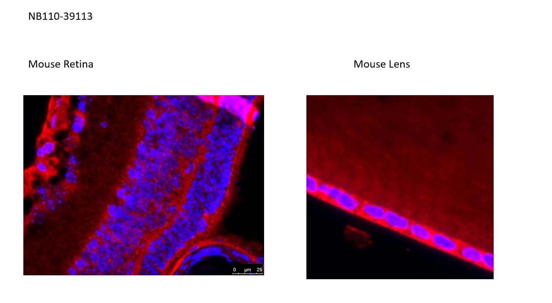
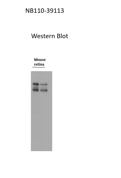
Application: ImmunocytochemistrySample Tested: mouse eyeSpecies: MouseVerified Customer | Posted 06/12/2017Mouse eye retinause at 1:100 concentration
-
Application: Western BlotSample Tested: Retina (inner nuclear layer)Species: Mouse and HumanVerified Customer | Posted 06/12/2017Use at a conc of 1:500 for 20ug sample protein loaded
-
Application: Western BlotSample Tested: membrane fraction from lung cancer cellsSpecies: HumanVerified Customer | Posted 03/09/2014GLUT 1 in A549 cells
-
Application: Western BlotSample Tested: HeLa cell lysates, Sample Amount: 20 ugSpecies: HumanVerified Customer | Posted 09/01/2010
There are no reviews that match your criteria.
Protocols
View specific protocols for Glut1 Antibody - BSA Free (NB110-39113):
Protocol for Flow Cytometry Intracellular Staining
Sample Preparation.
1. Grow cells to 60-85% confluency. Flow cytometry requires between 2 x 10⁵ and 1 x 10⁶ cells for optimal performance.
2. If cells are adherent, harvest gently by washing once with staining buffer and then scraping. Avoid using trypsin as this can disrupt certain epitopes of interest. If enzymatic harvest is required, use Accutase, Collagenase, or TrypLE Express for a less damaging option.
3. Reserve 100 uL for counting, then transfer cell volume into a 50 mL conical tube and centrifuge for 8 minutes at 400 RCF.
a. Count cells using a hemocytometer and a 1:1 trypan blue exclusion stain to determine cell viability before starting the flow protocol. If cells appear blue, do not proceed.
4. Re-suspend cells to a concentration of 1 x 10⁶ cells/mL in staining buffer (NBP2-26247).
5. Aliquot out 100 uL samples in accordance with your experimental samples.
Tip: When cell surface and intracellular staining are required in the same sample, it is advisable that the cell surface staining be performed first since the fixation and permeabilization steps might reduce the availability of surface antigens.
Intracellular Staining.
Tip: When performing intracellular staining, it is important to use appropriate fixation and permeabilization reagents based upon the target and its subcellular location. Generally, our Intracellular Flow Assay Kit (NBP2-29450) is a good place to start as it contains an optimized combination of reagents for intracellular staining as well as an inhibitor of intracellular protein transport (necessary if staining secreted proteins). Certain targets may require more gentle or transient permeabilization protocols such as the commonly employed methanol or saponin-based methods.
Protocol for Cytoplasmic Targets:
1. Fix the cells by adding 100 uL fixation solution (such as 4% PFA) to each sample for 10-15 minutes.
2. Permeabilize cells by adding 100 uL of a permeabilization buffer to every 1 x 10⁶ cells present in the sample. Mix well and incubate at room temperature for 15 minutes.
a. For cytoplasmic targets, use a gentle permeabilization solution such as 1X PBS + 0.5% Saponin or 1X PBS + 0.5% Tween-20.
b. To maintain the permeabilized state throughout your experiment, use staining buffer + 0.1% of the permeabilization reagent (i.e. 0.1% Tween-20 or 0.1% Saponin).
3. Following the 15 minute incubation, add 2 mL of the staining buffer + 0.1% permeabilizer to each sample.
4. Centrifuge for 1 minute at 400 RCF.
5. Discard supernatant and re-suspend in 100 uL of staining buffer + 0.1% permeabilizer.
6. Add appropriate amount of each antibody (eg. 1 test or 1 ug per sample, as experimentally determined).
7. Mix well and incubate at room temperature for 30 minutes- 1 hour. Gently mix samples every 10-15 minutes.
8. Following the primary/conjugate incubation, add 1-2 mL/sample of staining buffer +0.1% permeabilizer and centrifuge for 1 minute at 400 RCF.
9. Wash twice by re-suspending cells in staining buffer (2 mL for tubes or 200 uL for wells) and centrifuging at 400 RCF for 5 minutes. Discard supernatant.
10. Add appropriate amount of secondary antibody (as experimentally determined) to each sample.
11. Incubate at room temperature in dark for 20 minutes.
12. Add 1-2 mL of staining buffer and centrifuge at 400 RCF for 1 minute and discard supernatant.
13. Wash twice by re-suspending cells in staining buffer (2 mL for tubes or 200 uL for wells) and centrifuging at 400 RCF for 5 minutes. Discard supernatant.
14. Resuspend in an appropriate volume of staining buffer (usually 500 uL per sample) and proceed with analysis on your flow cytometer.
Immunocytochemistry Protocol
Culture cells to appropriate density in 35 mm culture dishes or 6-well plates.
1. Remove culture medium and wash the cells briefly in PBS. Add 10% formalin to the dish and fix at room temperature for 10 minutes.
2. Remove the formalin and wash the cells in PBS.
3. Permeablize the cells with 0.1% Triton X100 or other suitable detergent for 10 min.
4. Remove the permeablization buffer and wash three times for 10 minutes each in PBS. Be sure to not let the specimen dry out.
5. To block nonspecific antibody binding, incubate in 10% normal goat serum from 1 hour to overnight at room temperature.
6. Add primary antibody at appropriate dilution and incubate overnight at 4C.
7. Remove primary antibody and replace with PBS. Wash three times for 10 minutes each.
8. Add secondary antibody at appropriate dilution. Incubate for 1 hour at room temperature.
9. Remove secondary antibody and replace with PBS. Wash three times for 10 minutes each.
10. Counter stain DNA with DAPi if required.
Immunohistochemistry-Paraffin Embedded Sections
Antigen Unmasking:
Bring slides to a boil in 10 mM sodium citrate buffer (pH 6.0) then maintain at a sub-boiling temperature for 10 minutes. Cool slides on bench-top for 30 minutes (keep slides in the sodium citrate buffer all the time).
Staining:
1. Wash sections in deionized water three times for 5 minutes each.
2. Wash sections in PBS for 5 minutes.
3. Block each section with 100-400 ul blocking solution (1% BSA in PBS) for 1 hour at room temperature.
4. Remove blocking solution and add 100-400 ul diluted primary antibody. Incubate overnight at 4 C.
5. Remove antibody solution and wash sections in wash buffer three times for 5 minutes each.
6. Add 100-400 ul HRP polymer conjugated secondary antibody. Incubate 30 minutes at room temperature.
7. Wash sections three times in wash buffer for 5 minutes each.
8. Add 100-400 ul DAB substrate to each section and monitor staining closely.
9. As soon as the sections develop, immerse slides in deionized water.
10. Counterstain sections in hematoxylin.
11. Wash sections in deionized water two times for 5 minutes each.
12. Dehydrate sections.
13. Mount coverslips.
Western Blot Protocol
1. Perform SDS-PAGE on samples to be analyzed, loading 40 ug of total protein per lane.
2. Transfer proteins to membrane according to the instructions provided by the manufacturer of the membrane and transfer apparatus.
3. Stain according to standard Ponceau S procedure (or similar product) to assess transfer success, and mark molecular weight standards where appropriate.
4. Rinse the blot.
5. Block the membrane using standard blocking buffer for at least 1 hour.
6. Wash the membrane in wash buffer three times for 10 minutes each.
7. Dilute primary antibody in blocking buffer and incubate 1 hour at room temperature.
8. Wash the membrane in wash buffer three times for 10 minutes each.
9. Apply the diluted HRP conjugated secondary antibody in blocking buffer (as per manufacturers instructions) and incubate 1 hour at room temperature.
10. Wash the blot in wash buffer three times for 10 minutes each (this step can be repeated as required to reduce background).
11. Apply the detection reagent of choice in accordance with the manufacturers instructions.
Note: Tween-20 can be added to the blocking or antibody dilution buffer at a final concentration of 0.05-0.2%.
Find general support by application which include: protocols, troubleshooting, illustrated assays, videos and webinars.
- 7-Amino Actinomycin D (7-AAD) Cell Viability Flow Cytometry Protocol
- Antigen Retrieval Protocol (PIER)
- Antigen Retrieval for Frozen Sections Protocol
- Appropriate Fixation of IHC/ICC Samples
- Cellular Response to Hypoxia Protocols
- ChIP Protocol Video
- Chromatin Immunoprecipitation (ChIP) Protocol
- Chromatin Immunoprecipitation Protocol
- Chromogenic IHC Staining of Formalin-Fixed Paraffin-Embedded (FFPE) Tissue Protocol
- Chromogenic Immunohistochemistry Staining of Frozen Tissue
- ClariTSA™ Fluorophore Kits
- Detection & Visualization of Antibody Binding
- Extracellular Membrane Flow Cytometry Protocol
- Flow Cytometry Protocol for Cell Surface Markers
- Flow Cytometry Protocol for Staining Membrane Associated Proteins
- Flow Cytometry Staining Protocols
- Flow Cytometry Troubleshooting Guide
- Fluorescent IHC Staining of Frozen Tissue Protocol
- Graphic Protocol for Heat-induced Epitope Retrieval
- Graphic Protocol for the Preparation and Fluorescent IHC Staining of Frozen Tissue Sections
- Graphic Protocol for the Preparation and Fluorescent IHC Staining of Paraffin-embedded Tissue Sections
- Graphic Protocol for the Preparation of Gelatin-coated Slides for Histological Tissue Sections
- ICC Cell Smear Protocol for Suspension Cells
- ICC Immunocytochemistry Protocol Videos
- ICC for Adherent Cells
- IHC Sample Preparation (Frozen sections vs Paraffin)
- Immunocytochemistry (ICC) Protocol
- Immunocytochemistry Troubleshooting
- Immunofluorescence of Organoids Embedded in Cultrex Basement Membrane Extract
- Immunofluorescent IHC Staining of Formalin-Fixed Paraffin-Embedded (FFPE) Tissue Protocol
- Immunohistochemistry (IHC) and Immunocytochemistry (ICC) Protocols
- Immunohistochemistry Frozen Troubleshooting
- Immunohistochemistry Paraffin Troubleshooting
- Intracellular Flow Cytometry Protocol Using Alcohol (Methanol)
- Intracellular Flow Cytometry Protocol Using Detergents
- Intracellular Nuclear Staining Flow Cytometry Protocol Using Detergents
- Intracellular Staining Flow Cytometry Protocol Using Alcohol Permeabilization
- Intracellular Staining Flow Cytometry Protocol Using Detergents to Permeabilize Cells
- Preparing Samples for IHC/ICC Experiments
- Preventing Non-Specific Staining (Non-Specific Binding)
- Primary Antibody Selection & Optimization
- Propidium Iodide Cell Viability Flow Cytometry Protocol
- Protocol for Heat-Induced Epitope Retrieval (HIER)
- Protocol for Liperfluo
- Protocol for Making a 4% Formaldehyde Solution in PBS
- Protocol for VisUCyte™ HRP Polymer Detection Reagent
- Protocol for the Characterization of Human Th22 Cells
- Protocol for the Characterization of Human Th9 Cells
- Protocol for the Fluorescent ICC Staining of Cell Smears - Graphic
- Protocol for the Fluorescent ICC Staining of Cultured Cells on Coverslips - Graphic
- Protocol for the Preparation & Fixation of Cells on Coverslips
- Protocol for the Preparation and Chromogenic IHC Staining of Frozen Tissue Sections
- Protocol for the Preparation and Chromogenic IHC Staining of Frozen Tissue Sections - Graphic
- Protocol for the Preparation and Chromogenic IHC Staining of Paraffin-embedded Tissue Sections
- Protocol for the Preparation and Chromogenic IHC Staining of Paraffin-embedded Tissue Sections - Graphic
- Protocol for the Preparation and Fluorescent ICC Staining of Cells on Coverslips
- Protocol for the Preparation and Fluorescent ICC Staining of Non-adherent Cells
- Protocol for the Preparation and Fluorescent ICC Staining of Stem Cells on Coverslips
- Protocol for the Preparation and Fluorescent IHC Staining of Frozen Tissue Sections
- Protocol for the Preparation and Fluorescent IHC Staining of Paraffin-embedded Tissue Sections
- Protocol for the Preparation of Gelatin-coated Slides for Histological Tissue Sections
- Protocol for the Preparation of a Cell Smear for Non-adherent Cell ICC - Graphic
- Protocol: Annexin V and PI Staining by Flow Cytometry
- Protocol: Annexin V and PI Staining for Apoptosis by Flow Cytometry
- R&D Systems Quality Control Western Blot Protocol
- TUNEL and Active Caspase-3 Detection by IHC/ICC Protocol
- The Importance of IHC/ICC Controls
- Troubleshooting Guide: Fluorokine Flow Cytometry Kits
- Troubleshooting Guide: Immunohistochemistry
- Troubleshooting Guide: Western Blot Figures
- Western Blot Conditions
- Western Blot Protocol
- Western Blot Protocol for Cell Lysates
- Western Blot Troubleshooting
- Western Blot Troubleshooting Guide
- View all Protocols, Troubleshooting, Illustrated assays and Webinars
FAQs for Glut1 Antibody - BSA Free
Showing
1
-
1 of
1 FAQ
Showing All
-
Q: Can you tell me what, if any, antigen retrieval method was used for the staining of paraffin tissue sections with GLUT1 Antibody (NB110-39113)?
A: We recommend the use of HIER (heat-induced epitope retrieval) with 0.01 M sodium citrate buffer, pH 6.0 at 100C for 20 minutes in a steamer or pressure cooker.


