Key Product Details
Species Reactivity
Validated:
Human, Mouse, Rat, Rabbit
Predicted:
Bovine (93%), Primate (100%). Backed by our 100% Guarantee.
Applications
Immunohistochemistry, Immunohistochemistry-Paraffin, Immunohistochemistry-Frozen, Western Blot, Flow Cytometry, Flow (Intracellular), Immunocytochemistry/ Immunofluorescence, Chromatin Immunoprecipitation, Chromatin Immunoprecipitation (ChIP), In vitro assay
Label
FITC (Excitation = 495 nm, Emission = 519 nm)
Antibody Source
Polyclonal Rabbit IgG
Loading...
Product Specifications
Immunogen
This Glut1 antibody is made against a synthetic peptide made to an N-terminal region of the human GLUT1 protein (between residues 1-100). [Swiss-Prot# P11166].
Reactivity Notes
Rabbit reactivity reported in scientific literature (PMID: 29456650). 100% sequence identity with primate, 93% sequence identity with bovine.
Localization
Cell membrane, Cytoplasm (near membranes), Melanosome
Marker
Plasma Membrane Marker
Clonality
Polyclonal
Host
Rabbit
Isotype
IgG
Theoretical MW
54.1 kDa.
Disclaimer note: The observed molecular weight of the protein may vary from the listed predicted molecular weight due to post translational modifications, post translation cleavages, relative charges, and other experimental factors.
Disclaimer note: The observed molecular weight of the protein may vary from the listed predicted molecular weight due to post translational modifications, post translation cleavages, relative charges, and other experimental factors.
Scientific Data Images for Glut1 Antibody [FITC]
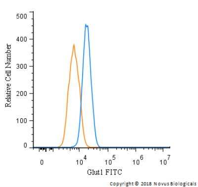
Flow Cytometry: Glut1 Antibody [FITC] [NB110-39113F] - An intracellular stain was performed on HepG cells with Glut1 Antibody NB110-39113F (blue) and a matched isotype control (orange). Cells were fixed with 4% PFA and then permeabilized with 0.1% saponin. Cells were incubated in an antibody dilution of 10 ug/mL for 30 minutes at room temperature. Both antibodies were conjugated to FITC.
Applications for Glut1 Antibody [FITC]
Application
Recommended Usage
Chromatin Immunoprecipitation
Optimal dilutions of this antibody should be experimentally determined.
Chromatin Immunoprecipitation (ChIP)
Optimal dilutions of this antibody should be experimentally determined.
Flow (Intracellular)
Optimal dilutions of this antibody should be experimentally determined.
Flow Cytometry
Optimal dilutions of this antibody should be experimentally determined.
Immunocytochemistry/ Immunofluorescence
Optimal dilutions of this antibody should be experimentally determined.
Immunohistochemistry
Optimal dilutions of this antibody should be experimentally determined.
Immunohistochemistry-Frozen
Optimal dilutions of this antibody should be experimentally determined.
Immunohistochemistry-Paraffin
Optimal dilutions of this antibody should be experimentally determined.
In vitro assay
Optimal dilutions of this antibody should be experimentally determined.
Western Blot
Optimal dilutions of this antibody should be experimentally determined.
Application Notes
Optimal dilution of this antibody should be experimentally determined.
Spectra Viewer
Plan Your Experiments
Use our spectra viewer to interactively plan your experiments, assessing multiplexing options. View the excitation and emission spectra for our fluorescent dye range and other commonly used dyes.
Spectra Viewer
Flow Cytometry Panel Builder
Bio-Techne Knows Flow Cytometry
Save time and reduce costly mistakes by quickly finding compatible reagents using the Panel Builder Tool.
Advanced Features
- Spectra Viewer - Custom analysis of spectra from multiple fluorochromes
- Spillover Popups - Visualize the spectra of individual fluorochromes
- Antigen Density Selector - Match fluorochrome brightness with antigen density

Formulation, Preparation, and Storage
Purification
Immunogen affinity purified
Formulation
PBS
Preservative
0.05% Sodium Azide
Concentration
Please see the vial label for concentration. If unlisted please contact technical services.
Shipping
The product is shipped with polar packs. Upon receipt, store it immediately at the temperature recommended below.
Stability & Storage
Store at 4C in the dark.
Background: Glut1
GLUT1 (Human glycosylated form theoretical molecular weight 55kDa) functions primarily as a glucose transporter but can transport other substrates including mannose, galactose and glucosamine across the membrane (3). Like other GLUT family members, GLUT1 is broadly expressed, nevertheless it is the predominant glucose transporter expressed in red blood cells and brain endothelial cells (1). SLC2A1 mutations underscore the autosomal dominant disorder GLUT1 deficiency syndrome (GLUTI-DS) which is characterized by low glucose levels in the brain or hypoglycorrhachia due to insufficient glucose transport across the blood brain barrier (2, 4, 5). Phenotypically, GLUT1-DS is characterized by early onset seizures, neurologic developmental delay, microcephaly, and ataxia (4). GLUT1 is highly expressed in the endothelium of cutaneous vascular lesions and serves as a marker for the diagnosis of juvenile or infantile hemangiomas (6).
References
1. Augustin, R. (2010). The protein family of glucose transport facilitators: It's not only about glucose after all. IUBMB Life. https://doi.org/10.1002/iub.315
2. Mueckler, M., & Thorens, B. (2013). The SLC2 (GLUT) family of membrane transporters. Molecular Aspects of Medicine. https://doi.org/10.1016/j.mam.2012.07.001
3. Stein, W. D., & Litman, T. (2015). Carrier-Mediated Transport. In Channels, Carriers, and Pumps. https://doi.org/10.1016/b978-0-12-416579-3.00004-6
4. Pearson, T. S., Akman, C., Hinton, V. J., Engelstad, K., & De Vivo, D. C. (2013). Phenotypic spectrum of glucose transporter type 1 deficiency syndrome (Glut1 DS). Current Neurology and Neuroscience Reports. https://doi.org/10.1007/s11910-013-0342-7
5. Messana, T., Russo, A., Vergaro, R., Boni, A., Santucci, M., & Pini, A. (2018). Glucose transporter type 1 deficiency syndrome: Developmental delay and early-onset ataxia in a novel mutation of the SLC2A1 gene. Journal of Pediatric Neurosciences. https://doi.org/10.4103/JPN.JPN_169_17
6. van Vugt, L. J., van der Vleuten, C. J. M., Flucke, U., & Blokx, W. A. M. (2017). The utility of GLUT1 as a diagnostic marker in cutaneous vascular anomalies: A review of literature and recommendations for daily practice. Pathology Research and Practice. https://doi.org/10.1016/j.prp.2017.04.023
Long Name
Glucose Transporter Type 1
Alternate Names
DYT17, DYT18, DYT9, EIG12, GLUT1DS, SLC2A1
Gene Symbol
SLC2A1
Additional Glut1 Products
Product Documents for Glut1 Antibody [FITC]
Certificate of Analysis
To download a Certificate of Analysis, please enter a lot or batch number in the search box below.
Product Specific Notices for Glut1 Antibody [FITC]
This product is for research use only and is not approved for use in humans or in clinical diagnosis. Primary Antibodies are guaranteed for 1 year from date of receipt.
Related Research Areas
Customer Reviews for Glut1 Antibody [FITC]
There are currently no reviews for this product. Be the first to review Glut1 Antibody [FITC] and earn rewards!
Have you used Glut1 Antibody [FITC]?
Submit a review and receive an Amazon gift card!
$25/€18/£15/$25CAN/¥2500 Yen for a review with an image
$10/€7/£6/$10CAN/¥1110 Yen for a review without an image
Submit a review
Protocols
Find general support by application which include: protocols, troubleshooting, illustrated assays, videos and webinars.
- 7-Amino Actinomycin D (7-AAD) Cell Viability Flow Cytometry Protocol
- Antigen Retrieval Protocol (PIER)
- Antigen Retrieval for Frozen Sections Protocol
- Appropriate Fixation of IHC/ICC Samples
- Cellular Response to Hypoxia Protocols
- ChIP Protocol Video
- Chromatin Immunoprecipitation (ChIP) Protocol
- Chromatin Immunoprecipitation Protocol
- Chromogenic IHC Staining of Formalin-Fixed Paraffin-Embedded (FFPE) Tissue Protocol
- Chromogenic Immunohistochemistry Staining of Frozen Tissue
- ClariTSA™ Fluorophore Kits
- Detection & Visualization of Antibody Binding
- Extracellular Membrane Flow Cytometry Protocol
- Flow Cytometry Protocol for Cell Surface Markers
- Flow Cytometry Protocol for Staining Membrane Associated Proteins
- Flow Cytometry Staining Protocols
- Flow Cytometry Troubleshooting Guide
- Fluorescent IHC Staining of Frozen Tissue Protocol
- Graphic Protocol for Heat-induced Epitope Retrieval
- Graphic Protocol for the Preparation and Fluorescent IHC Staining of Frozen Tissue Sections
- Graphic Protocol for the Preparation and Fluorescent IHC Staining of Paraffin-embedded Tissue Sections
- Graphic Protocol for the Preparation of Gelatin-coated Slides for Histological Tissue Sections
- ICC Cell Smear Protocol for Suspension Cells
- ICC Immunocytochemistry Protocol Videos
- ICC for Adherent Cells
- IHC Sample Preparation (Frozen sections vs Paraffin)
- Immunocytochemistry (ICC) Protocol
- Immunocytochemistry Troubleshooting
- Immunofluorescence of Organoids Embedded in Cultrex Basement Membrane Extract
- Immunofluorescent IHC Staining of Formalin-Fixed Paraffin-Embedded (FFPE) Tissue Protocol
- Immunohistochemistry (IHC) and Immunocytochemistry (ICC) Protocols
- Immunohistochemistry Frozen Troubleshooting
- Immunohistochemistry Paraffin Troubleshooting
- Intracellular Flow Cytometry Protocol Using Alcohol (Methanol)
- Intracellular Flow Cytometry Protocol Using Detergents
- Intracellular Nuclear Staining Flow Cytometry Protocol Using Detergents
- Intracellular Staining Flow Cytometry Protocol Using Alcohol Permeabilization
- Intracellular Staining Flow Cytometry Protocol Using Detergents to Permeabilize Cells
- Preparing Samples for IHC/ICC Experiments
- Preventing Non-Specific Staining (Non-Specific Binding)
- Primary Antibody Selection & Optimization
- Propidium Iodide Cell Viability Flow Cytometry Protocol
- Protocol for Heat-Induced Epitope Retrieval (HIER)
- Protocol for Liperfluo
- Protocol for Making a 4% Formaldehyde Solution in PBS
- Protocol for VisUCyte™ HRP Polymer Detection Reagent
- Protocol for the Characterization of Human Th22 Cells
- Protocol for the Characterization of Human Th9 Cells
- Protocol for the Fluorescent ICC Staining of Cell Smears - Graphic
- Protocol for the Fluorescent ICC Staining of Cultured Cells on Coverslips - Graphic
- Protocol for the Preparation & Fixation of Cells on Coverslips
- Protocol for the Preparation and Chromogenic IHC Staining of Frozen Tissue Sections
- Protocol for the Preparation and Chromogenic IHC Staining of Frozen Tissue Sections - Graphic
- Protocol for the Preparation and Chromogenic IHC Staining of Paraffin-embedded Tissue Sections
- Protocol for the Preparation and Chromogenic IHC Staining of Paraffin-embedded Tissue Sections - Graphic
- Protocol for the Preparation and Fluorescent ICC Staining of Cells on Coverslips
- Protocol for the Preparation and Fluorescent ICC Staining of Non-adherent Cells
- Protocol for the Preparation and Fluorescent ICC Staining of Stem Cells on Coverslips
- Protocol for the Preparation and Fluorescent IHC Staining of Frozen Tissue Sections
- Protocol for the Preparation and Fluorescent IHC Staining of Paraffin-embedded Tissue Sections
- Protocol for the Preparation of Gelatin-coated Slides for Histological Tissue Sections
- Protocol for the Preparation of a Cell Smear for Non-adherent Cell ICC - Graphic
- Protocol: Annexin V and PI Staining by Flow Cytometry
- Protocol: Annexin V and PI Staining for Apoptosis by Flow Cytometry
- R&D Systems Quality Control Western Blot Protocol
- TUNEL and Active Caspase-3 Detection by IHC/ICC Protocol
- The Importance of IHC/ICC Controls
- Troubleshooting Guide: Fluorokine Flow Cytometry Kits
- Troubleshooting Guide: Immunohistochemistry
- Troubleshooting Guide: Western Blot Figures
- Western Blot Conditions
- Western Blot Protocol
- Western Blot Protocol for Cell Lysates
- Western Blot Troubleshooting
- Western Blot Troubleshooting Guide
- View all Protocols, Troubleshooting, Illustrated assays and Webinars


