Glutamate Dehydrogenase Antibody - BSA Free
Novus Biologicals | Catalog # NB600-853
![Western Blot: Glutamate Dehydrogenase Antibody [NB600-853]](https://resources.rndsystems.com/images/products/Glutamate-Dehydrogenase-Antibody-Western-Blot-NB600-853-img0006.jpg "Western Blot: Glutamate Dehydrogenase Antibody [NB600-853]")
Loading...
Key Product Details
Species Reactivity
Validated:
Human, Bovine
Cited:
Human, Rat
Applications
Validated:
Immunohistochemistry, Western Blot, ELISA, Immunoprecipitation
Cited:
Western Blot, IF/IHC
Label
Unconjugated
Antibody Source
Polyclonal Rabbit Serum
Format
BSA Free
Loading...
Product Specifications
Immunogen
This Glutamate Dehydrogenase Antibody was prepared from whole rabbit serum produced by repeated immunizations with a full length Glutamate Dehydrogenase protein isolated from Bovine Liver. (Uniprot: P00366)
Reactivity Notes
BLAST analysis was used to determine that cross reactivity is suggested for both mitochondrial and brain isoforms (GDH1 and GDH2), from both bovine and human sources. Additionally similar reactivity is suggested for most primate species including green monkey, white gibbon, chimpanzee orangutan, and gorilla. A high degree of sequence homology is also noted for Glutamate Dehydrogenase from chicken, mouse, rat, dog, and other mammals as well as Xenopus tropicalis, zebrafish, rainbow trout and Atlantic salmon.
Human reactivity reported in scientific literature (PMID: 27623078).
Human reactivity reported in scientific literature (PMID: 27623078).
Localization
Mitochondrial matrix
Marker
Mitochondria Marker
Clonality
Polyclonal
Host
Rabbit
Isotype
Serum
Description
This product was prepared from monospecific antiserum by a delipidation and defibrination. Assay by immunoelectrophoresis resulted in a single precipitin arc against anti-rabbit serum, purified and partially purified Glutamate Dehydrogenase [Bovine Liver]
Store vial at 4C prior to restoration. For extended storage aliquot contents and freeze at -20C or below. Avoid cycles of freezing and thawing. Centrifuge product if not completely clear after standing at room temperature. This product is stable for several weeks at 4C as an undiluted liquid. Dilute only prior to immediate use.
Store vial at 4C prior to restoration. For extended storage aliquot contents and freeze at -20C or below. Avoid cycles of freezing and thawing. Centrifuge product if not completely clear after standing at room temperature. This product is stable for several weeks at 4C as an undiluted liquid. Dilute only prior to immediate use.
Scientific Data Images for Glutamate Dehydrogenase Antibody - BSA Free
Western Blot: Glutamate Dehydrogenase Antibody [NB600-853]
Western Blot: Glutamate Dehydrogenase Antibody [NB600-853] - Western blot analysis is shown using Glutamate Dehydrogenase antibody to detect the enzyme from bovine liver preparations. Comparison to a molecular weight marker indicates a predominant band of ~62 kDa. The higher molecular weight band may represent a subunit dimer. A 4-20% gradient gel was used to separate proteins prior to transfer to 0.2 um nitrocellulose. The blot was incubated with a 1:1,000 dilution of the antibody for 2 h at room temperature followed by detection using IRDye(TM)800 labeled Goat-a-Rabbit IgG [H&L] diluted 1:5,000 for 45 min at room temperature. IRDye(TM)800 fluorescence image was captured using the Odyssey(R) Infrared Imaging System developed by LI-COR. IRDye is a trademark of LI-COR, Inc. Other detection systems will yield similar results.
N/A: Glutamate Dehydrogenase Antibody [NB600-853]
Applications for Glutamate Dehydrogenase Antibody - BSA Free
Application
Recommended Usage
ELISA
1:4000-1:16000
Immunoprecipitation
1:100
Western Blot
1:1000-1:3000
Application Notes
This antibody has been tested by western blot. Specific conditions for reactivity should be optimized by the end user. Bovine glutamate dehydrogenase exists as a homohexamer located within the mitochondrial matrix. Expect a band approximately 56 kDa in size corresponding to glutamate dehydrogenase monomer subunit by western blotting in the appropriate cell or tissue extract. Anti-Glutamate Dehydrogenase Antibody is suitable for use in ELISA.
Use in Immunohistochemistry reported in scientific literature (PMID 27623078).
Use in Immunohistochemistry reported in scientific literature (PMID 27623078).
Reviewed Applications
Read 1 review rated 4 using NB600-853 in the following applications:
Formulation, Preparation, and Storage
Purification
Delipidation and Defibrination
Formulation
0.02 M Potassium Phosphate, 0.15 M Sodium Chloride, pH 7.2
Format
BSA Free
Preservative
0.01% Sodium Azide
Concentration
Please see the vial label for concentration. If unlisted please contact technical services.
Shipping
The product is shipped with polar packs. Upon receipt, store it immediately at the temperature recommended below.
Stability & Storage
Store at -20C. Avoid freeze-thaw cycles.
Background: Glutamate Dehydrogenase 1/GLUD1
Additional Glutamate Dehydrogenase 1/GLUD1 Products
Product Documents for Glutamate Dehydrogenase Antibody - BSA Free
Certificate of Analysis
To download a Certificate of Analysis, please enter a lot or batch number in the search box below.
Product Specific Notices for Glutamate Dehydrogenase Antibody - BSA Free
This product is for research use only and is not approved for use in humans or in clinical diagnosis. Primary Antibodies are guaranteed for 1 year from date of receipt.
Related Research Areas
Citations for Glutamate Dehydrogenase Antibody - BSA Free
 Powered by Bioz
Customer Reviews for Glutamate Dehydrogenase Antibody - BSA Free (1)
4 out of 5
1 Customer Rating
Have you used Glutamate Dehydrogenase Antibody - BSA Free?
Submit a review and receive an Amazon gift card!
$25/€18/£15/$25CAN/¥2500 Yen for a review with an image
$10/€7/£6/$10CAN/¥1110 Yen for a review without an image
Submit a review
Customer Images
Showing
1
-
1 of
1 review
Showing All
Filter By:
-
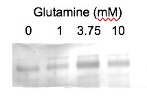
Application: Western BlotSample Tested: Porcine embryoSpecies: PigVerified Customer | Posted 01/09/2019Analysis of glutamate dehydrogenase expression in day 6 porcine blastocysts after culture for 6 days in 0, 1, 3.75, or 10 mM glutamine.A 4-20% gradient gel was used to separate proteins from pools of 40 blastocysts before transfer to PVDF membranes. The membrane was incubated in 1:1000 anti-glutamate dehydrogenase primary overnight at 4 degrees Celsius. The next day, membranes were incubated in 1:10000 anti-rabbit IgG HRP secondary for 1 hour.

There are no reviews that match your criteria.
Protocols
Find general support by application which include: protocols, troubleshooting, illustrated assays, videos and webinars.
- Antigen Retrieval Protocol (PIER)
- Antigen Retrieval for Frozen Sections Protocol
- Appropriate Fixation of IHC/ICC Samples
- Cellular Response to Hypoxia Protocols
- Chromogenic IHC Staining of Formalin-Fixed Paraffin-Embedded (FFPE) Tissue Protocol
- Chromogenic Immunohistochemistry Staining of Frozen Tissue
- ClariTSA™ Fluorophore Kits
- Detection & Visualization of Antibody Binding
- ELISA Sample Preparation & Collection Guide
- ELISA Troubleshooting Guide
- Fluorescent IHC Staining of Frozen Tissue Protocol
- Graphic Protocol for Heat-induced Epitope Retrieval
- Graphic Protocol for the Preparation and Fluorescent IHC Staining of Frozen Tissue Sections
- Graphic Protocol for the Preparation and Fluorescent IHC Staining of Paraffin-embedded Tissue Sections
- Graphic Protocol for the Preparation of Gelatin-coated Slides for Histological Tissue Sections
- How to Run an R&D Systems DuoSet ELISA
- How to Run an R&D Systems Quantikine ELISA
- How to Run an R&D Systems Quantikine™ QuicKit™ ELISA
- IHC Sample Preparation (Frozen sections vs Paraffin)
- Immunofluorescent IHC Staining of Formalin-Fixed Paraffin-Embedded (FFPE) Tissue Protocol
- Immunohistochemistry (IHC) and Immunocytochemistry (ICC) Protocols
- Immunohistochemistry Frozen Troubleshooting
- Immunohistochemistry Paraffin Troubleshooting
- Immunoprecipitation Protocol
- Preparing Samples for IHC/ICC Experiments
- Preventing Non-Specific Staining (Non-Specific Binding)
- Primary Antibody Selection & Optimization
- Protocol for Heat-Induced Epitope Retrieval (HIER)
- Protocol for Making a 4% Formaldehyde Solution in PBS
- Protocol for VisUCyte™ HRP Polymer Detection Reagent
- Protocol for the Preparation & Fixation of Cells on Coverslips
- Protocol for the Preparation and Chromogenic IHC Staining of Frozen Tissue Sections
- Protocol for the Preparation and Chromogenic IHC Staining of Frozen Tissue Sections - Graphic
- Protocol for the Preparation and Chromogenic IHC Staining of Paraffin-embedded Tissue Sections
- Protocol for the Preparation and Chromogenic IHC Staining of Paraffin-embedded Tissue Sections - Graphic
- Protocol for the Preparation and Fluorescent IHC Staining of Frozen Tissue Sections
- Protocol for the Preparation and Fluorescent IHC Staining of Paraffin-embedded Tissue Sections
- Protocol for the Preparation of Gelatin-coated Slides for Histological Tissue Sections
- Quantikine HS ELISA Kit Assay Principle, Alkaline Phosphatase
- Quantikine HS ELISA Kit Principle, Streptavidin-HRP Polymer
- R&D Systems Quality Control Western Blot Protocol
- Sandwich ELISA (Colorimetric) – Biotin/Streptavidin Detection Protocol
- Sandwich ELISA (Colorimetric) – Direct Detection Protocol
- TUNEL and Active Caspase-3 Detection by IHC/ICC Protocol
- The Importance of IHC/ICC Controls
- Troubleshooting Guide: ELISA
- Troubleshooting Guide: Immunohistochemistry
- Troubleshooting Guide: Western Blot Figures
- Western Blot Conditions
- Western Blot Protocol
- Western Blot Protocol for Cell Lysates
- Western Blot Troubleshooting
- Western Blot Troubleshooting Guide
- View all Protocols, Troubleshooting, Illustrated assays and Webinars
Loading...