GOLGA4 Antibody - BSA Free
Novus Biologicals | Catalog # NBP3-04456
![Western Blot: GOLGA4 AntibodyBSA Free [NBP3-04456]](https://resources.rndsystems.com/images/products/GOLGA4-Antibody-Western-Blot-NBP3-04456-img0004.jpg "Western Blot: GOLGA4 AntibodyBSA Free [NBP3-04456]")
Loading...
Key Product Details
Species Reactivity
Validated:
Human, Mouse, Rat
Cited:
Human, Mouse
Applications
Validated:
Immunohistochemistry, Immunohistochemistry-Paraffin, Western Blot, ELISA, Immunocytochemistry/ Immunofluorescence, Proximity Ligation Assay
Cited:
Immunocytochemistry/Immunofluorescence
Label
Unconjugated
Antibody Source
Polyclonal Rabbit IgG
Format
BSA Free
Loading...
Product Specifications
Immunogen
Recombinant fusion protein containing a sequence corresponding to amino acids 1-150 of human GOLGA4 (NP_002069.2). MFKKLKQKISEEQQQLQQALAPAQASSNSSTPTRMRSRTSSFTEQLDEGTPNRESGDTQSFAQKLQLRVPSVESLFRSPIKESLFRSSSKESLVRTSSRESLNRLDLDSSTASFDPPSDMDSEAEDLVGNSDSLNKEQLIQRLRRMERSL
Clonality
Polyclonal
Host
Rabbit
Isotype
IgG
Theoretical MW
261 kDa.
Disclaimer note: The observed molecular weight of the protein may vary from the listed predicted molecular weight due to post translational modifications, post translation cleavages, relative charges, and other experimental factors.
Disclaimer note: The observed molecular weight of the protein may vary from the listed predicted molecular weight due to post translational modifications, post translation cleavages, relative charges, and other experimental factors.
Scientific Data Images for GOLGA4 Antibody - BSA Free
Western Blot: GOLGA4 AntibodyBSA Free [NBP3-04456]
Western Blot: GOLGA4 Antibody [NBP3-04456] - Analysis of extracts of various cell lines, using GOLGA4 antibody at 1:1000 dilution. Secondary antibody: HRP Goat Anti-Rabbit IgG (H+L) at 1:10000 dilution. Lysates/proteins: 25ug per lane. Blocking buffer: 3% nonfat dry milk in TBST. Detection: ECL Basic Kit![Immunocytochemistry/ Immunofluorescence: GOLGA4 Antibody - BSA Free [NBP3-04456]](https://resources.rndsystems.com/images/products/GOLGA4-Antibody-Immunocytochemistry-Immunofluorescence-NBP3-04456-img0003.jpg "Immunocytochemistry/ Immunofluorescence: GOLGA4 Antibody - BSA Free [NBP3-04456]")
Immunocytochemistry/ Immunofluorescence: GOLGA4 Antibody - BSA Free [NBP3-04456]
Immunocytochemistry/Immunofluorescence: GOLGA4 Antibody [NBP3-04456] - Analysis of L929 cells using GOLGA4 Rabbit pAb at dilution of 1:100 (40x lens). Blue: DAPI for nuclear staining. GOLGA4 antibody
Immunocytochemistry/Immunofluorescence: GOLGA4 Antibody [NBP3-04456] -
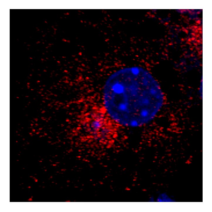
Immunocytochemistry/Immunofluorescence: GOLGA4 Antibody [NBP3-04456] - Rat hepatocytes were stained with anti-GOLGA4 Antibody. Image from verified customer review.
Immunocytochemistry/Immunofluorescence: GOLGA4 Antibody [NBP3-04456] -
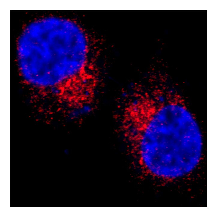
Immunocytochemistry/Immunofluorescence: GOLGA4 Antibody [NBP3-04456] - Mice hepatocytes were stained with anti-GOLGA4 Antibody. Image from verified customer review.
Immunocytochemistry/Immunofluorescence: GOLGA4 Antibody [NBP3-04456] -
Immunocytochemistry/Immunofluorescence: GOLGA4 Antibody [NBP3-04456] - HepG2 cells stained with GOLGA4 Antibody. Image from verified customer review.
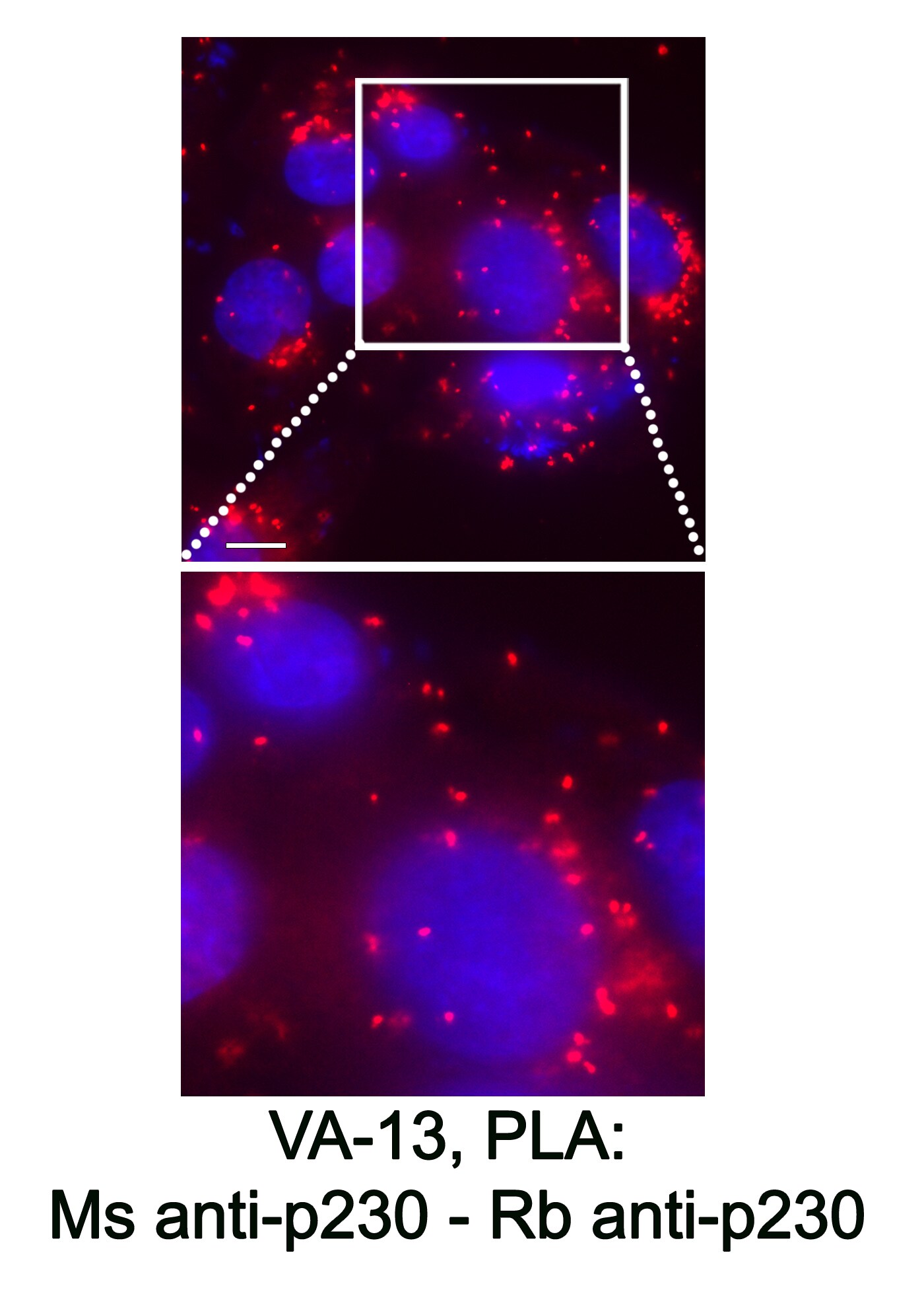
Proximity Ligation Assay: Rabbit Polyclonal GOLGA4 Antibody [NBP3-04456]
Proximity Ligation Assay: Rabbit Polyclonal GOLGA4 Antibody [NBP3-04456] - PLA using a Rabbit anti-p230 Ab (recognizing amino acids 1-150 in the protein's N-terminus) (NBP3-04456) and Mouse Ab recognizing the central regions of p230 located within aa 1482-1584. Image from a verified customer review.
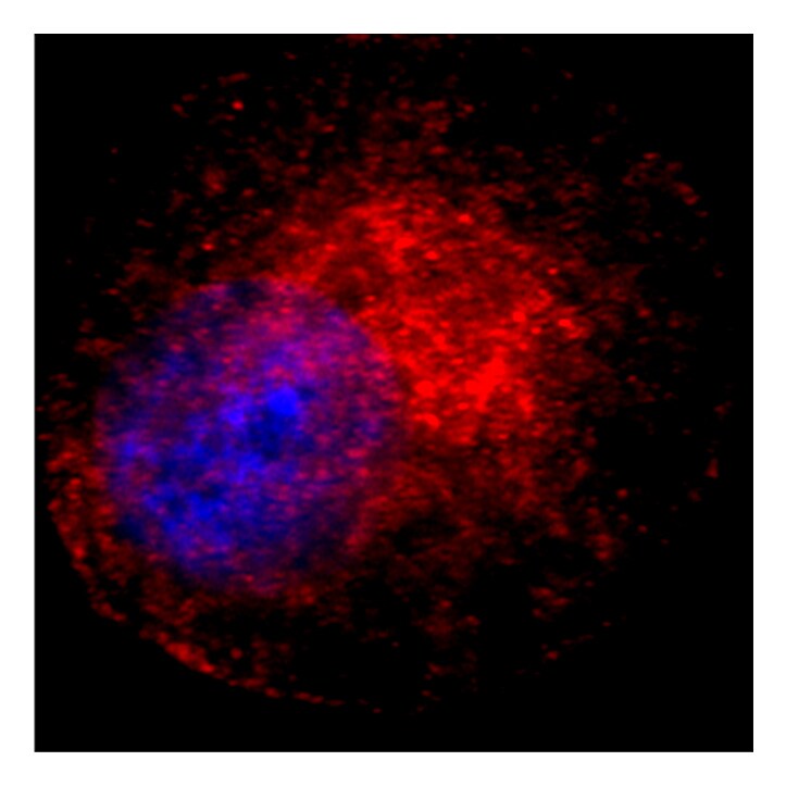
Immunocytochemistry/ Immunofluorescence: GOLGA4 Antibody - BSA Free [NBP3-04456] -
Immunocytochemistry/ Immunofluorescence: GOLGA4 Antibody - BSA Free [NBP3-04456] - Immunofluorescence analysis of C6 cells using GOLGA4 Rabbit pAb (A10216) at dilution of 1:100 (40x lens). Blue: DAPI for nuclear staining.
Immunocytochemistry/ Immunofluorescence: GOLGA4 Antibody - BSA Free [NBP3-04456] -
Immunocytochemistry/ Immunofluorescence: GOLGA4 Antibody - BSA Free [NBP3-04456] - Immunofluorescence analysis of HeLa cells using GOLGA4 Rabbit pAb (A10216) at dilution of 1:100 (40x lens). Blue: DAPI for nuclear staining.Applications for GOLGA4 Antibody - BSA Free
Application
Recommended Usage
Immunocytochemistry/ Immunofluorescence
1:50-1:200
Immunohistochemistry
1:50-1:200
Proximity Ligation Assay
Valifated for Proximity Ligation Assay from a verified customer review.
Western Blot
1:1000-1:2000
Reviewed Applications
Read 4 reviews rated 4.8 using NBP3-04456 in the following applications:
Formulation, Preparation, and Storage
Purification
Affinity purified
Formulation
PBS (pH 7.3), 50% glycerol
Format
BSA Free
Preservative
0.02% Sodium Azide
Concentration
Please see the vial label for concentration. If unlisted please contact technical services.
Shipping
The product is shipped with polar packs. Upon receipt, store it immediately at the temperature recommended below.
Stability & Storage
Store at -20C. Avoid freeze-thaw cycles.
Background: GOLGA4
Alternate Names
72.1 protein, GCP2, GOLG, golgin A4, golgin subfamily a, 4, golgin-240, golgin-245, MU-RMS-40.18, p230, protein 72.1
Gene Symbol
GOLGA4
Additional GOLGA4 Products
Product Documents for GOLGA4 Antibody - BSA Free
Certificate of Analysis
To download a Certificate of Analysis, please enter a lot or batch number in the search box below.
Product Specific Notices for GOLGA4 Antibody - BSA Free
This product is for research use only and is not approved for use in humans or in clinical diagnosis. Primary Antibodies are guaranteed for 1 year from date of receipt.
Citations for GOLGA4 Antibody - BSA Free
 Powered by Bioz
Customer Reviews for GOLGA4 Antibody - BSA Free (4)
4.8 out of 5
4 Customer Ratings
Have you used GOLGA4 Antibody - BSA Free?
Submit a review and receive an Amazon gift card!
$25/€18/£15/$25CAN/¥2500 Yen for a review with an image
$10/€7/£6/$10CAN/¥1110 Yen for a review without an image
Submit a review
Customer Images
Showing
1
-
4 of
4 reviews
Showing All
Filter By:
-
Application: Proximity Ligation AssaySample Tested: HepG2 and HepG2 CELLSSpecies: HumanVerified Customer | Posted 11/07/2024PLA using a Rabbit anti-p230 Ab (recognizing amino acids 1-150 in the protein's N-terminus) (Novus Biologicals, NBP3-04456) and Mouse Ab recognizing the central regions of p230 located within aa 1482-1584.The examination of the spatial relationship between p230's C- and N-termini using PLA.
 Bio-Techne ResponseThis review was submitted through the legacy Novus Innovators Program, reflecting a new species or application tested on a primary antibody.
 -
Application: ImmunofluorescenceSample Tested: Rat hepatocytesSpecies: RatVerified Customer | Posted 06/27/2023Rat hepatocytes were stained with anti-GolgA4 Ab
-
Application: ImmunofluorescenceSample Tested: mouse primary hepatocytesSpecies: MouseVerified Customer | Posted 06/27/2023Mice hepatocytes were stained with anti-GolgA4 Ab.
-
Application: ImmunofluorescenceSample Tested: HepG2 hepatoma cellsSpecies: HumanVerified Customer | Posted 06/27/2023HepG2 cells stained with GolgA4 antibody
There are no reviews that match your criteria.
Protocols
Find general support by application which include: protocols, troubleshooting, illustrated assays, videos and webinars.
- Antigen Retrieval Protocol (PIER)
- Antigen Retrieval for Frozen Sections Protocol
- Appropriate Fixation of IHC/ICC Samples
- Cellular Response to Hypoxia Protocols
- Chromogenic IHC Staining of Formalin-Fixed Paraffin-Embedded (FFPE) Tissue Protocol
- Chromogenic Immunohistochemistry Staining of Frozen Tissue
- ClariTSA™ Fluorophore Kits
- Detection & Visualization of Antibody Binding
- ELISA Sample Preparation & Collection Guide
- ELISA Troubleshooting Guide
- Fluorescent IHC Staining of Frozen Tissue Protocol
- Graphic Protocol for Heat-induced Epitope Retrieval
- Graphic Protocol for the Preparation and Fluorescent IHC Staining of Frozen Tissue Sections
- Graphic Protocol for the Preparation and Fluorescent IHC Staining of Paraffin-embedded Tissue Sections
- Graphic Protocol for the Preparation of Gelatin-coated Slides for Histological Tissue Sections
- How to Run an R&D Systems DuoSet ELISA
- How to Run an R&D Systems Quantikine ELISA
- How to Run an R&D Systems Quantikine™ QuicKit™ ELISA
- ICC Cell Smear Protocol for Suspension Cells
- ICC Immunocytochemistry Protocol Videos
- ICC for Adherent Cells
- IHC Sample Preparation (Frozen sections vs Paraffin)
- Immunocytochemistry (ICC) Protocol
- Immunocytochemistry Troubleshooting
- Immunofluorescence of Organoids Embedded in Cultrex Basement Membrane Extract
- Immunofluorescent IHC Staining of Formalin-Fixed Paraffin-Embedded (FFPE) Tissue Protocol
- Immunohistochemistry (IHC) and Immunocytochemistry (ICC) Protocols
- Immunohistochemistry Frozen Troubleshooting
- Immunohistochemistry Paraffin Troubleshooting
- Preparing Samples for IHC/ICC Experiments
- Preventing Non-Specific Staining (Non-Specific Binding)
- Primary Antibody Selection & Optimization
- Protocol for Heat-Induced Epitope Retrieval (HIER)
- Protocol for Making a 4% Formaldehyde Solution in PBS
- Protocol for VisUCyte™ HRP Polymer Detection Reagent
- Protocol for the Fluorescent ICC Staining of Cell Smears - Graphic
- Protocol for the Fluorescent ICC Staining of Cultured Cells on Coverslips - Graphic
- Protocol for the Preparation & Fixation of Cells on Coverslips
- Protocol for the Preparation and Chromogenic IHC Staining of Frozen Tissue Sections
- Protocol for the Preparation and Chromogenic IHC Staining of Frozen Tissue Sections - Graphic
- Protocol for the Preparation and Chromogenic IHC Staining of Paraffin-embedded Tissue Sections
- Protocol for the Preparation and Chromogenic IHC Staining of Paraffin-embedded Tissue Sections - Graphic
- Protocol for the Preparation and Fluorescent ICC Staining of Cells on Coverslips
- Protocol for the Preparation and Fluorescent ICC Staining of Non-adherent Cells
- Protocol for the Preparation and Fluorescent ICC Staining of Stem Cells on Coverslips
- Protocol for the Preparation and Fluorescent IHC Staining of Frozen Tissue Sections
- Protocol for the Preparation and Fluorescent IHC Staining of Paraffin-embedded Tissue Sections
- Protocol for the Preparation of Gelatin-coated Slides for Histological Tissue Sections
- Protocol for the Preparation of a Cell Smear for Non-adherent Cell ICC - Graphic
- Quantikine HS ELISA Kit Assay Principle, Alkaline Phosphatase
- Quantikine HS ELISA Kit Principle, Streptavidin-HRP Polymer
- R&D Systems Quality Control Western Blot Protocol
- Sandwich ELISA (Colorimetric) – Biotin/Streptavidin Detection Protocol
- Sandwich ELISA (Colorimetric) – Direct Detection Protocol
- TUNEL and Active Caspase-3 Detection by IHC/ICC Protocol
- The Importance of IHC/ICC Controls
- Troubleshooting Guide: ELISA
- Troubleshooting Guide: Immunohistochemistry
- Troubleshooting Guide: Western Blot Figures
- Western Blot Conditions
- Western Blot Protocol
- Western Blot Protocol for Cell Lysates
- Western Blot Troubleshooting
- Western Blot Troubleshooting Guide
- View all Protocols, Troubleshooting, Illustrated assays and Webinars
Loading...