GR/NR3C1 Antibody (BuGR2) - BSA Free
Novus Biologicals | Catalog # NB300-731
![Western Blot: GR/NR3C1 Antibody (BuGR2)BSA Free [NB300-731]](https://resources.rndsystems.com/images/products/GR-NR3C1-Antibody-BuGR2-Western-Blot-NB300-731-img0017.jpg "Western Blot: GR/NR3C1 Antibody (BuGR2)BSA Free [NB300-731]")
Key Product Details
Species Reactivity
Validated:
Human, Mouse, Rat, Guinea Pig, Rabbit, Sheep, Yeast
Cited:
Human, Mouse
Applications
Validated:
Immunohistochemistry, Immunohistochemistry-Paraffin, Western Blot, Block/Neutralize, Flow Cytometry, Immunocytochemistry/ Immunofluorescence, Immunoprecipitation, Chromatin Immunoprecipitation (ChIP), Gel Super Shift Assays
Cited:
Western Blot, Flow Cytometry, Immunocytochemistry/ Immunofluorescence, Chemotaxis
Label
Unconjugated
Antibody Source
Monoclonal Mouse IgG2A Clone # BuGR2
Format
BSA Free
Loading...
Product Specifications
Immunogen
Partially purified rat GR.
Reactivity Notes
Please note that this antibody is reactive to Mouse and derived from the same host, Mouse. Additional Mouse on Mouse blocking steps may be required for IHC and ICC experiments. Please contact Technical Support for more information.
Localization
Cytoplasmic in the absence of ligand; nuclear after ligand-binding.
Specificity
Using enzymatic digestion analysis, has been shown to react with the undigested 97 kDa GR, a 17 kDa DNA-binding trypsin fragment, and a 45 kDa steroid- and DNA-binding chymotrypsin fragment.
Clonality
Monoclonal
Host
Mouse
Isotype
IgG2A
Scientific Data Images for GR/NR3C1 Antibody (BuGR2) - BSA Free
Western Blot: GR/NR3C1 Antibody (BuGR2)BSA Free [NB300-731]
Western Blot: GR/NR3C1 Antibody (BuGR2) [NB300-731] - Analysis of glucocorticoid receptor on mouse liver extract.![Immunocytochemistry/ Immunofluorescence: GR/NR3C1 Antibody (BuGR2) - BSA Free [NB300-731]](https://resources.rndsystems.com/images/products/GR-NR3C1-Antibody-BuGR2-Immunocytochemistry-Immunofluorescence-NB300-731-img0007.jpg "Immunocytochemistry/ Immunofluorescence: GR/NR3C1 Antibody (BuGR2) - BSA Free [NB300-731]")
Immunocytochemistry/ Immunofluorescence: GR/NR3C1 Antibody (BuGR2) - BSA Free [NB300-731]
Immunocytochemistry/Immunofluorescence: GR/NR3C1 Antibody (BuGR2) [NB300-731] - Analysis of Glucocorticoid Receptor using Glucocorticoid Receptor Monoclonal Antibody (BuGR2) shows staining in U251 Cells. Glucocorticoid Receptor (green), F-Actin staining with Phalloidin (red) and nuclei with DAPI (blue) is shown. Cells were grown on chamber slides and fixed with formaldehyde prior to staining. Cells were probed without (control) or with an antibody recognizing Glucocorticoid Receptor at a dilution of 1:100 over night at 4C, washed with PBS and incubated with a DyLight-488 conjugated.![Flow Cytometry: GR/NR3C1 Antibody (BuGR2) - BSA Free [NB300-731]](https://resources.rndsystems.com/images/products/GR-NR3C1-Antibody-BuGR2-Flow-Cytometry-NB300-731-img0018.jpg "Flow Cytometry: GR/NR3C1 Antibody (BuGR2) - BSA Free [NB300-731]")
Flow Cytometry: GR/NR3C1 Antibody (BuGR2) - BSA Free [NB300-731]
Flow Cytometry: GR/NR3C1 Antibody (BuGR2) [NB300-731] - Using the Alexa Fluor 647 direct conjugate An intracellular stain was performed on HeLa cells with GR/NR3C1 (BuGR2) antibody NB300-731AF647 (blue) and a matched isotype control NB600-986AF647 (orange). Cells were fixed with 4% PFA and then permeablized with 0.1% saponin. Cells were incubated in an antibody dilution of 2 ug/mL for 30 minutes at room temperature. Both antibodies were conjugated to Alexa Fluor 647.![Immunocytochemistry/ Immunofluorescence: GR/NR3C1 Antibody (BuGR2) - BSA Free [NB300-731]](https://resources.rndsystems.com/images/products/GR-NR3C1-Antibody-BuGR2-Immunocytochemistry-Immunofluorescence-NB300-731-img0014.jpg "Immunocytochemistry/ Immunofluorescence: GR/NR3C1 Antibody (BuGR2) - BSA Free [NB300-731]")
Immunocytochemistry/ Immunofluorescence: GR/NR3C1 Antibody (BuGR2) - BSA Free [NB300-731]
Immunocytochemistry/Immunofluorescence: GR/NR3C1 Antibody (BuGR2) [NB300-731] - Analysis of Glucocorticoid Receptor using Glucocorticoid Receptor Monoclonal Antibody (BuGR2) shows staining in A549 Cells. Glucocorticoid Receptor (green), F-Actin staining with Phalloidin (red) and nuclei with DAPI (blue) is shown. Cells were grown on chamber slides and fixed with formaldehyde prior to staining. Cells were probed without (control) or with an antibody recognizing Glucocorticoid Receptor at a dilution of 1:100 over night at 4C, washed with PBS and incubated with a DyLight-488 conjugated.![Immunocytochemistry/ Immunofluorescence: GR/NR3C1 Antibody (BuGR2) - BSA Free [NB300-731]](https://resources.rndsystems.com/images/products/GR-NR3C1-Antibody-BuGR2-Immunocytochemistry-Immunofluorescence-NB300-731-img0006.jpg "Immunocytochemistry/ Immunofluorescence: GR/NR3C1 Antibody (BuGR2) - BSA Free [NB300-731]")
Immunocytochemistry/ Immunofluorescence: GR/NR3C1 Antibody (BuGR2) - BSA Free [NB300-731]
Immunocytochemistry/Immunofluorescence: GR/NR3C1 Antibody (BuGR2) [NB300-731] - Analysis of Glucocorticoid Receptor using Glucocorticoid Receptor Monoclonal Antibody (BuGR2) shows staining in HeLa Cells. Glucocorticoid Receptor (green), F-Actin staining with Phalloidin (red) and nuclei with DAPI (blue) is shown. Cells were grown on chamber slides and fixed with formaldehyde prior to staining. Cells were probed without (control) or with an antibody recognizing Glucocorticoid Receptor at a dilution of 1:100 over night at 4C, washed with PBS and incubated with a DyLight-488 conjugated.![Flow Cytometry: GR/NR3C1 Antibody (BuGR2) - BSA Free [NB300-731]](https://resources.rndsystems.com/images/products/GR-NR3C1-Antibody-BuGR2-Flow-Cytometry-NB300-731-img0012.jpg "Flow Cytometry: GR/NR3C1 Antibody (BuGR2) - BSA Free [NB300-731]")
Flow Cytometry: GR/NR3C1 Antibody (BuGR2) - BSA Free [NB300-731]
Flow Cytometry: GR/NR3C1 Antibody (BuGR2) [NB300-731] - Analysis of GR in Jurkat cells compared to an isotype control (blue).![Flow Cytometry: GR/NR3C1 Antibody (BuGR2) - BSA Free [NB300-731]](https://resources.rndsystems.com/images/products/GR-NR3C1-Antibody-BuGR2-Flow-Cytometry-NB300-731-img0015.jpg "Flow Cytometry: GR/NR3C1 Antibody (BuGR2) - BSA Free [NB300-731]")
Flow Cytometry: GR/NR3C1 Antibody (BuGR2) - BSA Free [NB300-731]
Flow Cytometry: GR/NR3C1 Antibody (BuGR2) [NB300-731] - Analysis of Glucocorticoid Receptor in NIH/3T3 cells compared to an isotype control (blue).![Flow Cytometry: GR/NR3C1 Antibody (BuGR2) - BSA Free [NB300-731]](https://resources.rndsystems.com/images/products/GR-NR3C1-Antibody-BuGR2-Flow-Cytometry-NB300-731-img0016.jpg "Flow Cytometry: GR/NR3C1 Antibody (BuGR2) - BSA Free [NB300-731]")
Flow Cytometry: GR/NR3C1 Antibody (BuGR2) - BSA Free [NB300-731]
Flow Cytometry: GR/NR3C1 Antibody (BuGR2) [NB300-731] - Analysis of Glucocorticoid Receptor in HeLa cells compared to an isotype control (blue).![Flow Cytometry: GR/NR3C1 Antibody (BuGR2) - BSA Free [NB300-731]](https://resources.rndsystems.com/images/products/GR-NR3C1-Antibody-BuGR2-Flow-Cytometry-NB300-731-img0019.jpg "Flow Cytometry: GR/NR3C1 Antibody (BuGR2) - BSA Free [NB300-731]")
Flow Cytometry: GR/NR3C1 Antibody (BuGR2) - BSA Free [NB300-731]
Flow Cytometry: GR/NR3C1 Antibody (BuGR2) [NB300-731] - An intracellular stain was performed on Jurkat cells with GR/NR3C1 (BuGR2) antibody NB300-731PE (blue) and a matched isotype control (orange). Cells were fixed with 4% PFA and then permeablized with 0.1% saponin. Cells were incubated in an antibody dilution of 5 ug/mL for 30 minutes at room temperature. Both antibodies were conjugated to Phycoerthrin.Applications for GR/NR3C1 Antibody (BuGR2) - BSA Free
Application
Recommended Usage
Chromatin Immunoprecipitation (ChIP)
1:10-1:500
Flow Cytometry
1:10 - 1:1000
Gel Super Shift Assays
1:10 - 1:100
Immunocytochemistry/ Immunofluorescence
1:10 - 1:500
Immunohistochemistry
1:10 - 1:500
Immunohistochemistry-Paraffin
5 ug/mL
Immunoprecipitation
1:10 - 1:500
Western Blot
5 ug/mL
Application Notes
Blocking, ChIP, and ELISA usages were reported in scientific literature. Use in ICC/IF was reported in scientific literature (PMID: 30402116)
Reviewed Applications
Read 3 reviews rated 3 using NB300-731 in the following applications:
Flow Cytometry Panel Builder
Bio-Techne Knows Flow Cytometry
Save time and reduce costly mistakes by quickly finding compatible reagents using the Panel Builder Tool.
Advanced Features
- Spectra Viewer - Custom analysis of spectra from multiple fluorochromes
- Spillover Popups - Visualize the spectra of individual fluorochromes
- Antigen Density Selector - Match fluorochrome brightness with antigen density

Formulation, Preparation, and Storage
Purification
Protein A purified
Reconstitution
Reconstitute with 0.1 ml sterilized water to desired concentration.
Formulation
PBS (pH 7.2)
Format
BSA Free
Preservative
0.05% Sodium Azide
Concentration
LYOPH mg/ml
Shipping
The product is shipped with polar packs. Upon receipt, store it immediately at the temperature recommended below.
Stability & Storage
Store at -20C. Avoid freeze-thaw cycles.
Calculators
Background: GR/NR3C1
Long Name
Glucocorticoid Receptor
Alternate Names
NR3C1
Gene Symbol
NR3C1
Additional GR/NR3C1 Products
Product Documents for GR/NR3C1 Antibody (BuGR2) - BSA Free
Certificate of Analysis
To download a Certificate of Analysis, please enter a lot or batch number in the search box below.
Product Specific Notices for GR/NR3C1 Antibody (BuGR2) - BSA Free
This product is for research use only and is not approved for use in humans or in clinical diagnosis. Primary Antibodies are guaranteed for 1 year from date of receipt.
Related Research Areas
Citations for GR/NR3C1 Antibody (BuGR2) - BSA Free
 Powered by Bioz
Customer Reviews for GR/NR3C1 Antibody (BuGR2) - BSA Free (3)
3 out of 5
3 Customer Ratings
Have you used GR/NR3C1 Antibody (BuGR2) - BSA Free?
Submit a review and receive an Amazon gift card!
$25/€18/£15/$25CAN/¥2500 Yen for a review with an image
$10/€7/£6/$10CAN/¥1110 Yen for a review without an image
Submit a review
Customer Images
-(01-mg)_NB300-731_9831.png)
-(01-mg)_NB300-731_9716.png)
Showing
1
-
3 of
3 reviews
Showing All
Filter By:
-
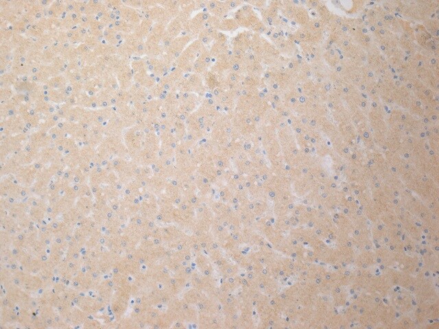
Application: Immunohistochemistry-ParaffinSample Tested: Liver tissue and LiverSpecies: EquineVerified Customer | Posted 02/01/2020Tried HIER ph 6 and HIER pH 9 with concentrations as high as 1:200. Never saw any nuclear staining in horse liver; only non-specific background staining.
 Bio-Techne ResponseThis review was submitted through the legacy Novus Innovators Program, reflecting a new species or application tested on a primary antibody.
 -
Application: Western BlotSample Tested: mouse hippocampal tissue lysateSpecies: MouseVerified Customer | Posted 09/03/2014Blot with anti-glucocorticoid receptor 2 (NB300-371), duplicated sample of mouse hippocampal protein extract
-
Application: Western BlotVerified Customer | Posted 08/29/2014Blot with anti-glucocorticoid receptor 2 (NB300-371), duplicated sample of mouse hippocampal protein extract
There are no reviews that match your criteria.
Protocols
Find general support by application which include: protocols, troubleshooting, illustrated assays, videos and webinars.
- 7-Amino Actinomycin D (7-AAD) Cell Viability Flow Cytometry Protocol
- Antigen Retrieval Protocol (PIER)
- Antigen Retrieval for Frozen Sections Protocol
- Appropriate Fixation of IHC/ICC Samples
- Cellular Response to Hypoxia Protocols
- ChIP Protocol Video
- Chromatin Immunoprecipitation (ChIP) Protocol
- Chromatin Immunoprecipitation Protocol
- Chromogenic IHC Staining of Formalin-Fixed Paraffin-Embedded (FFPE) Tissue Protocol
- Chromogenic Immunohistochemistry Staining of Frozen Tissue
- ClariTSA™ Fluorophore Kits
- Detection & Visualization of Antibody Binding
- Extracellular Membrane Flow Cytometry Protocol
- Flow Cytometry Protocol for Cell Surface Markers
- Flow Cytometry Protocol for Staining Membrane Associated Proteins
- Flow Cytometry Staining Protocols
- Flow Cytometry Troubleshooting Guide
- Fluorescent IHC Staining of Frozen Tissue Protocol
- Graphic Protocol for Heat-induced Epitope Retrieval
- Graphic Protocol for the Preparation and Fluorescent IHC Staining of Frozen Tissue Sections
- Graphic Protocol for the Preparation and Fluorescent IHC Staining of Paraffin-embedded Tissue Sections
- Graphic Protocol for the Preparation of Gelatin-coated Slides for Histological Tissue Sections
- ICC Cell Smear Protocol for Suspension Cells
- ICC Immunocytochemistry Protocol Videos
- ICC for Adherent Cells
- IHC Sample Preparation (Frozen sections vs Paraffin)
- Immunocytochemistry (ICC) Protocol
- Immunocytochemistry Troubleshooting
- Immunofluorescence of Organoids Embedded in Cultrex Basement Membrane Extract
- Immunofluorescent IHC Staining of Formalin-Fixed Paraffin-Embedded (FFPE) Tissue Protocol
- Immunohistochemistry (IHC) and Immunocytochemistry (ICC) Protocols
- Immunohistochemistry Frozen Troubleshooting
- Immunohistochemistry Paraffin Troubleshooting
- Immunoprecipitation Protocol
- Intracellular Flow Cytometry Protocol Using Alcohol (Methanol)
- Intracellular Flow Cytometry Protocol Using Detergents
- Intracellular Nuclear Staining Flow Cytometry Protocol Using Detergents
- Intracellular Staining Flow Cytometry Protocol Using Alcohol Permeabilization
- Intracellular Staining Flow Cytometry Protocol Using Detergents to Permeabilize Cells
- Preparing Samples for IHC/ICC Experiments
- Preventing Non-Specific Staining (Non-Specific Binding)
- Primary Antibody Selection & Optimization
- Propidium Iodide Cell Viability Flow Cytometry Protocol
- Protocol for Heat-Induced Epitope Retrieval (HIER)
- Protocol for Liperfluo
- Protocol for Making a 4% Formaldehyde Solution in PBS
- Protocol for VisUCyte™ HRP Polymer Detection Reagent
- Protocol for the Characterization of Human Th22 Cells
- Protocol for the Characterization of Human Th9 Cells
- Protocol for the Fluorescent ICC Staining of Cell Smears - Graphic
- Protocol for the Fluorescent ICC Staining of Cultured Cells on Coverslips - Graphic
- Protocol for the Preparation & Fixation of Cells on Coverslips
- Protocol for the Preparation and Chromogenic IHC Staining of Frozen Tissue Sections
- Protocol for the Preparation and Chromogenic IHC Staining of Frozen Tissue Sections - Graphic
- Protocol for the Preparation and Chromogenic IHC Staining of Paraffin-embedded Tissue Sections
- Protocol for the Preparation and Chromogenic IHC Staining of Paraffin-embedded Tissue Sections - Graphic
- Protocol for the Preparation and Fluorescent ICC Staining of Cells on Coverslips
- Protocol for the Preparation and Fluorescent ICC Staining of Non-adherent Cells
- Protocol for the Preparation and Fluorescent ICC Staining of Stem Cells on Coverslips
- Protocol for the Preparation and Fluorescent IHC Staining of Frozen Tissue Sections
- Protocol for the Preparation and Fluorescent IHC Staining of Paraffin-embedded Tissue Sections
- Protocol for the Preparation of Gelatin-coated Slides for Histological Tissue Sections
- Protocol for the Preparation of a Cell Smear for Non-adherent Cell ICC - Graphic
- Protocol: Annexin V and PI Staining by Flow Cytometry
- Protocol: Annexin V and PI Staining for Apoptosis by Flow Cytometry
- R&D Systems Quality Control Western Blot Protocol
- TUNEL and Active Caspase-3 Detection by IHC/ICC Protocol
- The Importance of IHC/ICC Controls
- Troubleshooting Guide: Fluorokine Flow Cytometry Kits
- Troubleshooting Guide: Immunohistochemistry
- Troubleshooting Guide: Western Blot Figures
- Western Blot Conditions
- Western Blot Protocol
- Western Blot Protocol for Cell Lysates
- Western Blot Troubleshooting
- Western Blot Troubleshooting Guide
- View all Protocols, Troubleshooting, Illustrated assays and Webinars
Loading...
Associated Pathways
