Hepatic Sinusoidal Endothelial Cells Antibody (SE-1) - BSA Free
Novus Biologicals | Catalog # NB110-68095
Key Product Details
Validated by
Biological Validation
Species Reactivity
Validated:
Human, Mouse, Rat
Cited:
Human, Mouse, Rat
Applications
Validated:
Immunohistochemistry, Immunohistochemistry-Paraffin, Immunohistochemistry-Frozen, Western Blot, Flow Cytometry, Immunocytochemistry/ Immunofluorescence
Cited:
Immunohistochemistry, Immunohistochemistry-Paraffin, Western Blot, Flow Cytometry, Immunocytochemistry/ Immunofluorescence, IF/IHC
Label
Unconjugated
Antibody Source
Monoclonal Mouse IgG2a Kappa Clone # SE-1
Format
BSA Free
Loading...
Product Specifications
Immunogen
Rat Hepatic Sinusoidal Endothelial Cells
Clonality
Monoclonal
Host
Mouse
Isotype
IgG2a Kappa
Scientific Data Images for Hepatic Sinusoidal Endothelial Cells Antibody (SE-1) - BSA Free
![Western Blot: Hepatic Sinusoidal Endothelial Cells Antibody (SE-1) [NB110-68095]](https://resources.rndsystems.com/images/products/Hepatic-Sinusoidal-Endothelial-Cells-Antibody-SE-1-Western-Blot-NB110-68095-img0009.jpg "Western Blot: Hepatic Sinusoidal Endothelial Cells Antibody (SE-1) [NB110-68095]")
![Immunocytochemistry/ Immunofluorescence: Hepatic Sinusoidal Endothelial Cells Antibody (SE-1) [NB110-68095]](https://resources.rndsystems.com/images/products/Hepatic-Sinusoidal-Endothelial-Cells-Antibody-SE-1-Immunocytochemistry-Immunofluorescence-NB110-68095-img0010.jpg "Immunocytochemistry/ Immunofluorescence: Hepatic Sinusoidal Endothelial Cells Antibody (SE-1) [NB110-68095]")
Immunocytochemistry/ Immunofluorescence: Hepatic Sinusoidal Endothelial Cells Antibody (SE-1) [NB110-68095]
Hepatic-Sinusoidal-Endothelial-Cells-Antibody-SE-1-Immunocytochemistry-Immunofluorescence-NB110-68095-img0010.jpg![Immunohistochemistry-Frozen: Hepatic Sinusoidal Endothelial Cells Antibody (SE-1) [NB110-68095]](https://resources.rndsystems.com/images/products/Hepatic-Sinusoidal-Endothelial-Cells-Antibody-SE-1-Immunohistochemistry-Frozen-NB110-68095-img0008.jpg "Immunohistochemistry-Frozen: Hepatic Sinusoidal Endothelial Cells Antibody (SE-1) [NB110-68095]")
Immunohistochemistry-Frozen: Hepatic Sinusoidal Endothelial Cells Antibody (SE-1) [NB110-68095]
Immunohistochemistry-Frozen: Hepatic Sinusoidal Endothelial Cells Antibody (SE-1) [NB110-68095] - Frozen rat liver tissue sections. DAPI (blue), SE1 (green), cytochrome P450 (red). Tissue sections were acetone fixed. SE1 antibody at 1:500, incubated at 4C overnight. Multiplexed with cytochrome P450 from another vendor. IHC-Fr image submitted by a verified customer review.![Flow Cytometry: Hepatic Sinusoidal Endothelial Cells Antibody (SE-1) [NB110-68095]](https://resources.rndsystems.com/images/products/Hepatic-Sinusoidal-Endothelial-Cells-Antibody-SE-1-Flow-Cytometry-NB110-68095-img0011.jpg "Flow Cytometry: Hepatic Sinusoidal Endothelial Cells Antibody (SE-1) [NB110-68095]")
Flow Cytometry: Hepatic Sinusoidal Endothelial Cells Antibody (SE-1) [NB110-68095]
Hepatic-Sinusoidal-Endothelial-Cells-Antibody-SE-1-Flow-Cytometry-NB110-68095-img0011.jpg![Western Blot: Hepatic Sinusoidal Endothelial Cells Antibody (SE-1) [NB110-68095]](https://resources.rndsystems.com/images/products/Hepatic-Sinusoidal-Endothelial-Cells-Antibody-SE-1-Western-Blot-NB110-68095-img0006.jpg "Western Blot: Hepatic Sinusoidal Endothelial Cells Antibody (SE-1) [NB110-68095]")
Western Blot: Hepatic Sinusoidal Endothelial Cells Antibody (SE-1) [NB110-68095]
Western Blot: Hepatic Sinusoidal Endothelial Cells Antibody (SE-1) [NB110-68095] - Total protein from human, mouse and rat liver was separated on a 12% gel by SDS-PAGE, transferred to PVDF membrane and blocked in 5% non-fat milk in TBST. The membrane was probed with 2.0 ug/ml anti-H.S.E.C in 1% non-fat milk in TBST and detected with an anti-mouse HRP secondary antibody using chemiluminescence.![Immunohistochemistry-Frozen: Hepatic Sinusoidal Endothelial Cells Antibody (SE-1) [NB110-68095]](https://resources.rndsystems.com/images/products/Hepatic-Sinusoidal-Endothelial-Cells-Antibody-SE-1-Immunohistochemistry-Frozen-NB110-68095-img0004.jpg "Immunohistochemistry-Frozen: Hepatic Sinusoidal Endothelial Cells Antibody (SE-1) [NB110-68095]")
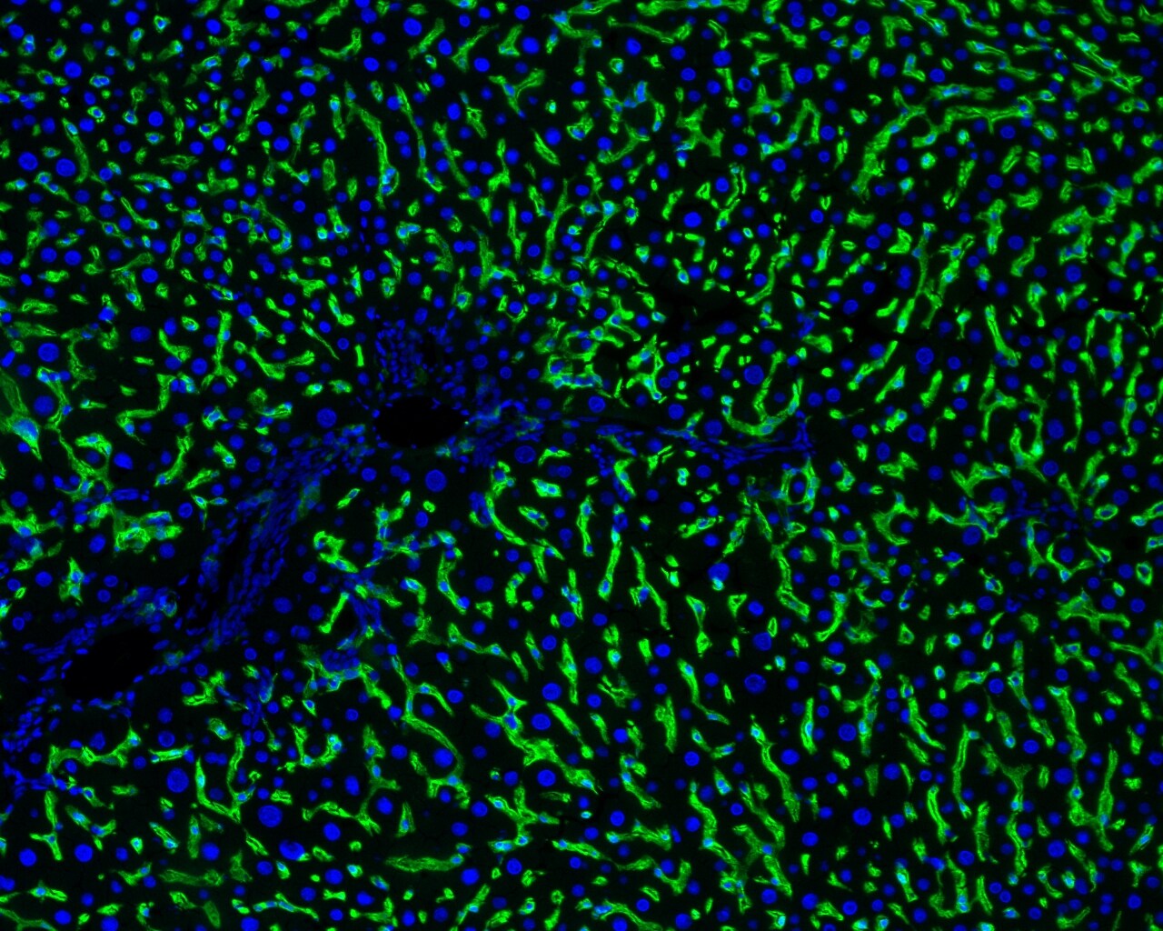
Immunohistochemistry-Frozen: Hepatic Sinusoidal Endothelial Cells Antibody (SE-1) [NB110-68095]
Immunohistochemistry-Frozen: Hepatic Sinusoidal Endothelial Cells Antibody (SE-1) [NB110-68095] - Analysis of frozen normal rat liver tissue sections using anti-Hepatic Sinusoidal Endothelial Cells antibody (green). Nuclei were counterstained with DAPI (blue). Image from verified customer review.![Flow Cytometry: Hepatic Sinusoidal Endothelial Cells Antibody (SE-1) [NB110-68095]](https://resources.rndsystems.com/images/products/Hepatic-Sinusoidal-Endothelial-Cells-Antibody-SE-1-Flow-Cytometry-NB110-68095-img0007.jpg "Flow Cytometry: Hepatic Sinusoidal Endothelial Cells Antibody (SE-1) [NB110-68095]")
Flow Cytometry: Hepatic Sinusoidal Endothelial Cells Antibody (SE-1) [NB110-68095]
Flow Cytometry: Hepatic Sinusoidal Endothelial Cells Antibody (SE-1) [NB110-68095] - Using the FITC direct conjugate Flow Cytometry: Surface staining of Rat Liver sinusoidal endothelial cells with Mouse anti-Rat Hepatic Sinusoidal Endothelial Cells [FITC] [NB110-68095F] and negative control. Total viable cells were used for analysis. Image courtesy of customer. [NB110-68095] -")
Immunocytochemistry/ Immunofluorescence: Hepatic Sinusoidal Endothelial Cells Antibody (SE-1) [NB110-68095] -
Immunocytochemistry/ Immunofluorescence: Hepatic Sinusoidal Endothelial Cells Antibody (SE-1) [NB110-68095] - Expression of scavenger receptors & immune lectins in rat LSECs & KCs. a. Unscaled heatmaps of normalized log2 expression values (log2 (RPKM+ 1), & log2 (iBAQ+ 1)) for scavenger receptors (SR) & C-type lectins in the KC & LSEC transcriptomes & proteomes. Underlined: Genes expressed in the transcriptome that were also present in the proteome. b. Absolute abundance of selected SR gene products in the KC & LSEC transcriptomes & proteomes. The bar height reflects good correlation between the transcriptome & proteome data for gene products of Clec4g, Clec4m, Stab1, & Stab2 in both cell types. The abundance of gene products of Marco & Cd5l were well correlated between the KC transcriptome & proteome, while LSECs showed high abundance of these gene products only at mRNA level. c. Immune labeling of non-parenchymal liver cell (NPC) cultures for selected SRs & C-type lectins. NPCs from the 25–45% interface on the Percoll gradient were incubated for 1 h, then fixed 15 min in 4% paraformaldehyde, & double immune-labeled with antibodies to Fc gamma RIIb2 (SE-1; red fluorescence; left column), or CD68 (red fluorescence; right column), & to either stabilin-2 (STAB2; green), mannose receptor (MRC1; green), SR-A1 (green), or SR-B1 (green). Overlap of green & red fluorescence is seen as yellow staining in the overlay images. Antibodies are listed in Table 1. Cell nuclei were stained with DAPI (blue). Arrow heads point to CD68 positive KCs. Antibodies to stabilin-2 & Fc gamma RIIb2 (SE-1) specifically labeled LSECs & the CD68-antibody specifically labeled KCs, whereas positive labeling for the mannose receptor, SR-A1, & SR-B1 was observed in both LSECs & KCs Image collected & cropped by CiteAb from the following publication (https://pubmed.ncbi.nlm.nih.gov/33246411), licensed under a CC-BY license. Not internally tested by Novus Biologicals. - BSA Free [NB110-68095] -")
Immunohistochemistry-Frozen: Hepatic Sinusoidal Endothelial Cells Antibody (SE-1) - BSA Free [NB110-68095] -
Expression of scavenger receptors and immune lectins in rat LSECs and KCs. a. Unscaled heatmaps of normalized log2 expression values (log2 (RPKM+ 1), and log2 (iBAQ+ 1)) for scavenger receptors (SR) and C-type lectins in the KC and LSEC transcriptomes and proteomes. Underlined: Genes expressed in the transcriptome that were also present in the proteome. b. Absolute abundance of selected SR gene products in the KC and LSEC transcriptomes and proteomes. The bar height reflects good correlation between the transcriptome and proteome data for gene products of Clec4g, Clec4m, Stab1, and Stab2 in both cell types. The abundance of gene products of Marco and Cd5l were well correlated between the KC transcriptome and proteome, while LSECs showed high abundance of these gene products only at mRNA level. c. Immune labeling of non-parenchymal liver cell (NPC) cultures for selected SRs and C-type lectins. NPCs from the 25–45% interface on the Percoll gradient were incubated for 1 h, then fixed 15 min in 4% paraformaldehyde, and double immune-labeled with antibodies to Fc gamma RIIb2 (SE-1; red fluorescence; left column), or CD68 (red fluorescence; right column), and to either stabilin-2 (STAB2; green), mannose receptor (MRC1; green), SR-A1 (green), or SR-B1 (green). Overlap of green and red fluorescence is seen as yellow staining in the overlay images. Antibodies are listed in Table 1. Cell nuclei were stained with DAPI (blue). Arrow heads point to CD68 positive KCs. Antibodies to stabilin-2 and Fc gamma RIIb2 (SE-1) specifically labeled LSECs and the CD68-antibody specifically labeled KCs, whereas positive labeling for the mannose receptor, SR-A1, and SR-B1 was observed in both LSECs and KCs Image collected and cropped by CiteAb from the following open publication (https://pubmed.ncbi.nlm.nih.gov/33246411), licensed under a CC-BY license. Not internally tested by Novus Biologicals.Applications for Hepatic Sinusoidal Endothelial Cells Antibody (SE-1) - BSA Free
Application
Recommended Usage
Flow Cytometry
reported in scientific literature (PMID 9428229)
Immunohistochemistry
1-5 ug/ml
Immunohistochemistry-Frozen
1-5 ug/ml
Immunohistochemistry-Paraffin
1-5 ug/ml
Western Blot
1-5 ug/ml
Application Notes
In WB, Hepatic Sinusoidal Endothelial Cells antibody (clone SE-1) generates a specific band around 45-50 kDa molecular weight position. For IHC use neutral buffered formalin fixated (perfusion fixation is recommended) paraffin embedded tissues after proteinase K treatment or acetone fixed frozen sections.
Reviewed Applications
Read 3 reviews rated 3.7 using NB110-68095 in the following applications:
Flow Cytometry Panel Builder
Bio-Techne Knows Flow Cytometry
Save time and reduce costly mistakes by quickly finding compatible reagents using the Panel Builder Tool.
Advanced Features
- Spectra Viewer - Custom analysis of spectra from multiple fluorochromes
- Spillover Popups - Visualize the spectra of individual fluorochromes
- Antigen Density Selector - Match fluorochrome brightness with antigen density

Formulation, Preparation, and Storage
Purification
Protein A purified
Formulation
PBS
Format
BSA Free
Preservative
0.02% Sodium Azide
Concentration
1 mg/ml
Shipping
The product is shipped with polar packs. Upon receipt, store it immediately at the temperature recommended below.
Stability & Storage
Store at 4C short term. Aliquot and store at -20C long term. Avoid freeze-thaw cycles.
Background: Hepatic Sinusoidal Endothelial Cells
Alternate Names
Hepatic cells, Hepatic Sinusoidal Endothelial Cells, HSE, SE1, Sinusoidal Endothelial Cells
Additional Hepatic Sinusoidal Endothelial Cells Products
Product Documents for Hepatic Sinusoidal Endothelial Cells Antibody (SE-1) - BSA Free
Certificate of Analysis
To download a Certificate of Analysis, please enter a lot or batch number in the search box below.
Product Specific Notices for Hepatic Sinusoidal Endothelial Cells Antibody (SE-1) - BSA Free
This product is for research use only and is not approved for use in humans or in clinical diagnosis. Primary Antibodies are guaranteed for 1 year from date of receipt.
Citations for Hepatic Sinusoidal Endothelial Cells Antibody (SE-1) - BSA Free
 Powered by Bioz
Customer Reviews for Hepatic Sinusoidal Endothelial Cells Antibody (SE-1) - BSA Free (3)
3.7 out of 5
3 Customer Ratings
Have you used Hepatic Sinusoidal Endothelial Cells Antibody (SE-1) - BSA Free?
Submit a review and receive an Amazon gift card!
$25/€18/£15/$25CAN/¥2500 Yen for a review with an image
$10/€7/£6/$10CAN/¥1110 Yen for a review without an image
Submit a review
Customer Images

Showing
1
-
3 of
3 reviews
Showing All
Filter By:
-
Application: Immunohistochemistry-FrozenSample Tested: Frozen Rat Liver Tissue sectionsSpecies: RatVerified Customer | Posted 10/10/2019DAPI (blue), SE1 (green), cytochrome P450 (red)Normal rat liver Acetone fixation SE1 antibody used at 1:500 dilution, incubated at 4C overnight Multiplexed with cytochrome P450 antibody from another vendor

-
Application: Immunohistochemistry-FrozenSample Tested: Frozen Rat Liver Tissue sectionsSpecies: RatVerified Customer | Posted 10/15/2015Normal Rat Liver - SE-1 (green) and DAPI (blue)
-
Application: Immunohistochemistry-ParaffinSample Tested: See PMID 20400698Species: MouseVerified Customer | Posted 12/29/2014
There are no reviews that match your criteria.
Protocols
Find general support by application which include: protocols, troubleshooting, illustrated assays, videos and webinars.
- 7-Amino Actinomycin D (7-AAD) Cell Viability Flow Cytometry Protocol
- Antigen Retrieval Protocol (PIER)
- Antigen Retrieval for Frozen Sections Protocol
- Appropriate Fixation of IHC/ICC Samples
- Cellular Response to Hypoxia Protocols
- Chromogenic IHC Staining of Formalin-Fixed Paraffin-Embedded (FFPE) Tissue Protocol
- Chromogenic Immunohistochemistry Staining of Frozen Tissue
- ClariTSA™ Fluorophore Kits
- Detection & Visualization of Antibody Binding
- Extracellular Membrane Flow Cytometry Protocol
- Flow Cytometry Protocol for Cell Surface Markers
- Flow Cytometry Protocol for Staining Membrane Associated Proteins
- Flow Cytometry Staining Protocols
- Flow Cytometry Troubleshooting Guide
- Fluorescent IHC Staining of Frozen Tissue Protocol
- Graphic Protocol for Heat-induced Epitope Retrieval
- Graphic Protocol for the Preparation and Fluorescent IHC Staining of Frozen Tissue Sections
- Graphic Protocol for the Preparation and Fluorescent IHC Staining of Paraffin-embedded Tissue Sections
- Graphic Protocol for the Preparation of Gelatin-coated Slides for Histological Tissue Sections
- ICC Cell Smear Protocol for Suspension Cells
- ICC Immunocytochemistry Protocol Videos
- ICC for Adherent Cells
- IHC Sample Preparation (Frozen sections vs Paraffin)
- Immunocytochemistry (ICC) Protocol
- Immunocytochemistry Troubleshooting
- Immunofluorescence of Organoids Embedded in Cultrex Basement Membrane Extract
- Immunofluorescent IHC Staining of Formalin-Fixed Paraffin-Embedded (FFPE) Tissue Protocol
- Immunohistochemistry (IHC) and Immunocytochemistry (ICC) Protocols
- Immunohistochemistry Frozen Troubleshooting
- Immunohistochemistry Paraffin Troubleshooting
- Intracellular Flow Cytometry Protocol Using Alcohol (Methanol)
- Intracellular Flow Cytometry Protocol Using Detergents
- Intracellular Nuclear Staining Flow Cytometry Protocol Using Detergents
- Intracellular Staining Flow Cytometry Protocol Using Alcohol Permeabilization
- Intracellular Staining Flow Cytometry Protocol Using Detergents to Permeabilize Cells
- Preparing Samples for IHC/ICC Experiments
- Preventing Non-Specific Staining (Non-Specific Binding)
- Primary Antibody Selection & Optimization
- Propidium Iodide Cell Viability Flow Cytometry Protocol
- Protocol for Heat-Induced Epitope Retrieval (HIER)
- Protocol for Liperfluo
- Protocol for Making a 4% Formaldehyde Solution in PBS
- Protocol for VisUCyte™ HRP Polymer Detection Reagent
- Protocol for the Characterization of Human Th22 Cells
- Protocol for the Characterization of Human Th9 Cells
- Protocol for the Fluorescent ICC Staining of Cell Smears - Graphic
- Protocol for the Fluorescent ICC Staining of Cultured Cells on Coverslips - Graphic
- Protocol for the Preparation & Fixation of Cells on Coverslips
- Protocol for the Preparation and Chromogenic IHC Staining of Frozen Tissue Sections
- Protocol for the Preparation and Chromogenic IHC Staining of Frozen Tissue Sections - Graphic
- Protocol for the Preparation and Chromogenic IHC Staining of Paraffin-embedded Tissue Sections
- Protocol for the Preparation and Chromogenic IHC Staining of Paraffin-embedded Tissue Sections - Graphic
- Protocol for the Preparation and Fluorescent ICC Staining of Cells on Coverslips
- Protocol for the Preparation and Fluorescent ICC Staining of Non-adherent Cells
- Protocol for the Preparation and Fluorescent ICC Staining of Stem Cells on Coverslips
- Protocol for the Preparation and Fluorescent IHC Staining of Frozen Tissue Sections
- Protocol for the Preparation and Fluorescent IHC Staining of Paraffin-embedded Tissue Sections
- Protocol for the Preparation of Gelatin-coated Slides for Histological Tissue Sections
- Protocol for the Preparation of a Cell Smear for Non-adherent Cell ICC - Graphic
- Protocol: Annexin V and PI Staining by Flow Cytometry
- Protocol: Annexin V and PI Staining for Apoptosis by Flow Cytometry
- R&D Systems Quality Control Western Blot Protocol
- TUNEL and Active Caspase-3 Detection by IHC/ICC Protocol
- The Importance of IHC/ICC Controls
- Troubleshooting Guide: Fluorokine Flow Cytometry Kits
- Troubleshooting Guide: Immunohistochemistry
- Troubleshooting Guide: Western Blot Figures
- Western Blot Conditions
- Western Blot Protocol
- Western Blot Protocol for Cell Lysates
- Western Blot Troubleshooting
- Western Blot Troubleshooting Guide
- View all Protocols, Troubleshooting, Illustrated assays and Webinars
Loading...