His Tag Antibody (33D10.D2.G8) [Biotin]
Novus Biologicals | Catalog # NBP1-42782
Key Product Details
Species Reactivity
Epitope Tag
Applications
Immunohistochemistry, Immunohistochemistry-Paraffin, Western Blot, ELISA
Label
Biotin
Antibody Source
Monoclonal Mouse IgG1 kappa Clone # 33D10.D2.G8
Loading...
Product Specifications
Immunogen
His Tag Antibody (33D10.D2.G8) was produced in mice by repeated immunizations with 6X His epitope tag peptide H-H-H-H-H-H conjugated to KLH using maleimide.
Reactivity Notes
6x His tag specific
Specificity
This protein-A purified antibody is directed against the 6X His motif and is useful in determining its presence in various assays. This monoclonal anti-6X His tag antibody detects over-expressed proteins containing the 6X His epitope tag. To date, this antibody has reacted with all His tagged proteins so far tested. In western blotting of bacterial extracts, the antibody does not cross-react with endogenous proteins. The antibody recognizes the His-tag (His-His-His-His-His-His) fused to either the amino- or carboxy-termini of targeted proteins in transfected or transformed cells.
Clonality
Monoclonal
Host
Mouse
Isotype
IgG1 kappa
Description
Store vial at 4C prior to restoration. For extended storage aliquot contents and freeze at -20C or below. Avoid cycles of freezing and thawing. Centrifuge product if not completely clear after standing at room temperature. This product is stable for several weeks at 4C as an undiluted liquid. Dilute only prior to immediate use.
This protein-A purified antibody is directed against the 6X His motif and is useful in determining its presence in various assays
This protein-A purified antibody is directed against the 6X His motif and is useful in determining its presence in various assays
Scientific Data Images for His Tag Antibody (33D10.D2.G8) [Biotin]
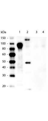
Western Blot: His Tag Antibody (33D10.D2.G8) [Biotin] [NBP1-42782] - 6-Histidine Epitope Tag Antibody (33D10.D2) [Biotin] [NBP1-42782] - Lane 1: 100 ng Purified histidine-tagged recombinant protein. Lane 2: 200 ng E. coli cell lysate containing histidine-tagged expression construct. Lane 3: 100ng Purified GST-tagged recombinant protein. Lane 4: 100 ng Purified FLAG-tagged recombinant protein. Primary antibody: Mouse anti-6xHIS Tag antibody at 1:5,000 overnight at 4C. Secondary antibody: Peroxidase mouse secondary antibody at 1:20,000 for 30 min at RT. Block: 5% BLOTTO for 1 hr at RT.
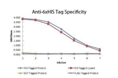
ELISA: His Tag Antibody (33D10.D2.G8) [Biotin] [NBP1-42782] - ELISA of Mouse anti-6xHIS Tag Antibody. Antigen: HIS-tagged purified protein and E. coli cell lysates expressing HIS-Tagged construct, GST- and RON-tagged purified proteins. Coating amount: 0.15 ug per well. Primary antibody: 6xHIS Tag antibody at 100 ug/mL. Dilution series: 2-fold. Mid-point concentration: 200 ng/mL. Secondary antibody: Peroxidase mouse secondary antibody at 1:10,000. Substrate: TMB
Applications for His Tag Antibody (33D10.D2.G8) [Biotin]
Application
Recommended Usage
ELISA
1:50000 - 1:175000
Immunohistochemistry
1:1000 - 1:5000
Immunohistochemistry-Paraffin
1:1000 - 1:5000
Western Blot
1:2000 - 1:10000
Application Notes
This product is optimally suited for monitoring expression of His-tagged fusion proteins. As such, anti-6X His/6X His can be used to identify fusion proteins containing the 6X His epitope. The antibody recognizes the His tag fused either to the amino- or carboxy- termini of targeted proteins. This antibody has been tested by ELISA and western blotting against both the immunizing peptide and His-containing recombinant proteins. Although not tested, this antibody is likely functional for immunoprecipitation and immunocytochemistry.
Formulation, Preparation, and Storage
Purification
Protein A purified
Reconstitution
Reconstitute with 100 ul deionized water (or equivalent)
Formulation
Lyophilized from 0.02 M Potassium Phosphate, 0.15 M Sodium Chloride, pH 7.2, 10 mg/mL Bovine Serum Albumin (BSA) - Immunoglobulin and Protease free
Preservative
0.01% Sodium Azide
Concentration
LYOPH mg/ml
Shipping
The product is shipped with polar packs. Upon receipt, store it immediately at the temperature recommended below.
Stability & Storage
Store lyophilized antibody at 4C in the dark. Aliquot reconstituted liquid and store at -20C. Avoid freeze-thaw cycles.
Calculators
Background: His Tag
References
1. Malhotra, A. (2009). Tagging for protein expression. Methods in Enzymology, Guide to Protein Purification, 2nd Edition, 463, 239-258. https://doi.org/10.1016/s0076-6879(09)63016-0
2. Terpe, K. (2003). Overview of tag protein fusions: from molecular and biochemical fundamentals to commercial systems. Applied Microbiology and Biotechnology, 60(5), 523-533. https://doi.org/10.1007/s00253-002-1158-6
3. Booth, W. T., Schlachter, C. R., Pote, S., Ussin, N., Mank, N. J., Klapper, V.,... Chruszcz, M. (2018). Impact of an N-terminal polyhistidine tag on protein thermal stability. ACS Omega, 3(1), 760-768. https://doi.org/10.1021/acsomega.7b01598
Long Name
Histidine Tag
Alternate Names
polyHistidine
Additional His Tag Products
Product Documents for His Tag Antibody (33D10.D2.G8) [Biotin]
Certificate of Analysis
To download a Certificate of Analysis, please enter a lot or batch number in the search box below.
Product Specific Notices for His Tag Antibody (33D10.D2.G8) [Biotin]
This product is for research use only and is not approved for use in humans or in clinical diagnosis. Primary Antibodies are guaranteed for 1 year from date of receipt.
Citations for His Tag Antibody (33D10.D2.G8) [Biotin]
 Powered by Bioz
Customer Reviews for His Tag Antibody (33D10.D2.G8) [Biotin]
There are currently no reviews for this product. Be the first to review His Tag Antibody (33D10.D2.G8) [Biotin] and earn rewards!
Have you used His Tag Antibody (33D10.D2.G8) [Biotin]?
Submit a review and receive an Amazon gift card!
$25/€18/£15/$25CAN/¥2500 Yen for a review with an image
$10/€7/£6/$10CAN/¥1110 Yen for a review without an image
Submit a review
Protocols
Find general support by application which include: protocols, troubleshooting, illustrated assays, videos and webinars.
- Antigen Retrieval Protocol (PIER)
- Antigen Retrieval for Frozen Sections Protocol
- Appropriate Fixation of IHC/ICC Samples
- Cellular Response to Hypoxia Protocols
- Chromogenic IHC Staining of Formalin-Fixed Paraffin-Embedded (FFPE) Tissue Protocol
- Chromogenic Immunohistochemistry Staining of Frozen Tissue
- ClariTSA™ Fluorophore Kits
- Detection & Visualization of Antibody Binding
- ELISA Sample Preparation & Collection Guide
- ELISA Troubleshooting Guide
- Fluorescent IHC Staining of Frozen Tissue Protocol
- Graphic Protocol for Heat-induced Epitope Retrieval
- Graphic Protocol for the Preparation and Fluorescent IHC Staining of Frozen Tissue Sections
- Graphic Protocol for the Preparation and Fluorescent IHC Staining of Paraffin-embedded Tissue Sections
- Graphic Protocol for the Preparation of Gelatin-coated Slides for Histological Tissue Sections
- How to Run an R&D Systems DuoSet ELISA
- How to Run an R&D Systems Quantikine ELISA
- How to Run an R&D Systems Quantikine™ QuicKit™ ELISA
- IHC Sample Preparation (Frozen sections vs Paraffin)
- Immunofluorescent IHC Staining of Formalin-Fixed Paraffin-Embedded (FFPE) Tissue Protocol
- Immunohistochemistry (IHC) and Immunocytochemistry (ICC) Protocols
- Immunohistochemistry Frozen Troubleshooting
- Immunohistochemistry Paraffin Troubleshooting
- Preparing Samples for IHC/ICC Experiments
- Preventing Non-Specific Staining (Non-Specific Binding)
- Primary Antibody Selection & Optimization
- Protocol for Heat-Induced Epitope Retrieval (HIER)
- Protocol for Making a 4% Formaldehyde Solution in PBS
- Protocol for VisUCyte™ HRP Polymer Detection Reagent
- Protocol for the Preparation & Fixation of Cells on Coverslips
- Protocol for the Preparation and Chromogenic IHC Staining of Frozen Tissue Sections
- Protocol for the Preparation and Chromogenic IHC Staining of Frozen Tissue Sections - Graphic
- Protocol for the Preparation and Chromogenic IHC Staining of Paraffin-embedded Tissue Sections
- Protocol for the Preparation and Chromogenic IHC Staining of Paraffin-embedded Tissue Sections - Graphic
- Protocol for the Preparation and Fluorescent IHC Staining of Frozen Tissue Sections
- Protocol for the Preparation and Fluorescent IHC Staining of Paraffin-embedded Tissue Sections
- Protocol for the Preparation of Gelatin-coated Slides for Histological Tissue Sections
- Quantikine HS ELISA Kit Assay Principle, Alkaline Phosphatase
- Quantikine HS ELISA Kit Principle, Streptavidin-HRP Polymer
- R&D Systems Quality Control Western Blot Protocol
- Sandwich ELISA (Colorimetric) – Biotin/Streptavidin Detection Protocol
- Sandwich ELISA (Colorimetric) – Direct Detection Protocol
- TUNEL and Active Caspase-3 Detection by IHC/ICC Protocol
- The Importance of IHC/ICC Controls
- Troubleshooting Guide: ELISA
- Troubleshooting Guide: Immunohistochemistry
- Troubleshooting Guide: Western Blot Figures
- Western Blot Conditions
- Western Blot Protocol
- Western Blot Protocol for Cell Lysates
- Western Blot Troubleshooting
- Western Blot Troubleshooting Guide
- View all Protocols, Troubleshooting, Illustrated assays and Webinars
Loading...