Histone H3 Antibody (1B1-B2) - Azide and BSA Free
Novus Biologicals | Catalog # NBP2-80770
Key Product Details
Validated by
Knockout/Knockdown
Species Reactivity
Human, Mouse, Rat
Applications
Immunohistochemistry, Immunohistochemistry-Paraffin, Western Blot, Flow Cytometry, Flow (Intracellular), Immunocytochemistry/ Immunofluorescence, Chromatin Immunoprecipitation (ChIP), Single Cell Western
Label
Unconjugated
Antibody Source
Monoclonal Mouse IgG3 Kappa Clone # 1B1-B2
Format
Azide and BSA Free
Loading...
Product Specifications
Immunogen
This Histone H3 antibody (1B1-B2) was raised against synthetic peptide corresponding to the C-terminus of human Histone H3.
Localization
Nucleus. Chromosome.
Clonality
Monoclonal
Host
Mouse
Isotype
IgG3 Kappa
Theoretical MW
15 kDa.
Disclaimer note: The observed molecular weight of the protein may vary from the listed predicted molecular weight due to post translational modifications, post translation cleavages, relative charges, and other experimental factors.
Disclaimer note: The observed molecular weight of the protein may vary from the listed predicted molecular weight due to post translational modifications, post translation cleavages, relative charges, and other experimental factors.
Scientific Data Images for Histone H3 Antibody (1B1-B2) - Azide and BSA Free
![Western Blot: Histone H3 Antibody (1B1-B2)Azide and BSA Free [NBP2-80770]](https://resources.rndsystems.com/images/products/Histone-H3-Antibody-1B1-B2-Azide-and-BSA-Free-Western-Blot-NBP2-80770-img0002.jpg "Western Blot: Histone H3 Antibody (1B1-B2)Azide and BSA Free [NBP2-80770]")
Western Blot: Histone H3 Antibody (1B1-B2)Azide and BSA Free [NBP2-80770]
Western Blot: Histone H3 Antibody (1B1-B2) - Azide and BSA Free [NBP2-80770] - Western blot image of Anti-Histone H3 antibody (clone 1B1-B2). Whole cell protein from HeLa (lane 1), NIH-3T3 (lane 2) and PC12 (lane 3) were separated by 4-15% SDS-PAGE and transfered to PVDF membrane. The membrane was probed with purified Anti-Histone H3 antibody at 2 ug/ml and detected with an HRP conjugated anti-mouse secondary using chemiluminescence. Observed molecular weight is ~15 kDa. Image from the standard format of this antibody.![Immunocytochemistry/ Immunofluorescence: Histone H3 Antibody (1B1-B2) - Azide and BSA Free [NBP2-80770]](https://resources.rndsystems.com/images/products/Histone-H3-Antibody-1B1-B2-Azide-and-BSA-Free-Immunocytochemistry-Immunofluorescence-NBP2-80770-img0003.jpg "Immunocytochemistry/ Immunofluorescence: Histone H3 Antibody (1B1-B2) - Azide and BSA Free [NBP2-80770]")
Immunocytochemistry/ Immunofluorescence: Histone H3 Antibody (1B1-B2) - Azide and BSA Free [NBP2-80770]
Immunocytochemistry/Immunofluorescence: Histone H3 Antibody (1B1-B2) - Azide and BSA Free [NBP2-80770] - HeLa cells were fixed for 10 minutes using 10% formalin and then permeabilized for 5 minutes using 1X TBS + 0.5% Triton X-100. The cells were incubated with anti-Histone H3 (1B1-B2) NBP2-36468 at a 1:200 dilution overnight at 4C and detected with an anti-mouse DyLight 488 (Green) at a 1:500 dilution. Actin was detected with Phalloidin 568 (Red) at a 1:200 dilution. Nuclei were counterstained with DAPI (Blue). Cells were imaged using a 40X objective. Image from the standard format of this antibody.![Immunohistochemistry: Histone H3 Antibody (1B1-B2) - Azide and BSA Free [NBP2-80770]](https://resources.rndsystems.com/images/products/Histone-H3-Antibody-1B1-B2-Azide-and-BSA-Free-Immunohistochemistry-NBP2-80770-img0004.jpg "Immunohistochemistry: Histone H3 Antibody (1B1-B2) - Azide and BSA Free [NBP2-80770]")
Immunohistochemistry: Histone H3 Antibody (1B1-B2) - Azide and BSA Free [NBP2-80770]
Immunohistochemistry: Histone H3 Antibody (1B1-B2) - Azide and BSA Free [NBP2-80770] - Analysis of Histone H3 in human breast cancer using DAB with hematoxylin counterstain. Image from the standard format of this antibody.![Flow Cytometry: Histone H3 Antibody (1B1-B2) - Azide and BSA Free [NBP2-80770]](https://resources.rndsystems.com/images/products/Histone-H3-Antibody-1B1-B2-Azide-and-BSA-Free-Flow-Cytometry-NBP2-80770-img0005.jpg "Flow Cytometry: Histone H3 Antibody (1B1-B2) - Azide and BSA Free [NBP2-80770]")
Flow Cytometry: Histone H3 Antibody (1B1-B2) - Azide and BSA Free [NBP2-80770]
Flow Cytometry: Histone H3 Antibody (1B1-B2) - Azide and BSA Free [NBP2-80770] - Analysis using Alexa Fluor (R) 647 conjugate of NBP2-36468. Jurkat cells stained with Alexa Fluor 647 conjugated Histone H3 antibody (red) or isotype control (blue). Flow cytometry image submitted by a verified customer review.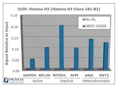
Chromatin Immunoprecipitation: Histone H3 Antibody (1B1-B2) - Azide and BSA Free [NBP2-80770] - Chromatin from one million formaldehyde cross-linked HeLa cells was precipitated using 2 ug of NBP2-36468 and 25 ul of magnetic IgG beads, using standard ChIP methods. A similar sample containing no antibody was included as a negative control. Immunoprecipitated DNA was quantified using quantitative real-time PCR and SYBR green dye, then normalized to the non-precipitated input chromatin. Representative target genes from active, inactive and heterochromatic regions of the genome show amplification, indicative of the presence of Histone H3. Image from the standard format of this antibody.
![Western Blot: Histone H3 Antibody (1B1-B2)Azide and BSA Free [NBP2-80770]](https://resources.rndsystems.com/images/products/Histone-H3-Antibody-1B1-B2-Azide-and-BSA-Free-Western-Blot-NBP2-80770-img0001.jpg "Western Blot: Histone H3 Antibody (1B1-B2)Azide and BSA Free [NBP2-80770]")
Western Blot: Histone H3 Antibody (1B1-B2)Azide and BSA Free [NBP2-80770]
Western Blot: Histone H3 Antibody (1B1-B2) - Azide and BSA Free [NBP2-80770] - Analysis in Hela cells using siRNA to knockdown Pol II, controls are GAPDH and Histone H3. Theoretical molecular weight of Histone H3 is 15 kDa. WB blot image submitted by a verified customer review. Image from the standard format of this antibody.Applications for Histone H3 Antibody (1B1-B2) - Azide and BSA Free
Application
Recommended Usage
Immunohistochemistry
1:200-1:500
Immunohistochemistry-Paraffin
1:200-1:500
Single Cell Western
100 ug/ml
Western Blot
1:1000. Use reported by customer review
Flow Cytometry Panel Builder
Bio-Techne Knows Flow Cytometry
Save time and reduce costly mistakes by quickly finding compatible reagents using the Panel Builder Tool.
Advanced Features
- Spectra Viewer - Custom analysis of spectra from multiple fluorochromes
- Spillover Popups - Visualize the spectra of individual fluorochromes
- Antigen Density Selector - Match fluorochrome brightness with antigen density

Formulation, Preparation, and Storage
Purification
Protein G purified
Formulation
PBS
Format
Azide and BSA Free
Preservative
No Preservative
Concentration
0.5 mg/ml
Shipping
The product is shipped with polar packs. Upon receipt, store it immediately at the temperature recommended below.
Stability & Storage
Store at 4C short term. Aliquot and store at -20C long term. Avoid freeze-thaw cycles.
Background: Histone H3
Histones are nuclear proteins responsible for the nucleosome structure of the chromosomal fiber in eukaryotes. Changes in chromatin structure play a large role in the regulation of transcription. The chromatin fibers are compacted through the interaction of a linker histone, H1, with the DNA between the nucleosomes to form higher order chromatin structures.
Common histone modifications include methylation of lysine and arginine, acetylation of lysine, phosphorylation of threonine and serine, and sumoylation, biotinylation, and ubiquitylation of lysine. Posttranslational modifications such as acetylation of core histones regulates gene expression, thus altering protein function and regulation (1). Histone H3 is primarily acetylated at lysines 9, 14, 18, and 23 and have a theoretical molecular weight of 15 kDa. Acetylation at lysine 9 and 14 appears to control histone deposition, chromatin assembly and active transcription. Methylation of arginine residues within histone H3 has also been linked to transcription regulation. Histone H3 has been linked to various types of cancer as a biomarker through the aberrant expression of histone deacetylase (HDAC) enzymes and changes to chromatins (2-4).
References
1. Zhang, Y. X., Akumuo, R. C., Espana, R. A., Yan, C. X., Gao, W. J., & Li, Y. C. (2018). The histone demethylase KDM6B in the medial prefrontal cortex epigenetically regulates cocaine reward memory. Neuropharmacology, 141, 113-125. doi:10.1016/j.neuropharm.2018.08.030
2. Nandakumar, V., Hansen, N., Glenn, H. L., Han, J. H., Helland, S., Hernandez, K,...Meldrum, D. R. (2016). Vorinostat differentially alters 3D nuclear structure of cancer and non-cancerous esophageal cells. Sci Rep, 6, 30593. doi:10.1038/srep30593
3. Zhou, M., Li, Y., Lin, S., Chen, Y., Qian, Y., Zhao, Z., & Fan, H. (2019). H3K9me3, H3K36me3, and H4K20me3 Expression Correlates with Patient Outcome in Esophageal Squamous Cell Carcinoma as Epigenetic Markers. Dig Dis Sci, 64(8), 2147-2157. doi:10.1007/s10620-019-05529-2
4. Li, Y., Guo, D., Sun, R., Chen, P., Qian, Q., & Fan, H. (2019). Methylation Patterns of Lys9 and Lys27 on Histone H3 Correlate with Patient Outcome in Gastric Cancer. Dig Dis Sci, 64(2), 439-446. doi:10.1007/s10620-018-5341-8
Alternate Names
H3F3A
Gene Symbol
H3C14
Additional Histone H3 Products
Product Documents for Histone H3 Antibody (1B1-B2) - Azide and BSA Free
Certificate of Analysis
To download a Certificate of Analysis, please enter a lot or batch number in the search box below.
Product Specific Notices for Histone H3 Antibody (1B1-B2) - Azide and BSA Free
This product is for research use only and is not approved for use in humans or in clinical diagnosis. Primary Antibodies are guaranteed for 1 year from date of receipt.
Related Research Areas
Customer Reviews for Histone H3 Antibody (1B1-B2) - Azide and BSA Free
There are currently no reviews for this product. Be the first to review Histone H3 Antibody (1B1-B2) - Azide and BSA Free and earn rewards!
Have you used Histone H3 Antibody (1B1-B2) - Azide and BSA Free?
Submit a review and receive an Amazon gift card!
$25/€18/£15/$25CAN/¥2500 Yen for a review with an image
$10/€7/£6/$10CAN/¥1110 Yen for a review without an image
Submit a review
Protocols
Find general support by application which include: protocols, troubleshooting, illustrated assays, videos and webinars.
- 7-Amino Actinomycin D (7-AAD) Cell Viability Flow Cytometry Protocol
- Antigen Retrieval Protocol (PIER)
- Antigen Retrieval for Frozen Sections Protocol
- Appropriate Fixation of IHC/ICC Samples
- Cellular Response to Hypoxia Protocols
- ChIP Protocol Video
- Chromatin Immunoprecipitation (ChIP) Protocol
- Chromatin Immunoprecipitation Protocol
- Chromogenic IHC Staining of Formalin-Fixed Paraffin-Embedded (FFPE) Tissue Protocol
- Chromogenic Immunohistochemistry Staining of Frozen Tissue
- ClariTSA™ Fluorophore Kits
- Detection & Visualization of Antibody Binding
- Extracellular Membrane Flow Cytometry Protocol
- Flow Cytometry Protocol for Cell Surface Markers
- Flow Cytometry Protocol for Staining Membrane Associated Proteins
- Flow Cytometry Staining Protocols
- Flow Cytometry Troubleshooting Guide
- Fluorescent IHC Staining of Frozen Tissue Protocol
- Graphic Protocol for Heat-induced Epitope Retrieval
- Graphic Protocol for the Preparation and Fluorescent IHC Staining of Frozen Tissue Sections
- Graphic Protocol for the Preparation and Fluorescent IHC Staining of Paraffin-embedded Tissue Sections
- Graphic Protocol for the Preparation of Gelatin-coated Slides for Histological Tissue Sections
- ICC Cell Smear Protocol for Suspension Cells
- ICC Immunocytochemistry Protocol Videos
- ICC for Adherent Cells
- IHC Sample Preparation (Frozen sections vs Paraffin)
- Immunocytochemistry (ICC) Protocol
- Immunocytochemistry Troubleshooting
- Immunofluorescence of Organoids Embedded in Cultrex Basement Membrane Extract
- Immunofluorescent IHC Staining of Formalin-Fixed Paraffin-Embedded (FFPE) Tissue Protocol
- Immunohistochemistry (IHC) and Immunocytochemistry (ICC) Protocols
- Immunohistochemistry Frozen Troubleshooting
- Immunohistochemistry Paraffin Troubleshooting
- Intracellular Flow Cytometry Protocol Using Alcohol (Methanol)
- Intracellular Flow Cytometry Protocol Using Detergents
- Intracellular Nuclear Staining Flow Cytometry Protocol Using Detergents
- Intracellular Staining Flow Cytometry Protocol Using Alcohol Permeabilization
- Intracellular Staining Flow Cytometry Protocol Using Detergents to Permeabilize Cells
- Preparing Samples for IHC/ICC Experiments
- Preventing Non-Specific Staining (Non-Specific Binding)
- Primary Antibody Selection & Optimization
- Propidium Iodide Cell Viability Flow Cytometry Protocol
- Protocol for Heat-Induced Epitope Retrieval (HIER)
- Protocol for Liperfluo
- Protocol for Making a 4% Formaldehyde Solution in PBS
- Protocol for VisUCyte™ HRP Polymer Detection Reagent
- Protocol for the Characterization of Human Th22 Cells
- Protocol for the Characterization of Human Th9 Cells
- Protocol for the Fluorescent ICC Staining of Cell Smears - Graphic
- Protocol for the Fluorescent ICC Staining of Cultured Cells on Coverslips - Graphic
- Protocol for the Preparation & Fixation of Cells on Coverslips
- Protocol for the Preparation and Chromogenic IHC Staining of Frozen Tissue Sections
- Protocol for the Preparation and Chromogenic IHC Staining of Frozen Tissue Sections - Graphic
- Protocol for the Preparation and Chromogenic IHC Staining of Paraffin-embedded Tissue Sections
- Protocol for the Preparation and Chromogenic IHC Staining of Paraffin-embedded Tissue Sections - Graphic
- Protocol for the Preparation and Fluorescent ICC Staining of Cells on Coverslips
- Protocol for the Preparation and Fluorescent ICC Staining of Non-adherent Cells
- Protocol for the Preparation and Fluorescent ICC Staining of Stem Cells on Coverslips
- Protocol for the Preparation and Fluorescent IHC Staining of Frozen Tissue Sections
- Protocol for the Preparation and Fluorescent IHC Staining of Paraffin-embedded Tissue Sections
- Protocol for the Preparation of Gelatin-coated Slides for Histological Tissue Sections
- Protocol for the Preparation of a Cell Smear for Non-adherent Cell ICC - Graphic
- Protocol: Annexin V and PI Staining by Flow Cytometry
- Protocol: Annexin V and PI Staining for Apoptosis by Flow Cytometry
- R&D Systems Quality Control Western Blot Protocol
- TUNEL and Active Caspase-3 Detection by IHC/ICC Protocol
- The Importance of IHC/ICC Controls
- Troubleshooting Guide: Fluorokine Flow Cytometry Kits
- Troubleshooting Guide: Immunohistochemistry
- Troubleshooting Guide: Western Blot Figures
- Western Blot Conditions
- Western Blot Protocol
- Western Blot Protocol for Cell Lysates
- Western Blot Troubleshooting
- Western Blot Troubleshooting Guide
- View all Protocols, Troubleshooting, Illustrated assays and Webinars
Loading...