Histone H3 Antibody - BSA Free
Novus Biologicals | Catalog # NB500-171
![Western Blot: Histone H3 AntibodyBSA Free [NB500-171]](https://resources.rndsystems.com/images/products/Histone-H3-Antibody-Western-Blot-NB500-171-img0013.jpg "Western Blot: Histone H3 AntibodyBSA Free [NB500-171]")
Key Product Details
Validated by
Biological Validation
Species Reactivity
Validated:
Human, Mouse, Rat, Insect, Xenopus, Yeast
Cited:
Human, Mouse, Rat, Frog - Xenopus (African Clawed Frog)
Predicted:
C. elegans (100%), Chicken (100%), Drosophila (100%), Plant (100%). Backed by our 100% Guarantee.
Applications
Validated:
Immunohistochemistry, Immunohistochemistry-Paraffin, Western Blot, Immunoblotting, Immunocytochemistry/ Immunofluorescence, Chromatin Immunoprecipitation (ChIP), Single Cell Western
Cited:
Western Blot, Immunocytochemistry/ Immunofluorescence, Chemotaxis, IF/IHC, Knockdown Validated
Label
Unconjugated
Antibody Source
Polyclonal Rabbit IgG
Format
BSA Free
Loading...
Product Specifications
Immunogen
This Histone H3 antibody was raised against a synthetic peptide made to an C-terminal portion of the human Histone H3 protein (between residues 100-136) [UniProt P68431]
Reactivity Notes
Predicted to react with many species based on 100% sequence homology including C. elegans, chicken, drosophila, and plant. Xenopus reactivity reported in scientific literature (PMID: 24048589). Insect (Aedes aegypti) reactivity reported from a verified customer review.
Localization
Nuclear
Clonality
Polyclonal
Host
Rabbit
Isotype
IgG
Theoretical MW
15 kDa.
Disclaimer note: The observed molecular weight of the protein may vary from the listed predicted molecular weight due to post translational modifications, post translation cleavages, relative charges, and other experimental factors.
Disclaimer note: The observed molecular weight of the protein may vary from the listed predicted molecular weight due to post translational modifications, post translation cleavages, relative charges, and other experimental factors.
Scientific Data Images for Histone H3 Antibody - BSA Free
Western Blot: Histone H3 AntibodyBSA Free [NB500-171]
Western Blot: Histone H3 Antibody [NB500-171] - Analysis of HeLa, COS, C6, and K562 cell lysate using histone H3 antibody [NB500-171] antibody at 1:100. Observed molecular weight is ~17 kDa.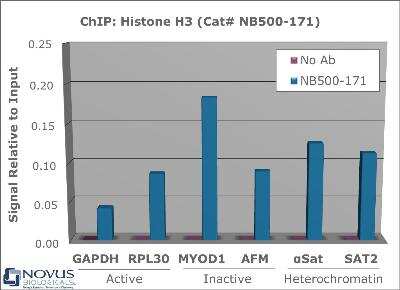
Chromatin Immunoprecipitation: Histone H3 Antibody [NB500-171] - Chromatin from one million formaldehyde cross-linked HeLa cells was precipitated using 2 ug of NB500-171 and 25 uL of magnetic IgG beads, using standard ChIP methods. A similar sample containing no antibody (No Ab) was included as a negative control. Immunoprecipitated DNA was quantified using quantitative real-time PCR and SYBR green dye, then normalized to the non-precipitated input chromatin. Representative target genes from active, inactive, and heterochromatic regions of the genome show amplification, indicative of the presence of Histone H3.
![Immunohistochemistry-Paraffin: Histone H3 Antibody - BSA Free [NB500-171]](https://resources.rndsystems.com/images/products/Histone-H3-Antibody-Immunohistochemistry-Paraffin-NB500-171-img0017.jpg "Immunohistochemistry-Paraffin: Histone H3 Antibody - BSA Free [NB500-171]")
Immunohistochemistry-Paraffin: Histone H3 Antibody - BSA Free [NB500-171]
Immunohistochemistry-Paraffin: Histone H3 Antibody [NB500-171] - Immunoperoxidase staining of Anti-Histone H3 in FFPE mouse testis.![Western Blot: Histone H3 AntibodyBSA Free [NB500-171]](https://resources.rndsystems.com/images/products/Histone-H3-Antibody-Western-Blot-NB500-171-img0020.jpg "Western Blot: Histone H3 AntibodyBSA Free [NB500-171]")
![Immunohistochemistry: Histone H3 Antibody - BSA Free [NB500-171]](https://resources.rndsystems.com/images/products/Histone-H3-Antibody-Immunohistochemistry-NB500-171-img0012.jpg "Immunohistochemistry: Histone H3 Antibody - BSA Free [NB500-171]")
Immunohistochemistry: Histone H3 Antibody - BSA Free [NB500-171]
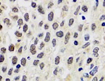
Immunohistochemistry: Histone H3 Antibody [NB500-171] - Analysis of Histone H3 in human breast cancer using DAB with hematoxylin counterstain.![Western Blot: Histone H3 AntibodyBSA Free [NB500-171]](https://resources.rndsystems.com/images/products/Histone-H3-Antibody-Western-Blot-NB500-171-img0024.jpg "Western Blot: Histone H3 AntibodyBSA Free [NB500-171]")
Western Blot: Histone H3 AntibodyBSA Free [NB500-171]
Histone-H3-Antibody-Western-Blot-NB500-171-img0024.jpg![Immunocytochemistry/ Immunofluorescence: Histone H3 Antibody - BSA Free [NB500-171]](https://resources.rndsystems.com/images/products/Histone-H3-Antibody-Immunocytochemistry-Immunofluorescence-NB500-171-img0021.jpg "Immunocytochemistry/ Immunofluorescence: Histone H3 Antibody - BSA Free [NB500-171]")
Immunocytochemistry/ Immunofluorescence: Histone H3 Antibody - BSA Free [NB500-171]
Immunocytochemistry/Immunofluorescence: Histone H3 Antibody [NB500-171] - HeLa cells were fixed in 4% paraformaldehyde for 10 minutes and permeabilized in 0.5% Triton X-100 in PBS for 5 minutes. The cells were incubated with anti- NB500-171 at 1 ug/ml overnight at 4C and detected with an anti-rabbit Dylight 488 (Green) at a 1:1000 dilution for 60 minutes. Nuclei were counterstained with DAPI (Blue). Cells were imaged using a 100X objective and digitally deconvolved.![Immunocytochemistry/ Immunofluorescence: Histone H3 Antibody - BSA Free [NB500-171]](https://resources.rndsystems.com/images/products/Histone-H3-Antibody-Immunocytochemistry-Immunofluorescence-NB500-171-img0022.jpg "Immunocytochemistry/ Immunofluorescence: Histone H3 Antibody - BSA Free [NB500-171]")
Immunocytochemistry/ Immunofluorescence: Histone H3 Antibody - BSA Free [NB500-171]
Immunocytochemistry/Immunofluorescence: Histone H3 Antibody [NB500-171] - PC12 cells were fixed in 4% paraformaldehyde for 10 minutes and permeabilized in 0.5% Triton X-100 in PBS for 5 minutes. The cells were incubated with anti- NB500-171 at 1 ug/ml overnight at 4C and detected with an anti-rabbit Dylight 488 (Green) at a 1:1000 dilution for 60 minutes. Nuclei were counterstained with DAPI (Blue). Cells were imaged using a 100X objective and digitally deconvolved.![Immunocytochemistry/ Immunofluorescence: Histone H3 Antibody - BSA Free [NB500-171]](https://resources.rndsystems.com/images/products/Histone-H3-Antibody-Immunocytochemistry-Immunofluorescence-NB500-171-img0023.jpg "Immunocytochemistry/ Immunofluorescence: Histone H3 Antibody - BSA Free [NB500-171]")
Immunocytochemistry/ Immunofluorescence: Histone H3 Antibody - BSA Free [NB500-171]
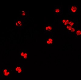
Immunocytochemistry/Immunofluorescence: Histone H3 Antibody [NB500-171] - Immunofluorescence staining to detect histone H3 in human blood. ICC/IF image submitted by a verified customer review.![Immunohistochemistry-Paraffin: Histone H3 Antibody - BSA Free [NB500-171]](https://resources.rndsystems.com/images/products/Histone-H3-Antibody-Immunohistochemistry-Paraffin-NB500-171-img0025.jpg "Immunohistochemistry-Paraffin: Histone H3 Antibody - BSA Free [NB500-171]")
Immunohistochemistry-Paraffin: Histone H3 Antibody - BSA Free [NB500-171]
Immunohistochemistry-Paraffin: Histone H3 Antibody [NB500-171] - Analysis of Histone H3 antibody on Mouse pancreas tissue. Histone H3 antibody dilution 1:200. Image from verified customer review.![Western Blot: Histone H3 AntibodyBSA Free [NB500-171]](https://resources.rndsystems.com/images/products/Histone-H3-Antibody-Western-Blot-NB500-171-img0014.jpg "Western Blot: Histone H3 AntibodyBSA Free [NB500-171]")
Western Blot: Histone H3 AntibodyBSA Free [NB500-171]
Western Blot: Histone H3 Antibody [NB500-171] - (1) HeLa, (2) S. cerevisiae whole cell lysates, (3) Histones purified from HeLa cells. Theoretical molecular weight is ~15 kDa.![Western Blot: Histone H3 AntibodyBSA Free [NB500-171]](https://resources.rndsystems.com/images/products/Histone-H3-Antibody-Western-Blot-NB500-171-img0015.jpg "Western Blot: Histone H3 AntibodyBSA Free [NB500-171]")
Western Blot: Histone H3 AntibodyBSA Free [NB500-171]
Western Blot: Histone H3 Antibody [NB500-171] - Total histone H3 levels in MCF-7 and HCT-116 cells. Observed molecular weight is ~17 kDa. WB image submitted by a verified customer review.![Immunocytochemistry/ Immunofluorescence: Histone H3 Antibody - BSA Free [NB500-171]](https://resources.rndsystems.com/images/products/Histone-H3-Antibody-Immunocytochemistry-Immunofluorescence-NB500-171-img0018.jpg "Immunocytochemistry/ Immunofluorescence: Histone H3 Antibody - BSA Free [NB500-171]")
Immunocytochemistry/ Immunofluorescence: Histone H3 Antibody - BSA Free [NB500-171]
Immunocytochemistry/Immunofluorescence: Histone H3 Antibody [NB500-171] - HeLa cells were fixed for 10 minutes using 10% formalin and then permeabilized for 5 minutes using 1X PBS + 0.5% Triton X-100. The cells were incubated with anti-Histone H3 at 5 ug/mL overnight at 4C and detected with an anti-rabbit DyLight 488 (Green) at a 1:500 dilution. Nuclei were counterstained with DAPI (Blue). Cells were imaged using a 40X objective.![Immunocytochemistry/ Immunofluorescence: Histone H3 Antibody - BSA Free [NB500-171]](https://resources.rndsystems.com/images/products/Histone-H3-Antibody-Immunocytochemistry-Immunofluorescence-NB500-171-img0019.jpg "Immunocytochemistry/ Immunofluorescence: Histone H3 Antibody - BSA Free [NB500-171]")
Immunocytochemistry/ Immunofluorescence: Histone H3 Antibody - BSA Free [NB500-171]
Immunocytochemistry/Immunofluorescence: Histone H3 Antibody [NB500-171] - NIH3T3 cells were fixed for 10 minutes using 10% formalin and then permeabilized for 5 minutes using 1X PBS + 0.5% Triton X-100. The cells were incubated with anti-Histone H3 at 5 ug/mL overnight at 4C and detected with an anti-rabbit DyLight 488 (Green) at a 1:500 dilution. Nuclei were counterstained with DAPI (Blue). Cells were imaged using a 40X objective.![Immunohistochemistry-Paraffin: Histone H3 Antibody - BSA Free [NB500-171]](https://resources.rndsystems.com/images/products/Histone-H3-Antibody-Immunohistochemistry-Paraffin-NB500-171-img0009.jpg "Immunohistochemistry-Paraffin: Histone H3 Antibody - BSA Free [NB500-171]")
Immunohistochemistry-Paraffin: Histone H3 Antibody - BSA Free [NB500-171]
Immunohistochemistry-Paraffin: Histone H3 Antibody [NB500-171] - Immunoperoxidase staining of Anti-Histone H3 in FFPE rat pancreas.![Immunohistochemistry-Paraffin: Histone H3 Antibody - BSA Free [NB500-171]](https://resources.rndsystems.com/images/products/Histone-H3-Antibody-Immunohistochemistry-Paraffin-NB500-171-img0010.jpg "Immunohistochemistry-Paraffin: Histone H3 Antibody - BSA Free [NB500-171]")
Immunohistochemistry-Paraffin: Histone H3 Antibody - BSA Free [NB500-171]
Immunohistochemistry-Paraffin: Histone H3 Antibody [NB500-171] - Immunoperoxidase staining of Anti-Histone H3 in FFPE rat colon.![Immunohistochemistry-Paraffin: Histone H3 Antibody - BSA Free [NB500-171]](https://resources.rndsystems.com/images/products/Histone-H3-Antibody-Immunohistochemistry-Paraffin-NB500-171-img0007.jpg "Immunohistochemistry-Paraffin: Histone H3 Antibody - BSA Free [NB500-171]")
Immunohistochemistry-Paraffin: Histone H3 Antibody - BSA Free [NB500-171]
Immunohistochemistry-Paraffin: Histone H3 Antibody [NB500-171] - Immunoperoxidase staining of Anti-Histone H3 in FFPE mouse brain.
Histone H3 in MCF7 Human Cell Line.
Histone H3 was detected in immersion fixed MCF7 human breast cancer cell line using Rabbit anti-Histone H3 Affinity Purified Polyclonal Antibody conjugated to FITC (Catalog # NB500-171F) (green) at 10 µg/mL overnight at 4C. Cells were stained counterstained with DAPI (blue). Cells were imaged using a 100X objective and digitally deconvolved.
Western Blot: Histone H3 Antibody - BSA Free [NB500-171] -
Western Blot: Histone H3 Antibody - BSA Free [NB500-171] - p53 but not ATM is required for chronic gamma -radiation-induced histone reductions. (a) Western blots of histone proteins in wild-type, TP53−/− & ATM−/− RPE-1 cells following exposures to 20 mGy/h chronic radiation for 7 days. (b) ATM activation, as shown by auto-phosphorylation on S1981, in response to chronic radiation in primary fibroblasts. Image collected & cropped by CiteAb from the following publication (https://pubmed.ncbi.nlm.nih.gov/32042076), licensed under a CC-BY license. Not internally tested by Novus Biologicals.
Western Blot: Histone H3 Antibody - BSA Free [NB500-171] -
Western Blot: Histone H3 Antibody - BSA Free [NB500-171] - Reduced histone levels in senescent cells is induced in vitro by different means, & in vivo from aged donors. (a) Phase-contrast images of primary fibroblasts induced into senescence by chronic gamma -radiation, oncogene over-expression or exhaustive replication (replicative senescence), & DNA damage from a single acute 4 Gy X-ray dose at an early time-point (1 hour) as a control for DNA damage without senescence. Scale bars 200 µm. (b) Western immunoblot analyses of histones in fibroblasts described in (a). (c) Histone levels in dermal fibroblasts isolated from human neonatal (age 0, donors a & b) & adult donors. Image collected & cropped by CiteAb from the following publication (https://pubmed.ncbi.nlm.nih.gov/32042076), licensed under a CC-BY license. Not internally tested by Novus Biologicals.
Western Blot: Histone H3 Antibody - BSA Free [NB500-171] -
Western Blot: Histone H3 Antibody - BSA Free [NB500-171] - Chronic gamma -radiation reduces histone levels. (a) Western blot analyses of histone H2AX & gamma H2AX in primary fibroblasts exposed to various dose-rates of chronic gamma -radiation for 7 days. Cells irradiated with a single acute dose of 4 Gy X-ray were included as control. (b) Effect of chronic gamma -irradiation on H2AX levels in three different isogenic primary cell types from a different donor to that used in (a) at the same dose rates as in (a). (c) Immunoblots of other histones in chronically irradiated primary fibroblasts. (d) All significant histone level changes detected by SILAC LC-MS/MS protein analyses of samples from primary fibroblasts exposed or mock-exposed to chronic gamma -radiation for 7 days. Image collected & cropped by CiteAb from the following publication (https://pubmed.ncbi.nlm.nih.gov/32042076), licensed under a CC-BY license. Not internally tested by Novus Biologicals.Applications for Histone H3 Antibody - BSA Free
Application
Recommended Usage
Immunoblotting
reported in scientific literature (PMID 24048589)
Immunocytochemistry/ Immunofluorescence
reported in scientific literature (PMID 24048589)
Immunohistochemistry
1:100-1:300
Immunohistochemistry-Paraffin
1:100-1:300
Western Blot
1:1000-1:4000
Application Notes
In Western Blot, a band is seen ~15 kDa. In IHC-P, nuclear staining was observed in human, mouse, and rat tissues. Prior to immunostaining paraffin tissues, antigen retrieval with sodium citrate buffer (pH 6.0) is recommended.
Reviewed Applications
Read 9 reviews rated 4.9 using NB500-171 in the following applications:
Formulation, Preparation, and Storage
Purification
Immunogen affinity purified
Formulation
PBS
Format
BSA Free
Preservative
0.01% Sodium Azide
Concentration
1.0 mg/ml
Shipping
The product is shipped with polar packs. Upon receipt, store it immediately at the temperature recommended below.
Stability & Storage
Aliquot and store at -20C or -80C. Avoid freeze-thaw cycles.
Background: Histone H3
Histones are nuclear proteins responsible for the nucleosome structure of the chromosomal fiber in eukaryotes. Changes in chromatin structure play a large role in the regulation of transcription. The chromatin fibers are compacted through the interaction of a linker histone, H1, with the DNA between the nucleosomes to form higher order chromatin structures.
Common histone modifications include methylation of lysine and arginine, acetylation of lysine, phosphorylation of threonine and serine, and sumoylation, biotinylation, and ubiquitylation of lysine. Posttranslational modifications such as acetylation of core histones regulates gene expression, thus altering protein function and regulation (1). Histone H3 is primarily acetylated at lysines 9, 14, 18, and 23 and have a theoretical molecular weight of 15 kDa. Acetylation at lysine 9 and 14 appears to control histone deposition, chromatin assembly and active transcription. Methylation of arginine residues within histone H3 has also been linked to transcription regulation. Histone H3 has been linked to various types of cancer as a biomarker through the aberrant expression of histone deacetylase (HDAC) enzymes and changes to chromatins (2-4).
References
1. Zhang, Y. X., Akumuo, R. C., Espana, R. A., Yan, C. X., Gao, W. J., & Li, Y. C. (2018). The histone demethylase KDM6B in the medial prefrontal cortex epigenetically regulates cocaine reward memory. Neuropharmacology, 141, 113-125. doi:10.1016/j.neuropharm.2018.08.030
2. Nandakumar, V., Hansen, N., Glenn, H. L., Han, J. H., Helland, S., Hernandez, K,...Meldrum, D. R. (2016). Vorinostat differentially alters 3D nuclear structure of cancer and non-cancerous esophageal cells. Sci Rep, 6, 30593. doi:10.1038/srep30593
3. Zhou, M., Li, Y., Lin, S., Chen, Y., Qian, Y., Zhao, Z., & Fan, H. (2019). H3K9me3, H3K36me3, and H4K20me3 Expression Correlates with Patient Outcome in Esophageal Squamous Cell Carcinoma as Epigenetic Markers. Dig Dis Sci, 64(8), 2147-2157. doi:10.1007/s10620-019-05529-2
4. Li, Y., Guo, D., Sun, R., Chen, P., Qian, Q., & Fan, H. (2019). Methylation Patterns of Lys9 and Lys27 on Histone H3 Correlate with Patient Outcome in Gastric Cancer. Dig Dis Sci, 64(2), 439-446. doi:10.1007/s10620-018-5341-8
Additional Histone H3 Products
Product Documents for Histone H3 Antibody - BSA Free
Certificate of Analysis
To download a Certificate of Analysis, please enter a lot or batch number in the search box below.
Product Specific Notices for Histone H3 Antibody - BSA Free
This product is for research use only and is not approved for use in humans or in clinical diagnosis. Primary Antibodies are guaranteed for 1 year from date of receipt.
Related Research Areas
Citations for Histone H3 Antibody - BSA Free
 Powered by Bioz
Customer Reviews for Histone H3 Antibody - BSA Free (9)
4.9 out of 5
9 Customer Ratings
Have you used Histone H3 Antibody - BSA Free?
Submit a review and receive an Amazon gift card!
$25/€18/£15/$25CAN/¥2500 Yen for a review with an image
$10/€7/£6/$10CAN/¥1110 Yen for a review without an image
Submit a review
Customer Images

-(01-ml)_NB500-171_8286.jpg)
-(01-ml)_NB500-171_8081.jpg)
Showing
1
-
5 of
9 reviews
Showing All
Filter By:
-
Application: Western BlotSample Tested: 293 whole cell lysateSpecies: HumanVerified Customer | Posted 01/26/2023first lane is 293T protein, second lane is kdm5c knockdown 293T protein

-
Application: Immunohistochemistry-ParaffinSample Tested: Pancreas tissueSpecies: MouseVerified Customer | Posted 02/13/2022Mouse pancreas tissueHistone H3 antibody dilution 1:200
-
Application: ImmunofluorescenceSample Tested: human bloodSpecies: HumanVerified Customer | Posted 07/30/2021Immunofluorescence staining to detect histone H3 in human blood.
-
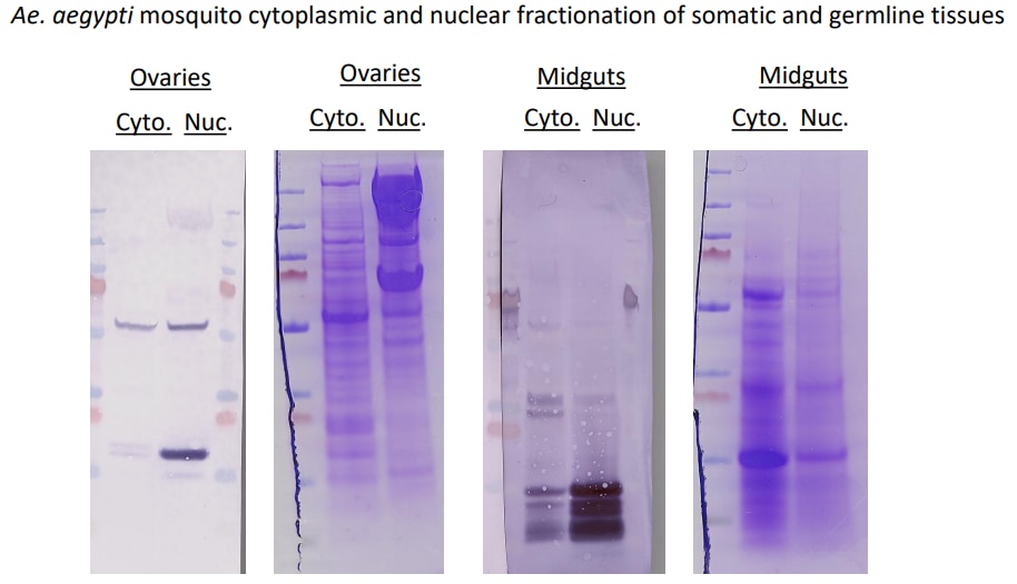
Application: Western BlotSample Tested: Ovary tissue, Midgut tissue and A549 Nuclear and cytoplasmic, Sample Amount: 30ugSpecies: Ae. aegyptiVerified Customer | Posted 05/22/2021Western blot and Commassie-stained SDS/PAGE gels of Ae. aegypti ovary and midgut tissues where cytoplasmic and nuclear fractions have been isolated.To test both anti-histone H3 and anti-beta tubulin as markers for nuclear or cytoplasmic fractions, respectively, in Ae. aegypti mosquitoes, a western blot was performed using midguts or ovaries dissected from mosquitoes 48h post-bloodmeal. The cytoplasmic or nuclear fractions from each tissue type were obtained and protein concentration was quantified by A260/280. Roughly 30ug of tissue was loaded onto SDS/PAGE gels that were either transferred to PVDF membranes or stained by Coomassie. Membranes were blocked in TBST 5% milk for 2h. Both anti-histone H3 and anti-beta tubulin were added 1:1000 in blocking buffer and incubated O/N shaking at 4C. Proteins were detected by Alkaline Phosphatase. In Ae. aegypti ovaries, anti-histone H3 bound to protein roughly 15kDa in the nuclear fraction of ovaries but not the cytoplasmic fraction, while anti-beta-tubulin bound to protein roughly 55kDa in both cytoplasmic and nuclear fractions. In Ae. aegypti midguts, anti-histone H3 bound to 3 proteins between 10-15kDa strongly in the nuclear fraction and faintly in the cytoplasmic fraction (perhaps due to nuclear contamination in the cytoplasmic fraction). anti-beta-tubulin did not react well in midgut fractions. For your reference, protein ladder = pageruler plus prestained ThermoFisher
-
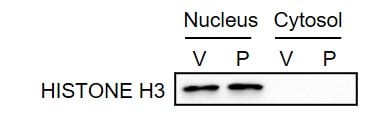
Application: Western BlotSample Tested: Breast cancer cellsSpecies: HumanVerified Customer | Posted 05/18/2020MDA-MB-231 cells were treated with vehicle (V) or paclitaxel (P). Cytosolic and nuclear lysates were prepared, and immunoblot assay was performed.
-
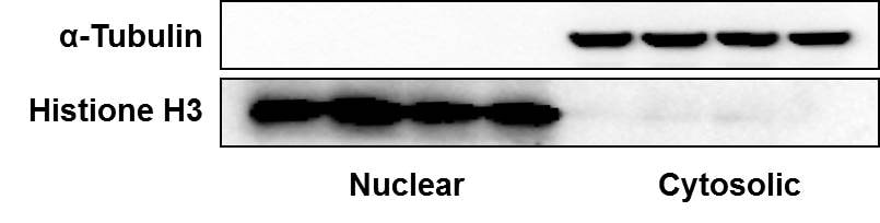
Application: Western BlotSample Tested: Human breast cancer cell linesSpecies: HumanVerified Customer | Posted 10/22/2018Lysates from human breast cancer cells were separated into nuclear and cytosolic proteins and antibodies against alpha-tubulin and Histone H3 were used as markers.
-
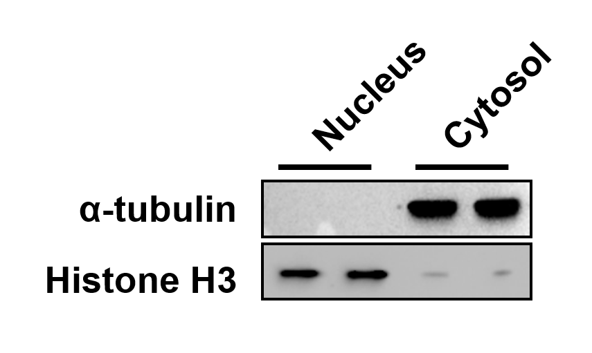
Application: Western BlotSample Tested: MDA-MB-231 cells (cytoplasmic and nuclear fractions)Species: HumanVerified Customer | Posted 12/27/2016Immunoblots of cytoplasmic and nuclear fractions of MDA-MB-231 cells. We used Novus anti-Histone H3 antibody NB500-171 to demonstrate purity of the fractions.
-
Application: Western BlotSample Tested:Species: HumanVerified Customer | Posted 06/13/2014Western blot analysis of extracts from HeLa, COS, C6 and K562 cells using Histone H3 antibody (NB500-171, 1:100).
-
Application: Western BlotSample Tested: Whole cell extract from MCF-7 and HCT-116 cellsSpecies: HumanVerified Customer | Posted 06/10/2014Total histone H3 levels in MCF-7 and HCT-116 cells
There are no reviews that match your criteria.
Protocols
View specific protocols for Histone H3 Antibody - BSA Free (NB500-171):
Immunohistochemistry-Paraffin Embedded Sections Protocol
Antigen Unmasking:
Bring slides to a boil in 10 mM sodium citrate buffer (pH 6.0) then maintain at a sub-boiling temperature for 10 minutes. Cool slides on bench-top for 30 minutes.
Staining:
1. Wash sections in deionized water three times for 5 minutes each.
2. Wash sections in wash buffer for 5 minutes.
3. Block each section with 100-400 ul blocking solution for 1 hour at room temperature.
4. Remove blocking solution and add 100-400 ul diluted primary antibody. Incubate overnight at 4 C.
5. Remove antibody solution and wash sections in wash buffer three times for 5 minutes each.
6. Add 100-400 ul biotinylated diluted secondary antibody. Incubate 30 minutes at room temperature.
7. Remove secondary antibody solution and wash sections three times with wash buffer for 5 minutes each.
8. Add 100-400 ul Streptavidin-HRP reagent to each section and incubate for 30 minutes at room temperature.
9. Wash sections three times in wash buffer for 5 minutes each.
10. Add 100-400 ul DAB substrate to each section and monitor staining closely.
11. As soon as the sections develop, immerse slides in deionized water.
12. Counterstain sections in hematoxylin.
13. Wash sections in deionized water two times for 5 minutes each.
14. Dehydrate sections.
15. Mount coverslips.
Western Blot Protocol
1. Perform SDS-PAGE on samples to be analyzed, loading 40 ug of total protein per lane.
2. Transfer proteins to membrane according to the instructions provided by the manufacturer of the membrane and transfer apparatus.
3. Stain according to standard Ponceau S procedure (or similar product) to assess transfer success, and mark molecular weight standards where appropriate.
4. Rinse the blot.
5. Block the membrane using standard blocking buffer for at least 1 hour.
6. Wash the membrane in wash buffer three times for 10 minutes each.
7. Dilute primary antibody in blocking buffer and incubate 1 hour at room temperature.
8. Wash the membrane in wash buffer three times for 10 minutes each.
9. Apply the diluted HRP conjugated secondary antibody in blocking buffer (as per manufacturers instructions) and incubate 1 hour at room temperature.
10. Wash the blot in wash buffer three times for 10 minutes each (this step can be repeated as required to reduce background).
11. Apply the detection reagent of choice in accordance with the manufacturers instructions.
Note: Tween-20 can be added to the blocking or antibody dilution buffer at a final concentration of 0.05-0.2%.
Find general support by application which include: protocols, troubleshooting, illustrated assays, videos and webinars.
- Antigen Retrieval Protocol (PIER)
- Antigen Retrieval for Frozen Sections Protocol
- Appropriate Fixation of IHC/ICC Samples
- Cellular Response to Hypoxia Protocols
- ChIP Protocol Video
- Chromatin Immunoprecipitation (ChIP) Protocol
- Chromatin Immunoprecipitation Protocol
- Chromogenic IHC Staining of Formalin-Fixed Paraffin-Embedded (FFPE) Tissue Protocol
- Chromogenic Immunohistochemistry Staining of Frozen Tissue
- ClariTSA™ Fluorophore Kits
- Detection & Visualization of Antibody Binding
- Fluorescent IHC Staining of Frozen Tissue Protocol
- Graphic Protocol for Heat-induced Epitope Retrieval
- Graphic Protocol for the Preparation and Fluorescent IHC Staining of Frozen Tissue Sections
- Graphic Protocol for the Preparation and Fluorescent IHC Staining of Paraffin-embedded Tissue Sections
- Graphic Protocol for the Preparation of Gelatin-coated Slides for Histological Tissue Sections
- ICC Cell Smear Protocol for Suspension Cells
- ICC Immunocytochemistry Protocol Videos
- ICC for Adherent Cells
- IHC Sample Preparation (Frozen sections vs Paraffin)
- Immunocytochemistry (ICC) Protocol
- Immunocytochemistry Troubleshooting
- Immunofluorescence of Organoids Embedded in Cultrex Basement Membrane Extract
- Immunofluorescent IHC Staining of Formalin-Fixed Paraffin-Embedded (FFPE) Tissue Protocol
- Immunohistochemistry (IHC) and Immunocytochemistry (ICC) Protocols
- Immunohistochemistry Frozen Troubleshooting
- Immunohistochemistry Paraffin Troubleshooting
- Preparing Samples for IHC/ICC Experiments
- Preventing Non-Specific Staining (Non-Specific Binding)
- Primary Antibody Selection & Optimization
- Protocol for Heat-Induced Epitope Retrieval (HIER)
- Protocol for Making a 4% Formaldehyde Solution in PBS
- Protocol for VisUCyte™ HRP Polymer Detection Reagent
- Protocol for the Fluorescent ICC Staining of Cell Smears - Graphic
- Protocol for the Fluorescent ICC Staining of Cultured Cells on Coverslips - Graphic
- Protocol for the Preparation & Fixation of Cells on Coverslips
- Protocol for the Preparation and Chromogenic IHC Staining of Frozen Tissue Sections
- Protocol for the Preparation and Chromogenic IHC Staining of Frozen Tissue Sections - Graphic
- Protocol for the Preparation and Chromogenic IHC Staining of Paraffin-embedded Tissue Sections
- Protocol for the Preparation and Chromogenic IHC Staining of Paraffin-embedded Tissue Sections - Graphic
- Protocol for the Preparation and Fluorescent ICC Staining of Cells on Coverslips
- Protocol for the Preparation and Fluorescent ICC Staining of Non-adherent Cells
- Protocol for the Preparation and Fluorescent ICC Staining of Stem Cells on Coverslips
- Protocol for the Preparation and Fluorescent IHC Staining of Frozen Tissue Sections
- Protocol for the Preparation and Fluorescent IHC Staining of Paraffin-embedded Tissue Sections
- Protocol for the Preparation of Gelatin-coated Slides for Histological Tissue Sections
- Protocol for the Preparation of a Cell Smear for Non-adherent Cell ICC - Graphic
- R&D Systems Quality Control Western Blot Protocol
- TUNEL and Active Caspase-3 Detection by IHC/ICC Protocol
- The Importance of IHC/ICC Controls
- Troubleshooting Guide: Immunohistochemistry
- Troubleshooting Guide: Western Blot Figures
- Western Blot Conditions
- Western Blot Protocol
- Western Blot Protocol for Cell Lysates
- Western Blot Troubleshooting
- Western Blot Troubleshooting Guide
- View all Protocols, Troubleshooting, Illustrated assays and Webinars
Loading...