Histone H4 [ac Lys8] Antibody
Novus Biologicals | Catalog # NBP2-42863
![Immunohistochemistry-Paraffin: Histone H4 [ac Lys8] Antibody [NBP2-42863]](https://resources.rndsystems.com/images/products/Histone-H4-[ac-Lys8]-Antibody-Immunohistochemistry-Paraffin-NBP2-42863-img0006.jpg "Immunohistochemistry-Paraffin: Histone H4 [ac Lys8] Antibody [NBP2-42863]")
Loading...
Key Product Details
Validated by
Biological Validation
Species Reactivity
Validated:
Human, Mouse, Rat
Predicted:
Xenopus (100%). Backed by our 100% Guarantee.
Applications
Immunohistochemistry, Immunohistochemistry-Paraffin, Immunohistochemistry Free-Floating, Western Blot, Immunocytochemistry/ Immunofluorescence, Immunoprecipitation, Chromatin Immunoprecipitation (ChIP), Dot Blot
Label
Unconjugated
Antibody Source
Polyclonal Rabbit IgG
Loading...
Product Specifications
Immunogen
Carrier-protein conjugated synthetic peptide surrounding acetyl Lys8 of human Histone H4. The exact sequence is proprietary.
Modification
ac Lys8
Clonality
Polyclonal
Host
Rabbit
Isotype
IgG
Scientific Data Images for Histone H4 [ac Lys8] Antibody
Immunohistochemistry-Paraffin: Histone H4 [ac Lys8] Antibody [NBP2-42863]
Immunohistochemistry-Paraffin: Histone H4 [ac Lys8] Antibody [NBP2-42863] - Analysis of rat hind brain. Histone H4K8ac (acetyl Lys8) antibody dilution: 1:500.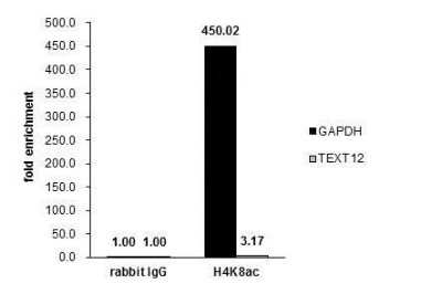
Chromatin Immunoprecipitation: Histone H4 [ac Lys8] Antibody [NBP2-42863] - Cross-linked ChIP was performed with HeLa chromatin extract treated with Trichostatin A (0.4 uM for 18 h) and 5 ug of either control rabbit IgG or anti-Histone H4K8ac (acetyl Lys8) antibody. The precipitated DNA was detected by PCR with primer set targeting to GAPDH or TEXT12.
![Immunohistochemistry-Paraffin: Histone H4 [ac Lys8] Antibody [NBP2-42863]](https://resources.rndsystems.com/images/products/Histone-H4-[ac-Lys8]-Antibody-Immunohistochemistry-Paraffin-NBP2-42863-img0004.jpg "Immunohistochemistry-Paraffin: Histone H4 [ac Lys8] Antibody [NBP2-42863]")
Immunohistochemistry-Paraffin: Histone H4 [ac Lys8] Antibody [NBP2-42863]
Immunohistochemistry-Paraffin: Histone H4 [ac Lys8] Antibody [NBP2-42863] - Analysis of mouse duodenum. Histone H4K8ac (acetyl Lys8) antibody dilution: 1:500.![Immunohistochemistry-Paraffin: Histone H4 [ac Lys8] Antibody [NBP2-42863]](https://resources.rndsystems.com/images/products/Histone-H4-[ac-Lys8]-Antibody-Immunohistochemistry-Paraffin-NBP2-42863-img0005.jpg "Immunohistochemistry-Paraffin: Histone H4 [ac Lys8] Antibody [NBP2-42863]")
Immunohistochemistry-Paraffin: Histone H4 [ac Lys8] Antibody [NBP2-42863]
Immunohistochemistry-Paraffin: Histone H4 [ac Lys8] Antibody [NBP2-42863] - Analysis of mouse kidney. Histone H4K8ac (acetyl Lys8) antibody dilution: 1:500.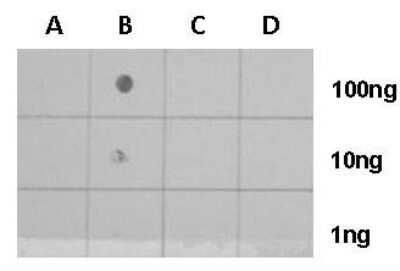
Dot Blot: Histone H4 [ac Lys8] Antibody [NBP2-42863] - Analysis of anti-Histone H4 (acetyl Lys9) antibody with peptide samples. Varied amount of peptide samples were spotted onto positively charged nylon membrane and blotted with Histone H4 (acetyl Lys9) antibody 1:5000 dilution. A: Peptide samples of H4K8 un B: Peptide samples of H4K8ac C: Peptide samples of H4K16un D: Peptide samples of H4K16ac.
![Immunoprecipitation: Histone H4 [ac Lys8] Antibody [NBP2-42863]](https://resources.rndsystems.com/images/products/Histone-H4-[ac-Lys8]-Antibody-Immunoprecipitation-NBP2-42863-img0007.jpg "Immunoprecipitation: Histone H4 [ac Lys8] Antibody [NBP2-42863]")
Immunoprecipitation: Histone H4 [ac Lys8] Antibody [NBP2-42863]
Immunoprecipitation: Histone H4 [ac Lys8] Antibody [NBP2-42863] - Analysis of HeLa whole cell lysate/extract:A. 30 ug whole cell lysate/extract of Histone H4 (acetyl K9) protein expressing HeLa cells (0.4uM TSA treatment for 18 hrs)
B. Control with 2.5 ug of pre-immune rabbit IgG
C. Immunoprecipitation of Histone H4 (acetyl K9) protein by 2.5 ug of Histone H4 (acetyl K9) antibody. 12% SDS-PAGE The immunoprecipitated Histone H4 (acetyl K9) protein was detected by Histone H4 (acetyl K9).
EasyBlot anti-rabbit IgG (HRP) was used as a secondary reagent.
![Histone H4 [ac Lys8] Antibody](https://resources.rndsystems.com/images/products/nbp2-42863_rabbit-polyclonal-histone-h4-ac-lys8-antibody-western-blot-261120251822436.jpg "Western Blot: Histone H4 [ac Lys8] Antibody [NBP2-42863] -")
Western Blot: Histone H4 [ac Lys8] Antibody [NBP2-42863] -
Untreated (-) and treated (+) HeLa whole cell extracts (30 ug) were separated by 15% SDS-PAGE, and the membrane was blotted with Histone H4K8ac (acetyl Lys8) antibody (NBP2-42863) diluted at 1:5000. The HRP-conjugated anti-rabbit IgG antibody was used to detect the primary antibody.![Histone H4 [ac Lys8] Antibody](https://resources.rndsystems.com/images/products/nbp2-42863_rabbit-polyclonal-histone-h4-ac-lys8-antibody-immunocytochemistry-immunofluorescence-261120251831543.jpg "Immunocytochemistry/ Immunofluorescence: Histone H4 [ac Lys8] Antibody [NBP2-42863] -")
Immunocytochemistry/ Immunofluorescence: Histone H4 [ac Lys8] Antibody [NBP2-42863] -
Histone H4 (acetyl Lys8) antibody detects Histone H4 (acetyl Lys8) protein at nucleus by immunofluorescent analysis.Sample: HeLa cells were fixed in 4% paraformaldehyde at RT for 15 min.
Green: Histone H4 (acetyl Lys8) protein stained by Histone H4 (acetyl Lys8) antibody (NBP2-42863) diluted at 1:500.
Red: alpha Tubulin antibody [GT114], a cytoskeleton marker, stained by diluted at 1:500.
Blue: Hoechst 33342 staining.
Applications for Histone H4 [ac Lys8] Antibody
Application
Recommended Usage
Immunocytochemistry/ Immunofluorescence
1:100-1:1000
Immunohistochemistry-Paraffin
1:100-1:1000
Immunoprecipitation
1:100-1:5000
Western Blot
1:1000-1:10000
Formulation, Preparation, and Storage
Purification
Antigen Affinity-purified
Formulation
PBS, 1% BSA, 20% Glycerol
Preservative
0.01% Thimerosal
Concentration
Concentrations vary lot to lot. See vial label for concentration. If unlisted please contact technical services.
Shipping
The product is shipped with polar packs. Upon receipt, store it immediately at the temperature recommended below.
Stability & Storage
Aliquot and store at -20C or -80C. Avoid freeze-thaw cycles.
Background: Histone H4
Alternate Names
H4, H4/M, H4FM, H4M, HIST1H4I, HIST4H4, Histone Cluster 1 H4, Histone Cluster 1 H4i, Histone Cluster 4 H4, H4K8ac
Gene Symbol
H4C16
Additional Histone H4 Products
Product Documents for Histone H4 [ac Lys8] Antibody
Certificate of Analysis
To download a Certificate of Analysis, please enter a lot or batch number in the search box below.
Product Specific Notices for Histone H4 [ac Lys8] Antibody
This product is for research use only and is not approved for use in humans or in clinical diagnosis. Primary Antibodies are guaranteed for 1 year from date of receipt.
⚠ WARNING: This product can expose you to chemicals including mercury, which is known to the State of California to cause reproductive toxicity with developmental effects. For more information go to www.P65Warnings.ca.gov.Related Research Areas
Customer Reviews for Histone H4 [ac Lys8] Antibody
There are currently no reviews for this product. Be the first to review Histone H4 [ac Lys8] Antibody and earn rewards!
Have you used Histone H4 [ac Lys8] Antibody?
Submit a review and receive an Amazon gift card!
$25/€18/£15/$25CAN/¥2500 Yen for a review with an image
$10/€7/£6/$10CAN/¥1110 Yen for a review without an image
Submit a review
Protocols
Find general support by application which include: protocols, troubleshooting, illustrated assays, videos and webinars.
- Antigen Retrieval Protocol (PIER)
- Antigen Retrieval for Frozen Sections Protocol
- Appropriate Fixation of IHC/ICC Samples
- Cellular Response to Hypoxia Protocols
- ChIP Protocol Video
- Chromatin Immunoprecipitation (ChIP) Protocol
- Chromatin Immunoprecipitation Protocol
- Chromogenic IHC Staining of Formalin-Fixed Paraffin-Embedded (FFPE) Tissue Protocol
- Chromogenic Immunohistochemistry Staining of Frozen Tissue
- ClariTSA™ Fluorophore Kits
- Detection & Visualization of Antibody Binding
- Fluorescent IHC Staining of Frozen Tissue Protocol
- Graphic Protocol for Heat-induced Epitope Retrieval
- Graphic Protocol for the Preparation and Fluorescent IHC Staining of Frozen Tissue Sections
- Graphic Protocol for the Preparation and Fluorescent IHC Staining of Paraffin-embedded Tissue Sections
- Graphic Protocol for the Preparation of Gelatin-coated Slides for Histological Tissue Sections
- ICC Cell Smear Protocol for Suspension Cells
- ICC Immunocytochemistry Protocol Videos
- ICC for Adherent Cells
- IHC Sample Preparation (Frozen sections vs Paraffin)
- Immunocytochemistry (ICC) Protocol
- Immunocytochemistry Troubleshooting
- Immunofluorescence of Organoids Embedded in Cultrex Basement Membrane Extract
- Immunofluorescent IHC Staining of Formalin-Fixed Paraffin-Embedded (FFPE) Tissue Protocol
- Immunohistochemistry (IHC) and Immunocytochemistry (ICC) Protocols
- Immunohistochemistry Frozen Troubleshooting
- Immunohistochemistry Paraffin Troubleshooting
- Immunoprecipitation Protocol
- Preparing Samples for IHC/ICC Experiments
- Preventing Non-Specific Staining (Non-Specific Binding)
- Primary Antibody Selection & Optimization
- Protocol for Heat-Induced Epitope Retrieval (HIER)
- Protocol for Making a 4% Formaldehyde Solution in PBS
- Protocol for VisUCyte™ HRP Polymer Detection Reagent
- Protocol for the Fluorescent ICC Staining of Cell Smears - Graphic
- Protocol for the Fluorescent ICC Staining of Cultured Cells on Coverslips - Graphic
- Protocol for the Preparation & Fixation of Cells on Coverslips
- Protocol for the Preparation and Chromogenic IHC Staining of Frozen Tissue Sections
- Protocol for the Preparation and Chromogenic IHC Staining of Frozen Tissue Sections - Graphic
- Protocol for the Preparation and Chromogenic IHC Staining of Paraffin-embedded Tissue Sections
- Protocol for the Preparation and Chromogenic IHC Staining of Paraffin-embedded Tissue Sections - Graphic
- Protocol for the Preparation and Fluorescent ICC Staining of Cells on Coverslips
- Protocol for the Preparation and Fluorescent ICC Staining of Non-adherent Cells
- Protocol for the Preparation and Fluorescent ICC Staining of Stem Cells on Coverslips
- Protocol for the Preparation and Fluorescent IHC Staining of Frozen Tissue Sections
- Protocol for the Preparation and Fluorescent IHC Staining of Paraffin-embedded Tissue Sections
- Protocol for the Preparation of Gelatin-coated Slides for Histological Tissue Sections
- Protocol for the Preparation of a Cell Smear for Non-adherent Cell ICC - Graphic
- R&D Systems Quality Control Western Blot Protocol
- TUNEL and Active Caspase-3 Detection by IHC/ICC Protocol
- The Importance of IHC/ICC Controls
- Troubleshooting Guide: Immunohistochemistry
- Troubleshooting Guide: Western Blot Figures
- Western Blot Conditions
- Western Blot Protocol
- Western Blot Protocol for Cell Lysates
- Western Blot Troubleshooting
- Western Blot Troubleshooting Guide
- View all Protocols, Troubleshooting, Illustrated assays and Webinars
Loading...
Associated Pathways
