HSD17B4 Antibody (OTI4C4)
Novus Biologicals | Catalog # NBP2-46005
Key Product Details
Species Reactivity
Validated:
Human, Mouse, Rat
Cited:
Mouse
Applications
Validated:
Immunohistochemistry, Immunohistochemistry-Paraffin, Western Blot, Immunocytochemistry/ Immunofluorescence, Immunoprecipitation, Proximity Ligation Assay
Cited:
Immunoprecipitation, Proximity Ligation Assay
Label
Unconjugated
Antibody Source
Monoclonal Mouse IgG1 Clone # OTI4C4
Loading...
Product Specifications
Immunogen
Full length human recombinant protein of human HSD17B4(NP_000405) produced in HEK293T cell.
Reactivity Notes
Mouse reactivity reported in scientific literature (PMID: 31176039). Please note that this antibody is reactive to Mouse and derived from the same host, Mouse. Mouse-On-Mouse blocking reagent may be needed for IHC and ICC experiments to reduce high background signal. You can find these reagents under catalog numbers PK-2200-NB and MP-2400-NB. Please contact Technical Support if you have any questions.
Clonality
Monoclonal
Host
Mouse
Isotype
IgG1
Scientific Data Images for HSD17B4 Antibody (OTI4C4)
![Western Blot: HSD17B4 Antibody (OTI4C4) [NBP2-46005]](https://resources.rndsystems.com/images/products/HSD17B4-Antibody-OTI4C4-Western-Blot-NBP2-46005-img0010.jpg "Western Blot: HSD17B4 Antibody (OTI4C4) [NBP2-46005]")
Western Blot: HSD17B4 Antibody (OTI4C4) [NBP2-46005]
Western Blot: HSD17B4 Antibody (OTI4C4) [NBP2-46005] - Analysis of HEK293T cells were transfected with the pCMV6-ENTRY control (Left lane) or pCMV6-ENTRY HSD17B4.![Immunohistochemistry: HSD17B4 Antibody (OTI4C4) [NBP2-46005]](https://resources.rndsystems.com/images/products/HSD17B4-Antibody-OTI4C4-Immunohistochemistry-NBP2-46005-img0009.jpg "Immunohistochemistry: HSD17B4 Antibody (OTI4C4) [NBP2-46005]")
Immunohistochemistry: HSD17B4 Antibody (OTI4C4) [NBP2-46005]
Immunohistochemistry: HSD17B4 Antibody (OTI4C4) [NBP2-46005] - Analysis of Carcinoma of Human liver tissue.![Immunohistochemistry: HSD17B4 Antibody (OTI4C4) [NBP2-46005]](https://resources.rndsystems.com/images/products/HSD17B4-Antibody-OTI4C4-Immunohistochemistry-NBP2-46005-img0003.jpg "Immunohistochemistry: HSD17B4 Antibody (OTI4C4) [NBP2-46005]")
Immunohistochemistry: HSD17B4 Antibody (OTI4C4) [NBP2-46005]
Immunohistochemistry: HSD17B4 Antibody (OTI4C4) [NBP2-46005] - Analysis of Human Ovary tissue.![Immunohistochemistry: HSD17B4 Antibody (OTI4C4) [NBP2-46005]](https://resources.rndsystems.com/images/products/HSD17B4-Antibody-OTI4C4-Immunohistochemistry-NBP2-46005-img0004.jpg "Immunohistochemistry: HSD17B4 Antibody (OTI4C4) [NBP2-46005]")
Immunohistochemistry: HSD17B4 Antibody (OTI4C4) [NBP2-46005]
Immunohistochemistry: HSD17B4 Antibody (OTI4C4) [NBP2-46005] - Analysis of Carcinoma of Human pancreas tissue.![Immunohistochemistry: HSD17B4 Antibody (OTI4C4) [NBP2-46005]](https://resources.rndsystems.com/images/products/HSD17B4-Antibody-OTI4C4-Immunohistochemistry-NBP2-46005-img0005.jpg "Immunohistochemistry: HSD17B4 Antibody (OTI4C4) [NBP2-46005]")
Immunohistochemistry: HSD17B4 Antibody (OTI4C4) [NBP2-46005]
Immunohistochemistry: HSD17B4 Antibody (OTI4C4) [NBP2-46005] - Analysis of Human prostate tissue.![Immunohistochemistry: HSD17B4 Antibody (OTI4C4) [NBP2-46005]](https://resources.rndsystems.com/images/products/HSD17B4-Antibody-OTI4C4-Immunohistochemistry-NBP2-46005-img0006.jpg "Immunohistochemistry: HSD17B4 Antibody (OTI4C4) [NBP2-46005]")
Immunohistochemistry: HSD17B4 Antibody (OTI4C4) [NBP2-46005]
Immunohistochemistry: HSD17B4 Antibody (OTI4C4) [NBP2-46005] - Analysis of Carcinoma of Human prostate tissue.![Immunohistochemistry: HSD17B4 Antibody (OTI4C4) [NBP2-46005]](https://resources.rndsystems.com/images/products/HSD17B4-Antibody-OTI4C4-Immunohistochemistry-NBP2-46005-img0007.jpg "Immunohistochemistry: HSD17B4 Antibody (OTI4C4) [NBP2-46005]")
Immunohistochemistry: HSD17B4 Antibody (OTI4C4) [NBP2-46005]
Immunohistochemistry: HSD17B4 Antibody (OTI4C4) [NBP2-46005] - Analysis of Human Kidney tissue.![Immunohistochemistry: HSD17B4 Antibody (OTI4C4) [NBP2-46005]](https://resources.rndsystems.com/images/products/HSD17B4-Antibody-OTI4C4-Immunohistochemistry-NBP2-46005-img0008.jpg "Immunohistochemistry: HSD17B4 Antibody (OTI4C4) [NBP2-46005]")
Immunohistochemistry: HSD17B4 Antibody (OTI4C4) [NBP2-46005]
Immunohistochemistry: HSD17B4 Antibody (OTI4C4) [NBP2-46005] - Analysis of Human liver tissue.![Immunofluorescence: HSD17B4 Antibody (OTI4C4) [NBP2-46005]](https://resources.rndsystems.com/images/products/HSD17B4-Antibody-OTI4C4-Immunofluorescence-NBP2-46005-img0001.jpg "Immunofluorescence: HSD17B4 Antibody (OTI4C4) [NBP2-46005]")
Immunofluorescence: HSD17B4 Antibody (OTI4C4) [NBP2-46005]
Immunofluorescence: HSD17B4 Antibody (OTI4C4) [NBP2-46005] - Analysis of COS7 cells transiently transfected by pCMV6-ENTRY HSD17B4.![Immunofluorescence: HSD17B4 Antibody (OTI4C4) [NBP2-46005]](https://resources.rndsystems.com/images/products/HSD17B4-Antibody-OTI4C4-Immunofluorescence-NBP2-46005-img0002.jpg "Immunofluorescence: HSD17B4 Antibody (OTI4C4) [NBP2-46005]")
Immunofluorescence: HSD17B4 Antibody (OTI4C4) [NBP2-46005]
Immunofluorescence: HSD17B4 Antibody (OTI4C4) [NBP2-46005] - Analysis of HeLa cells.Applications for HSD17B4 Antibody (OTI4C4)
Application
Recommended Usage
Immunocytochemistry/ Immunofluorescence
1:100
Immunohistochemistry
1:150
Immunoprecipitation
Reported in scientific literature (PMID: 31176039).
Proximity Ligation Assay
Reported in scientific literature (PMID: 31176039).
Western Blot
1:4000
Reviewed Applications
Read 1 review rated 4 using NBP2-46005 in the following applications:
Formulation, Preparation, and Storage
Purification
Immunogen affinity purified
Formulation
PBS (pH 7.3), 1.0% BSA and 50% Glycerol
Preservative
0.02% Sodium Azide
Concentration
1 mg/ml
Shipping
The product is shipped with polar packs. Upon receipt, store it immediately at the temperature recommended below.
Stability & Storage
Store at -20C. Avoid freeze-thaw cycles.
Background: HSD17B4
Alternate Names
12-alpha-trihydroxy-5-beta-cholest-24-enoyl-CoA hydratase, 7-alpha, D-bifunctional protein, EDH17B4, hydroxysteroid (17-beta) dehydrogenase 4, MPF-2, peroxisomal, peroxisomal multifunctional enzyme type 2,17-beta-HSD IV, peroxisomal multifunctional protein 2,17beta-estradiol dehydrogenase type IV, short chain dehydrogenase/reductase family 8C, member 1,3-alpha
Gene Symbol
HSD17B4
Additional HSD17B4 Products
Product Documents for HSD17B4 Antibody (OTI4C4)
Certificate of Analysis
To download a Certificate of Analysis, please enter a lot or batch number in the search box below.
Product Specific Notices for HSD17B4 Antibody (OTI4C4)
This product is for research use only and is not approved for use in humans or in clinical diagnosis. Primary Antibodies are guaranteed for 1 year from date of receipt.
Citations for HSD17B4 Antibody (OTI4C4)
 Powered by Bioz
Customer Reviews for HSD17B4 Antibody (OTI4C4) (1)
4 out of 5
1 Customer Rating
Have you used HSD17B4 Antibody (OTI4C4)?
Submit a review and receive an Amazon gift card!
$25/€18/£15/$25CAN/¥2500 Yen for a review with an image
$10/€7/£6/$10CAN/¥1110 Yen for a review without an image
Submit a review
Customer Images
Showing
1
-
1 of
1 review
Showing All
Filter By:
-
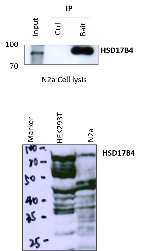
Application: Western BlotSample Tested: N2a and HEK293 T cell lysatesSpecies: MouseVerified Customer | Posted 02/01/2017We first test this antibody in HEK293T (Human Embryonic Kidney Cells) and N2a (mouse neuroblastoma cell line) cell lysis. Both of them show the positive bands when NBP2-46005 is used. Then we do the IP experiments, the bait is the interacting protein of HSD17B4 coated beads which are incubated with N2a cell lysates, we also set the control which is free of any interacting protein of HSD17B4. It can be detected in the blot that input and bait both show the positive bands of HSD17B4 but not control.

There are no reviews that match your criteria.
Protocols
Find general support by application which include: protocols, troubleshooting, illustrated assays, videos and webinars.
- Antigen Retrieval Protocol (PIER)
- Antigen Retrieval for Frozen Sections Protocol
- Appropriate Fixation of IHC/ICC Samples
- Cellular Response to Hypoxia Protocols
- Chromogenic IHC Staining of Formalin-Fixed Paraffin-Embedded (FFPE) Tissue Protocol
- Chromogenic Immunohistochemistry Staining of Frozen Tissue
- ClariTSA™ Fluorophore Kits
- Detection & Visualization of Antibody Binding
- Fluorescent IHC Staining of Frozen Tissue Protocol
- Graphic Protocol for Heat-induced Epitope Retrieval
- Graphic Protocol for the Preparation and Fluorescent IHC Staining of Frozen Tissue Sections
- Graphic Protocol for the Preparation and Fluorescent IHC Staining of Paraffin-embedded Tissue Sections
- Graphic Protocol for the Preparation of Gelatin-coated Slides for Histological Tissue Sections
- ICC Cell Smear Protocol for Suspension Cells
- ICC Immunocytochemistry Protocol Videos
- ICC for Adherent Cells
- IHC Sample Preparation (Frozen sections vs Paraffin)
- Immunocytochemistry (ICC) Protocol
- Immunocytochemistry Troubleshooting
- Immunofluorescence of Organoids Embedded in Cultrex Basement Membrane Extract
- Immunofluorescent IHC Staining of Formalin-Fixed Paraffin-Embedded (FFPE) Tissue Protocol
- Immunohistochemistry (IHC) and Immunocytochemistry (ICC) Protocols
- Immunohistochemistry Frozen Troubleshooting
- Immunohistochemistry Paraffin Troubleshooting
- Immunoprecipitation Protocol
- Preparing Samples for IHC/ICC Experiments
- Preventing Non-Specific Staining (Non-Specific Binding)
- Primary Antibody Selection & Optimization
- Protocol for Heat-Induced Epitope Retrieval (HIER)
- Protocol for Making a 4% Formaldehyde Solution in PBS
- Protocol for VisUCyte™ HRP Polymer Detection Reagent
- Protocol for the Fluorescent ICC Staining of Cell Smears - Graphic
- Protocol for the Fluorescent ICC Staining of Cultured Cells on Coverslips - Graphic
- Protocol for the Preparation & Fixation of Cells on Coverslips
- Protocol for the Preparation and Chromogenic IHC Staining of Frozen Tissue Sections
- Protocol for the Preparation and Chromogenic IHC Staining of Frozen Tissue Sections - Graphic
- Protocol for the Preparation and Chromogenic IHC Staining of Paraffin-embedded Tissue Sections
- Protocol for the Preparation and Chromogenic IHC Staining of Paraffin-embedded Tissue Sections - Graphic
- Protocol for the Preparation and Fluorescent ICC Staining of Cells on Coverslips
- Protocol for the Preparation and Fluorescent ICC Staining of Non-adherent Cells
- Protocol for the Preparation and Fluorescent ICC Staining of Stem Cells on Coverslips
- Protocol for the Preparation and Fluorescent IHC Staining of Frozen Tissue Sections
- Protocol for the Preparation and Fluorescent IHC Staining of Paraffin-embedded Tissue Sections
- Protocol for the Preparation of Gelatin-coated Slides for Histological Tissue Sections
- Protocol for the Preparation of a Cell Smear for Non-adherent Cell ICC - Graphic
- R&D Systems Quality Control Western Blot Protocol
- TUNEL and Active Caspase-3 Detection by IHC/ICC Protocol
- The Importance of IHC/ICC Controls
- Troubleshooting Guide: Immunohistochemistry
- Troubleshooting Guide: Western Blot Figures
- Western Blot Conditions
- Western Blot Protocol
- Western Blot Protocol for Cell Lysates
- Western Blot Troubleshooting
- Western Blot Troubleshooting Guide
- View all Protocols, Troubleshooting, Illustrated assays and Webinars
Loading...