Coagulation Factor III/Tissue Factor (TF), also known as thromboplastin and CD142, is a type I transmembrane protein found in a variety of cell types. It functions as a protein cofactor/receptor of Coagulation Factor VII, which is synthesized in the liver and circulated in the plasma (1). Upon binding of TF, the inactive factor VII is rapidly converted into activated VIIa. The resulting 1:1 complex of VIIa and TF initiates the coagulation pathway and has also important coagulation-independent functions such as angiogenesis (2). TF is synthesized as a 295 amino acid (aa) precursor, with a signal peptide (aa 1-32), an extracellular domain (aa 33-251), a transmembrane region (aa 252-274) and a cytoplasmic tail (aa 275-295) (3‑6).
Human Coagulation Factor III/Tissue Factor Antibody (323514)
R&D Systems | Catalog # MAB2339

Key Product Details
Validated by
Biological Validation
Species Reactivity
Validated:
Human
Cited:
Human
Applications
Validated:
Western Blot, ELISA Capture (Matched Antibody Pair), Immunoprecipitation
Cited:
Immunocytochemistry, Immunoprecipitation, ELISA Development
Label
Unconjugated
Antibody Source
Monoclonal Mouse IgG2A Clone # 323514
Loading...
Product Specifications
Immunogen
Mouse myeloma cell line NS0-derived recombinant human Coagulation Factor III
Gly34-Glu251
Accession # P13726
Gly34-Glu251
Accession # P13726
Specificity
Detects human coagulation factor III/Tissue Factor in direct ELISAs and Western blots. In direct ELISAs, no cross-reactivity with recombinant mouse Tissue Factor is observed.
Clonality
Monoclonal
Host
Mouse
Isotype
IgG2A
Scientific Data Images for Human Coagulation Factor III/Tissue Factor Antibody (323514)
Detection of Human Coagulation Factor III by Western Blot.
Western blot shows lysates of THP-1 human acute monocytic leukemia cell line treated (+) with TNF-a. PVDF membrane was probed with 2 µg/mL of Mouse Anti-Human Coagulation Factor III/Tissue Factor Monoclonal Antibody (Catalog # MAB2339) followed by HRP-conjugated Anti-Mouse IgG Secondary Antibody (Catalog # HAF007). A specific band was detected for Coagulation Factor III at approximately 50 and 54 kDa (as indicated). This experiment was conducted under reducing conditions and using Immunoblot Buffer Group 1.
Detection of Human Coagulation Factor III/Tissue Factor by Western Blot.
Western blot shows lysates of A431 human epithelial carcinoma cell line and A549 human lung carcinoma cell line (negative control). PVDF membrane was probed with 1 µg/mL of Mouse Anti-Human Coagulation Factor III/Tissue Factor Monoclonal Antibody (Catalog # MAB2339) followed by HRP-conjugated Anti-Mouse IgG Secondary Antibody (HAF018). A specific band was detected for Coagulation Factor III/Tissue Factor at approximately 48 kDa (as indicated). GAPDH (MAB5718) is shown as a loading control. This experiment was conducted under reducing conditions and using Western Blot Buffer Group 1.
Detection of Coagulation Factor III/Tissue Factor by Western Blot
SC1 inhibits TF-dependent tumor cell migration(A-C) MDA-MB-231 and BxPC3 cells transfected with TRIPZ non-targeting ShRNA (Sh-NT) or TF ShRNA (Sh-TF) were induced with doxycycline (Dox) for 5-7 days to deplete TF. The cells were subjected to immunoblotting (A) or cell migration assay. Migrated cells were stained with crystal violet (B) and the quantified results are plotted (C). (D-F) The indicated cell lines were assayed similarly for cell migration with various doses of SC1 or IgG (D, E) or 100 nM SC1 (F). Quantified results are plotted. *, P<0.05; **, P<0.01; ***, P<0.001. Image collected and cropped by CiteAb from the following open publication (https://pubmed.ncbi.nlm.nih.gov/28938620), licensed under a CC-BY license. Not internally tested by R&D Systems.
Detection of Coagulation Factor III/Tissue Factor by Western Blot
Mechanistic target of rapamycin (mTOR) inhibitor differentially targets TF-mRNA transcription or TF protein degradation in a cellular context-dependent manner. (A,B) U87MG and HCC827 (A), H1975 and PC9 (B) cells were treated as indicated, subjected to qPCR analysis of TF mRNA as described in section “Materials and Methods.” The relative mRNA levels are shown. (C,D) H1975 (C) and PC9 (D) cells were treated with 5 μmol/L MTI-31 alone or in combination with 10 μmol/L CQ or 0.01 μmol/L PS-341 for 24 h, then immunoblotted (left panels) and quantified (right panels). *P < 0.05; **P < 0.01, ***P < 0.001. (E) The amino acid sequence of TF was download from https://www.ncbi.nlm.nih.gov/to set up the model of TF structure using Pymol software. The KFERQ-motif is marked in yellow and its sequence is marked in red. Image collected and cropped by CiteAb from the following open publication (https://pubmed.ncbi.nlm.nih.gov/32923403), licensed under a CC-BY license. Not internally tested by R&D Systems.
Detection of Coagulation Factor III/Tissue Factor by Western Blot
Mechanistic target of rapamycin (mTOR) promotes TF overexpression in EGFR-mut tumors. (A) mTOR inhibitor WYE-132 and AZD8055 decreased TF expression in four indicated cell lines. The indicated cells were treated for 24 h in full growth medium followed by immunoblotting. (B) The indicated cells were serum-starved then stimulated with 50 ng/mL EGF without or with inhibitor for 24 h. (C) Serum-starved U87MG cells were stimulated with 10% serum without or with WYE-132 for 12 h and immunoblotted. (D,E) The indicated cell lines were infected with pGIPZ-shRNA encoding lentivirus, incubated for 6–7 days in full-growth medium and immunoblotted. (F) Cells as in D and E were serum-starved, stimulated with EGF and immunoblotted. Image collected and cropped by CiteAb from the following open publication (https://pubmed.ncbi.nlm.nih.gov/32923403), licensed under a CC-BY license. Not internally tested by R&D Systems.
Detection of Coagulation Factor III/Tissue Factor by Western Blot
Mechanistic target of rapamycin (mTOR) promotes TF overexpression in EGFR-mut tumors. (A) mTOR inhibitor WYE-132 and AZD8055 decreased TF expression in four indicated cell lines. The indicated cells were treated for 24 h in full growth medium followed by immunoblotting. (B) The indicated cells were serum-starved then stimulated with 50 ng/mL EGF without or with inhibitor for 24 h. (C) Serum-starved U87MG cells were stimulated with 10% serum without or with WYE-132 for 12 h and immunoblotted. (D,E) The indicated cell lines were infected with pGIPZ-shRNA encoding lentivirus, incubated for 6–7 days in full-growth medium and immunoblotted. (F) Cells as in D and E were serum-starved, stimulated with EGF and immunoblotted. Image collected and cropped by CiteAb from the following open publication (https://pubmed.ncbi.nlm.nih.gov/32923403), licensed under a CC-BY license. Not internally tested by R&D Systems.
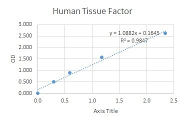
Human Coagulation Factor III / Tissue Factor ELISA Standard Curve
Recombinant Human Coagulation Factor III/Tissue Factor (Catalog # 2339-PA) was serially diluted and captured by Mouse Anti-Human Coagulation Factor III/Tissue Factor Monoclonal Antibody (Catalog # MAB2339) coated on a Clear Polystyrene Microplate (Catalog # DY990). Goat Anti-Human Coagulation Factor III/Tissue Factor Antigen Affinity-purified Polyclonal Antibody (Catalog # AF2339) was biotinylated and incubated with the protein captured on the plate. Detection of the standard curve was achieved by incubating Streptavidin-HRP (Catalog # DY998)Applications for Human Coagulation Factor III/Tissue Factor Antibody (323514)
Application
Recommended Usage
Immunoprecipitation
25 µg/mL
Sample: Conditioned cell culture medium spiked with Recombinant Human Coagulation Factor III/Tissue Factor (Catalog # 2339-PA), see our available Western blot detection antibodies
Sample: Conditioned cell culture medium spiked with Recombinant Human Coagulation Factor III/Tissue Factor (Catalog # 2339-PA), see our available Western blot detection antibodies
Western Blot
1-2 µg/mL
Sample: THP‑1 human acute monocytic leukemia cell line treated with TNF-alpha and A431 human epithelial carcinoma cell line
Sample: THP‑1 human acute monocytic leukemia cell line treated with TNF-alpha and A431 human epithelial carcinoma cell line
Human Coagulation Factor III/Tissue Factor Sandwich Immunoassay
ELISA Capture (Matched Antibody Pair)
Please Note: Optimal dilutions of this antibody should be experimentally determined.
Reviewed Applications
Read 2 reviews rated 5 using MAB2339 in the following applications:
Formulation, Preparation, and Storage
Purification
Protein A or G purified from hybridoma culture supernatant
Reconstitution
Reconstitute at 0.5 mg/mL in sterile PBS. For liquid material, refer to CoA for concentration.
Loading...
Formulation
Lyophilized from a 0.2 μm filtered solution in PBS with Trehalose. *Small pack size (SP) is supplied either lyophilized or as a 0.2 µm filtered solution in PBS.
Shipping
Lyophilized product is shipped at ambient temperature. Liquid small pack size (-SP) is shipped with polar packs. Upon receipt, store immediately at the temperature recommended below.
Stability & Storage
Use a manual defrost freezer and avoid repeated freeze-thaw cycles.
- 12 months from date of receipt, -20 to -70 °C as supplied.
- 1 month, 2 to 8 °C under sterile conditions after reconstitution.
- 6 months, -20 to -70 °C under sterile conditions after reconstitution.
Calculators
Background: Coagulation Factor III/Tissue Factor
References
- Morrissey, J.H. (2004) in Handbook of Proteolytic Enzymes. Barrett, A.J. et al. (ed) San Diego, Academic Press, p. 1659.
- Versteeg, H.H. et al. (2003) Carcinogenesis 24:1009.
- Scarpati, E.M. et al. (1987) Biochemistry 26:5234.
- Fisher, K.L. et al. (1987) Thromb. Res. 48:89.
- Morrissey, J.H. et al. (1987) Cell 50:129.
- Spicer, E.K. (1987) Proc. Natl. Acad. Sci. USA 84:5148.
Alternate Names
CD142, F3, Thromboplastin, Tissue Factor
Gene Symbol
F3
UniProt
Additional Coagulation Factor III/Tissue Factor Products
Product Documents for Human Coagulation Factor III/Tissue Factor Antibody (323514)
Certificate of Analysis
To download a Certificate of Analysis, please enter a lot or batch number in the search box below.
Note: Certificate of Analysis not available for kit components.
Product Specific Notices for Human Coagulation Factor III/Tissue Factor Antibody (323514)
For research use only
Related Research Areas
Citations for Human Coagulation Factor III/Tissue Factor Antibody (323514)
 Powered by Bioz
Customer Reviews for Human Coagulation Factor III/Tissue Factor Antibody (323514) (2)
5 out of 5
2 Customer Ratings
Have you used Human Coagulation Factor III/Tissue Factor Antibody (323514)?
Submit a review and receive an Amazon gift card!
$25/€18/£15/$25CAN/¥2500 Yen for a review with an image
$10/€7/£6/$10CAN/¥1110 Yen for a review without an image
Submit a review
Customer Images

Showing
1
-
2 of
2 reviews
Showing All
Filter By:
-
Application: ELISASample Tested: Serum and PlasmaSpecies: HumanVerified Customer | Posted 07/06/2021

-
Application: Meso Scale DiscoverySample Tested: EDTA PlasmaSpecies: HumanVerified Customer | Posted 06/22/2018After biotinylation, used as a capture reagent according to the manufacturer’s protocol (Meso Scale Diagnostics LLC). Dynamic range for human TF was 10-25,000 pg/mL.
There are no reviews that match your criteria.
Protocols
Find general support by application which include: protocols, troubleshooting, illustrated assays, videos and webinars.
- Cellular Response to Hypoxia Protocols
- Immunoprecipitation Protocol
- R&D Systems Quality Control Western Blot Protocol
- Troubleshooting Guide: Western Blot Figures
- Western Blot Conditions
- Western Blot Protocol
- Western Blot Protocol for Cell Lysates
- Western Blot Troubleshooting
- Western Blot Troubleshooting Guide
- View all Protocols, Troubleshooting, Illustrated assays and Webinars
Loading...
Associated Pathways
