Coagulation Factor III/Tissue Factor (TF), also known as thromboplastin and CD142, is an integral membrane protein found in a variety of cell types. It functions as a protein cofactor/receptor of Coagulation Factor VII, which is synthesized in the liver and circulated in the plasma (1). Upon binding of TF, the inactive factor VII is rapidly converted into activated VIIa. The resulting 1:1 complex of VIIa and TF initiates the coagulation pathway and has also important coagulation-independent functions such as angiognesis (2). Synthesized as a 295 amino acid precursor, TF consists of a signal peptide (residues 1-32) and the mature chain (residues 33-295). As a type I membrane protein, it contains a transmembrane region (residues 252-274) and a cytoplasmic tail (residues 275-295) (3-6).
Human Coagulation Factor III/Tissue Factor Antibody
R&D Systems | Catalog # AF2339

Key Product Details
Species Reactivity
Validated:
Human
Cited:
Human
Applications
Validated:
Immunohistochemistry, Western Blot, Flow Cytometry, Immunoprecipitation, CyTOF-ready
Cited:
Immunohistochemistry, Immunohistochemistry-Paraffin, Immunohistochemistry-Frozen, Western Blot, Flow Cytometry, Immunocytochemistry, ELISA Capture, ELISA Development, Electron Microscopy
Label
Unconjugated
Antibody Source
Polyclonal Goat IgG
Loading...
Product Specifications
Immunogen
Mouse myeloma cell line NS0-derived recombinant human Coagulation Factor III/Tissue Factor
Gly34-Glu251
Accession # P13726
Gly34-Glu251
Accession # P13726
Specificity
Detects human Coagulation Factor III/Tissue Factor in direct ELISAs and Western blots. In direct ELISAs, less than 5% cross‑reactivity with recombinant mouse Coagulation Factor III/Tissue Factor is observed.
Clonality
Polyclonal
Host
Goat
Isotype
IgG
Scientific Data Images for Human Coagulation Factor III/Tissue Factor Antibody
Detection of Human Coagulation Factor III/Tissue Factor by Western Blot.
Western blot shows lysates of A431 human epithelial carcinoma cell line and A549 human lung carcinoma cell line (negative control). PVDF membrane was probed with 0.5 µg/mL of Goat Anti-Human Coagulation Factor III/Tissue Factor Antigen Affinity-purified Polyclonal Antibody (Catalog # AF2339) followed by HRP-conjugated Anti-Goat IgG Secondary Antibody (HAF019). A specific band was detected for Coagulation Factor III/Tissue Factor at approximately 48 kDa (as indicated). GAPDH (AF5718) is shown as a loading control. This experiment was conducted under reducing conditions and using Western Blot Buffer Group 1.
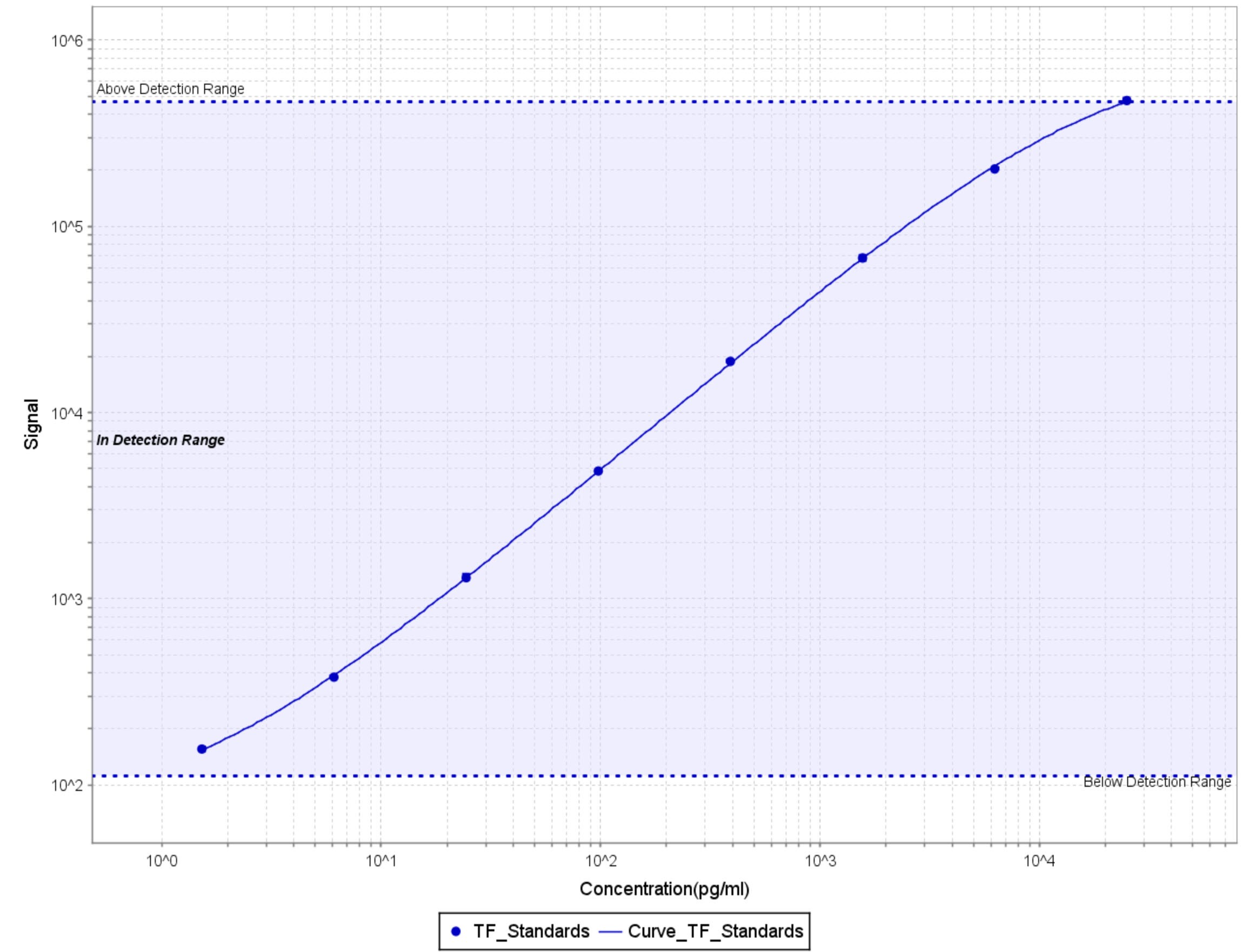
Human Coagulation Factor III / Tissue Factor ELISA Standard Curve
Recombinant Human Coagulation Factor III/Tissue Factor (Catalog # 2339-PA) was serially diluted and captured by Mouse Anti-Human Coagulation Factor III/Tissue Factor Monoclonal Antibody (Catalog # MAB2339) coated on a Clear Polystyrene Microplate (Catalog # DY990). Goat Anti-Human Coagulation Factor III/Tissue Factor Antigen Affinity-purified Polyclonal Antibody (Catalog # AF2339) was biotinylated and incubated with the protein captured on the plate. Detection of the standard curve was achieved by incubating Streptavidin-HRP (Catalog # DY998)Applications for Human Coagulation Factor III/Tissue Factor Antibody
Application
Recommended Usage
CyTOF-ready
Ready to be labeled using established conjugation methods. No BSA or other carrier proteins that could interfere with conjugation.
Flow Cytometry
2.5 µg/106 cells
Sample: U937 human histiocytic lymphoma cell line treated with LPS
Sample: U937 human histiocytic lymphoma cell line treated with LPS
Immunohistochemistry
5-15 µg/mL
Sample: Immersion fixed paraffin-embedded sections of human brain (cerebellum)
Sample: Immersion fixed paraffin-embedded sections of human brain (cerebellum)
Immunoprecipitation
25 µg/mL
Sample: Conditioned cell culture medium spiked with Recombinant Human Coagulation Factor III/Tissue Factor (Catalog # 2339-PA), see our available Western blot detection antibodies
Sample: Conditioned cell culture medium spiked with Recombinant Human Coagulation Factor III/Tissue Factor (Catalog # 2339-PA), see our available Western blot detection antibodies
Western Blot
0.5 µg/mL
Sample: A431 human epithelial carcinoma cell line
Sample: A431 human epithelial carcinoma cell line
Reviewed Applications
Read 3 reviews rated 4.3 using AF2339 in the following applications:
Flow Cytometry Panel Builder
Bio-Techne Knows Flow Cytometry
Save time and reduce costly mistakes by quickly finding compatible reagents using the Panel Builder Tool.
Advanced Features
- Spectra Viewer - Custom analysis of spectra from multiple fluorochromes
- Spillover Popups - Visualize the spectra of individual fluorochromes
- Antigen Density Selector - Match fluorochrome brightness with antigen density

Formulation, Preparation, and Storage
Purification
Antigen Affinity-purified
Reconstitution
Reconstitute at 0.2 mg/mL in sterile PBS. For liquid material, refer to CoA for concentration.
Loading...
Formulation
Lyophilized from a 0.2 μm filtered solution in PBS with Trehalose. *Small pack size (SP) is supplied either lyophilized or as a 0.2 µm filtered solution in PBS.
Shipping
Lyophilized product is shipped at ambient temperature. Liquid small pack size (-SP) is shipped with polar packs. Upon receipt, store immediately at the temperature recommended below.
Stability & Storage
Use a manual defrost freezer and avoid repeated freeze-thaw cycles.
- 12 months from date of receipt, -20 to -70 °C as supplied.
- 1 month, 2 to 8 °C under sterile conditions after reconstitution.
- 6 months, -20 to -70 °C under sterile conditions after reconstitution.
Calculators
Background: Coagulation Factor III/Tissue Factor
References
- Morrissey, J.H. (2004) in Handbook of Proteolytic Enzymes. Barrett, A.J. et al. (ed) San Diego, Academic Press, p. 1659.
- Versteeg, H.H. et al. (2003) Carcinogenesis 24:1009.
- Scarpati, E.M. et al. (1987) Biochemistry 26:5234.
- Fisher, K.L. et al. (1987) Thromb. Res. 48:89.
- Morrissey, J.H. et al. (1987) Cell 50:129.
- Spicer, E.K. (1987) Proc. Natl. Acad. Sci. USA 84:5148.
Alternate Names
CD142, F3, Thromboplastin, Tissue Factor
Gene Symbol
F3
UniProt
Additional Coagulation Factor III/Tissue Factor Products
Product Documents for Human Coagulation Factor III/Tissue Factor Antibody
Certificate of Analysis
To download a Certificate of Analysis, please enter a lot or batch number in the search box below.
Note: Certificate of Analysis not available for kit components.
Product Specific Notices for Human Coagulation Factor III/Tissue Factor Antibody
For research use only
Related Research Areas
Citations for Human Coagulation Factor III/Tissue Factor Antibody
 Powered by Bioz
Customer Reviews for Human Coagulation Factor III/Tissue Factor Antibody (3)
4.3 out of 5
3 Customer Ratings
Have you used Human Coagulation Factor III/Tissue Factor Antibody?
Submit a review and receive an Amazon gift card!
$25/€18/£15/$25CAN/¥2500 Yen for a review with an image
$10/€7/£6/$10CAN/¥1110 Yen for a review without an image
Submit a review
Customer Images
Showing
1
-
3 of
3 reviews
Showing All
Filter By:
-
Application: MSD assaySample Tested: EDTA Plasma, Citrate Plasma and SerumSpecies: Human and Cynomolgus MonkeyVerified Customer | Posted 05/30/2018Used as detection antibody after labeling with Sulfo-Tag according to manufacturer's protocol (Meso Scale Diagnostics LLC). Detection range – 1.5-25,000 pg/ml

-
Application: Western BlotSample Tested: See PMID 22828416Species: HumanVerified Customer | Posted 01/06/2015
-
Application: ImmunofluorescenceSample Tested: See PMID 23468018Species: HumanVerified Customer | Posted 01/06/2015
There are no reviews that match your criteria.
Protocols
Find general support by application which include: protocols, troubleshooting, illustrated assays, videos and webinars.
- 7-Amino Actinomycin D (7-AAD) Cell Viability Flow Cytometry Protocol
- Antigen Retrieval Protocol (PIER)
- Antigen Retrieval for Frozen Sections Protocol
- Appropriate Fixation of IHC/ICC Samples
- Cellular Response to Hypoxia Protocols
- Chromogenic IHC Staining of Formalin-Fixed Paraffin-Embedded (FFPE) Tissue Protocol
- Chromogenic Immunohistochemistry Staining of Frozen Tissue
- ClariTSA™ Fluorophore Kits
- Detection & Visualization of Antibody Binding
- Extracellular Membrane Flow Cytometry Protocol
- Flow Cytometry Protocol for Cell Surface Markers
- Flow Cytometry Protocol for Staining Membrane Associated Proteins
- Flow Cytometry Staining Protocols
- Flow Cytometry Troubleshooting Guide
- Fluorescent IHC Staining of Frozen Tissue Protocol
- Graphic Protocol for Heat-induced Epitope Retrieval
- Graphic Protocol for the Preparation and Fluorescent IHC Staining of Frozen Tissue Sections
- Graphic Protocol for the Preparation and Fluorescent IHC Staining of Paraffin-embedded Tissue Sections
- Graphic Protocol for the Preparation of Gelatin-coated Slides for Histological Tissue Sections
- IHC Sample Preparation (Frozen sections vs Paraffin)
- Immunofluorescent IHC Staining of Formalin-Fixed Paraffin-Embedded (FFPE) Tissue Protocol
- Immunohistochemistry (IHC) and Immunocytochemistry (ICC) Protocols
- Immunohistochemistry Frozen Troubleshooting
- Immunohistochemistry Paraffin Troubleshooting
- Immunoprecipitation Protocol
- Intracellular Flow Cytometry Protocol Using Alcohol (Methanol)
- Intracellular Flow Cytometry Protocol Using Detergents
- Intracellular Nuclear Staining Flow Cytometry Protocol Using Detergents
- Intracellular Staining Flow Cytometry Protocol Using Alcohol Permeabilization
- Intracellular Staining Flow Cytometry Protocol Using Detergents to Permeabilize Cells
- Preparing Samples for IHC/ICC Experiments
- Preventing Non-Specific Staining (Non-Specific Binding)
- Primary Antibody Selection & Optimization
- Propidium Iodide Cell Viability Flow Cytometry Protocol
- Protocol for Heat-Induced Epitope Retrieval (HIER)
- Protocol for Liperfluo
- Protocol for Making a 4% Formaldehyde Solution in PBS
- Protocol for VisUCyte™ HRP Polymer Detection Reagent
- Protocol for the Characterization of Human Th22 Cells
- Protocol for the Characterization of Human Th9 Cells
- Protocol for the Preparation & Fixation of Cells on Coverslips
- Protocol for the Preparation and Chromogenic IHC Staining of Frozen Tissue Sections
- Protocol for the Preparation and Chromogenic IHC Staining of Frozen Tissue Sections - Graphic
- Protocol for the Preparation and Chromogenic IHC Staining of Paraffin-embedded Tissue Sections
- Protocol for the Preparation and Chromogenic IHC Staining of Paraffin-embedded Tissue Sections - Graphic
- Protocol for the Preparation and Fluorescent IHC Staining of Frozen Tissue Sections
- Protocol for the Preparation and Fluorescent IHC Staining of Paraffin-embedded Tissue Sections
- Protocol for the Preparation of Gelatin-coated Slides for Histological Tissue Sections
- Protocol: Annexin V and PI Staining by Flow Cytometry
- Protocol: Annexin V and PI Staining for Apoptosis by Flow Cytometry
- R&D Systems Quality Control Western Blot Protocol
- TUNEL and Active Caspase-3 Detection by IHC/ICC Protocol
- The Importance of IHC/ICC Controls
- Troubleshooting Guide: Fluorokine Flow Cytometry Kits
- Troubleshooting Guide: Immunohistochemistry
- Troubleshooting Guide: Western Blot Figures
- Western Blot Conditions
- Western Blot Protocol
- Western Blot Protocol for Cell Lysates
- Western Blot Troubleshooting
- Western Blot Troubleshooting Guide
- View all Protocols, Troubleshooting, Illustrated assays and Webinars
Loading...
Associated Pathways
