Epithelial (E) - Cadherin (ECAD), also known as cell-CAM120/80 in the human, uvomorulin in the mouse, Arc-1 in the dog, andL-CAM in the chicken, is a member of the Cadherin family of cell adhesion molecules. Cadherins are calcium-dependent transmembrane proteins which bind to one another in a homophilic manner. On their cytoplasmic side, they associate with the three catenins, alpha, beta, and gamma (plakoglobin). This association links the cadherin protein to the cytoskeleton. Without association with the catenins, the cadherins are non-adhesive. Cadherins play a role in development, specifically in tissue formation. They may also help to maintain tissue architecture in the adult. E-Cadherin may also play a role in tumor development, as loss of E-Cadherin has been associated with tumor invasiveness. E-Cadherin is a classical cadherin molecule. Classical cadherins consist of a large extracellular domain which contains DXD and DXNDN repeats responsible for mediating calcium-dependent adhesion, a single-pass transmembrane domain, and a short carboxy-terminal cytoplasmic domain responsible for interacting with the catenins. E-Cadherin contains five extracellular calcium-binding domains of approximately 110 amino acids each.
Human E-Cadherin Antibody (180215)
R&D Systems | Catalog # MAB1838

Key Product Details
Species Reactivity
Validated:
Human
Cited:
Human, Mouse
Applications
Validated:
Immunohistochemistry, Western Blot, Simple Western
Cited:
Immunohistochemistry, Immunohistochemistry-Paraffin, Immunohistochemistry-Frozen, Western Blot, Flow Cytometry, Immunocytochemistry, Simple Western
Label
Unconjugated
Antibody Source
Monoclonal Mouse IgG2B Clone # 180215
Loading...
Product Specifications
Immunogen
Mouse myeloma cell line NS0-derived recombinant human E-Cadherin
Asp155-Ile707
Accession # P12830
Asp155-Ile707
Accession # P12830
Specificity
Detects human E-Cadherin in direct ELISAs and Western blots. In direct ELISAs and Western blots, no cross‑reactivity with recombinant human (rh) Cadherin-8, rhCadherin-17, recombinant mouse E-Cadherin, rhN-Cadherin, rhP-Cadherin, or rhVE‑Cadherin is observed.
Clonality
Monoclonal
Host
Mouse
Isotype
IgG2B
Scientific Data Images for Human E-Cadherin Antibody (180215)
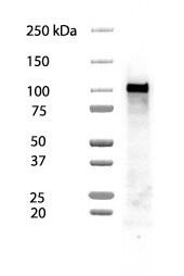
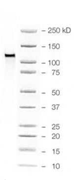
Detection of Human E‑Cadherin by Western Blot.
Western blot shows lysates of A549 human lung carcinoma cell line and HepG2 human hepatocellular carcinoma cell line. PVDF membrane was probed with 0.5 µg/mL of Mouse Anti-Human E-Cadherin Monoclonal Antibody (Catalog # MAB1838) followed by HRP-conjugated Anti-Mouse IgG Secondary Antibody (Catalog # HAF018). A specific band was detected for E-Cadherin at approximately 110 kDa (as indicated). This experiment was conducted under reducing conditions and using Immunoblot Buffer Group 1.
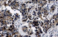
E-Cadherin in Human Colon.
E-Cadherin was detected in immersion fixed paraffin-embedded sections of human colon using Mouse Anti-Human E-Cadherin Monoclonal Antibody (Catalog # MAB1838) at 2 µg/mL overnight at 4 °C. Tissue was stained using the Anti-Mouse HRP-DAB Cell & Tissue Staining Kit (brown; Catalog # CTS002) and counterstained with hematoxylin (blue). Specific labeling was localized to the plasma membrane of epithelial cells. View our protocol for Chromogenic IHC Staining of Paraffin-embedded Tissue Sections.
Detection of Human E‑Cadherin by Simple WesternTM.
Simple Western lane view shows lysates of A549 human lung carcinoma cell line, loaded at 0.2 mg/mL. A specific band was detected for E‑Cadherin at approximately 166 kDa (as indicated) using 5 µg/mL of Mouse Anti-Human E‑Cadherin Monoclonal Antibody (Catalog # MAB1838). This experiment was conducted under reducing conditions and using the 12-230 kDa separation system.
Detection of Human E-Cadherin by Simple Western
D492 and D492M cells carry an epithelial and a mesenchymal phenotype, respectively. (A, B) Relative protein levels of phenotype‐specific markers in D492 and D492M cells as measured by simple western immunoassay (A; representative electropherograms, where the x‐axis shows the protein size (kDa), and the y‐axis indicates signal intensity, reflecting the amount of the protein), and the RPPA (B; average ± SD from three technical replicates). (C) Cell growth shown as increase in confluence (y‐axis) during time after seeding (x‐axis) tracked by the Incucyte (average ± SEM, n ≥ 3). (D) D492 and D492M cell colonies formed during 8‐day growth in 3D Matrigel and stained with phalloidin (red) for labeling F‐actin; scale bar, 50 µm. Image collected and cropped by CiteAb from the following open publication (https://pubmed.ncbi.nlm.nih.gov/33759347), licensed under a CC-BY license. Not internally tested by R&D Systems.
Detection of E-Cadherin by Western Blot
Targeting PCa/macrophage AR leads to increased macrophage recruitment and enhanced PCa migration through CCL2 inductionqPCR of CCL2 mRNA in THP-1 scramble (scr) and THP-1 silenced AR (siAR) cells/different PCa cell lines as indicated (left) and qPCR of CCL2 mRNA in C4-2 scr and C4-2 siAR cells (right).qPCR of CCL2 mRNA in THP-1 (scr or siAR) cells co-cultured with C4-2 scr or siAR cells (left) and in C4-2 (scr or siAR) cells co-cultured with THP-1 scr or siAR cells (right).ELISA of CCL2 in 24 h CM of C4-2 scr and C4-2 siAR cells (left) and in 24 h co-cultured CM of C4-2 scr or C4-2 siAR cells/THP-1 scr or siAR cells (right).ELISA of CCL2 in 24 h co-cultured CM of parental LNCaP cells/THP-1 scr or siAR cells (left) and in 24 h co-cultured CM of parental LAPC4 cells/THP-1 scr or siAR cells (right).Migration assay of C4-2 scr and C4-2 siAR cells incubated for 24 h (upper left), parental THP-1 cells/C4-2 scr or siAR cells after co-cultured for 16 h (upper right), parental C4-2 cells/THP-1 scr cells or siAR cells after co-cultured for 24 h (lower left), and C4-2 scr or C4-2 siAR cells/THP-1 scr or siAR cells after co-cultured for 24 h (lower right), (n = 3); bars in graphs (A–E), Mean ± SEM; bars in pictures, 400 μm (magnification 100×).Western blot of CCL2, EMT markers, AR, and PSA in parental C4-2 cells treated with CM of THP-1 scr and siAR, or co-cultured with THP-1 scr and siAR cells for 24 h (left), and in C4-2 scr and siAR cells (right). Image collected and cropped by CiteAb from the following open publication (https://pubmed.ncbi.nlm.nih.gov/23982944), licensed under a CC-BY license. Not internally tested by R&D Systems.Applications for Human E-Cadherin Antibody (180215)
Application
Recommended Usage
Immunohistochemistry
2-25 µg/mL
Sample: Immersion fixed paraffin-embedded sections of human colon and lung
Sample: Immersion fixed paraffin-embedded sections of human colon and lung
Simple Western
5 µg/mL
Sample: A549 human lung carcinoma cell line
Sample: A549 human lung carcinoma cell line
Western Blot
0.5 µg/mL
Sample: A549 human lung carcinoma cell line and HepG2 human hepatocellular carcinoma cell line
Sample: A549 human lung carcinoma cell line and HepG2 human hepatocellular carcinoma cell line
Reviewed Applications
Read 3 reviews rated 5 using MAB1838 in the following applications:
Formulation, Preparation, and Storage
Purification
Protein A or G purified from hybridoma culture supernatant
Reconstitution
Reconstitute at 0.5 mg/mL in sterile PBS. For liquid material, refer to CoA for concentration.
Loading...
Formulation
Lyophilized from a 0.2 μm filtered solution in PBS with Trehalose. *Small pack size (SP) is supplied either lyophilized or as a 0.2 µm filtered solution in PBS.
Shipping
Lyophilized product is shipped at ambient temperature. Liquid small pack size (-SP) is shipped with polar packs. Upon receipt, store immediately at the temperature recommended below.
Stability & Storage
Use a manual defrost freezer and avoid repeated freeze-thaw cycles.
- 12 months from date of receipt, -20 to -70 °C as supplied.
- 1 month, 2 to 8 °C under sterile conditions after reconstitution.
- 6 months, -20 to -70 °C under sterile conditions after reconstitution.
Calculators
Background: E-Cadherin
References
- Bussemakers, M.J.G. et al. (1993) Mol. Biol. Reports 17:123.
- Overduin, M. et al. (1995) Science 267:386.
- Takeichi, M. (1991) Science 251:1451.
Alternate Names
Arc-1, CAD1, Cadherin-1, CD324, CDH1, Cell-CAM120/80, ECAD, ECadherin, L-CAM, Uvomorulin
Gene Symbol
CDH1
UniProt
Additional E-Cadherin Products
Product Documents for Human E-Cadherin Antibody (180215)
Certificate of Analysis
To download a Certificate of Analysis, please enter a lot or batch number in the search box below.
Note: Certificate of Analysis not available for kit components.
Product Specific Notices for Human E-Cadherin Antibody (180215)
For research use only
Citations for Human E-Cadherin Antibody (180215)
 Powered by Bioz
Customer Reviews for Human E-Cadherin Antibody (180215) (3)
5 out of 5
3 Customer Ratings
Have you used Human E-Cadherin Antibody (180215)?
Submit a review and receive an Amazon gift card!
$25/€18/£15/$25CAN/¥2500 Yen for a review with an image
$10/€7/£6/$10CAN/¥1110 Yen for a review without an image
Submit a review
Customer Images
Showing
1
-
3 of
3 reviews
Showing All
Filter By:
-
Application: Western BlotSample Tested: MCF-7 human breast cancer cell lineSpecies: HumanVerified Customer | Posted 01/01/2026Whole cell lysate of human adipocytes and MCF7 cocultureWhole cell lysate of human adipocytes and MCF7 coculture

-
Application: Western BlotSample Tested: HCT-116 human colorectal carcinoma cell lineSpecies: HumanVerified Customer | Posted 01/01/2026Used at 0.5 ug/ml
-
Application: ImmunohistochemistrySample Tested: Cardiac carcinomaSpecies: HumanVerified Customer | Posted 10/05/2021
There are no reviews that match your criteria.
Protocols
Find general support by application which include: protocols, troubleshooting, illustrated assays, videos and webinars.
- Antigen Retrieval Protocol (PIER)
- Antigen Retrieval for Frozen Sections Protocol
- Appropriate Fixation of IHC/ICC Samples
- Cellular Response to Hypoxia Protocols
- Chromogenic IHC Staining of Formalin-Fixed Paraffin-Embedded (FFPE) Tissue Protocol
- Chromogenic Immunohistochemistry Staining of Frozen Tissue
- ClariTSA™ Fluorophore Kits
- Detection & Visualization of Antibody Binding
- Fluorescent IHC Staining of Frozen Tissue Protocol
- Graphic Protocol for Heat-induced Epitope Retrieval
- Graphic Protocol for the Preparation and Fluorescent IHC Staining of Frozen Tissue Sections
- Graphic Protocol for the Preparation and Fluorescent IHC Staining of Paraffin-embedded Tissue Sections
- Graphic Protocol for the Preparation of Gelatin-coated Slides for Histological Tissue Sections
- IHC Sample Preparation (Frozen sections vs Paraffin)
- Immunofluorescent IHC Staining of Formalin-Fixed Paraffin-Embedded (FFPE) Tissue Protocol
- Immunohistochemistry (IHC) and Immunocytochemistry (ICC) Protocols
- Immunohistochemistry Frozen Troubleshooting
- Immunohistochemistry Paraffin Troubleshooting
- Preparing Samples for IHC/ICC Experiments
- Preventing Non-Specific Staining (Non-Specific Binding)
- Primary Antibody Selection & Optimization
- Protocol for Heat-Induced Epitope Retrieval (HIER)
- Protocol for Making a 4% Formaldehyde Solution in PBS
- Protocol for VisUCyte™ HRP Polymer Detection Reagent
- Protocol for the Preparation & Fixation of Cells on Coverslips
- Protocol for the Preparation and Chromogenic IHC Staining of Frozen Tissue Sections
- Protocol for the Preparation and Chromogenic IHC Staining of Frozen Tissue Sections - Graphic
- Protocol for the Preparation and Chromogenic IHC Staining of Paraffin-embedded Tissue Sections
- Protocol for the Preparation and Chromogenic IHC Staining of Paraffin-embedded Tissue Sections - Graphic
- Protocol for the Preparation and Fluorescent IHC Staining of Frozen Tissue Sections
- Protocol for the Preparation and Fluorescent IHC Staining of Paraffin-embedded Tissue Sections
- Protocol for the Preparation of Gelatin-coated Slides for Histological Tissue Sections
- R&D Systems Quality Control Western Blot Protocol
- TUNEL and Active Caspase-3 Detection by IHC/ICC Protocol
- The Importance of IHC/ICC Controls
- Troubleshooting Guide: Immunohistochemistry
- Troubleshooting Guide: Western Blot Figures
- Western Blot Conditions
- Western Blot Protocol
- Western Blot Protocol for Cell Lysates
- Western Blot Troubleshooting
- Western Blot Troubleshooting Guide
- View all Protocols, Troubleshooting, Illustrated assays and Webinars
Loading...
Associated Pathways
