Endocan, also known as endothelial-cell specific molecule-1 (ESM-1), is a secreted cysteine-rich dermatan sulfate (DS) proteoglycan primarily expressed by endothelial cells within the vascular capillary network in kidney and in the alveolar walls of the lung (1). Endocan expression has also been detected in different epithelia and in adipocytes (2, 3). The expression of endocan is upregulated by TNF‑ alpha, IL-1 beta, or lipopolysaccharide and down-regulated by IFN‑ gamma (1). The human Endocan gene encodes a 184 amino acid (aa) residues precursor protein with a 19 aa hydrophobic signal peptide and a 165 aa mature region with 18 Cysteine residues (1). The DS chain is covalently attached to serine 137 (4). Endocan has been shown to bind CD11a/CD18 integrin (also known as lymphocyte function-associated antigen-1, LFA-1) on human lymphocytes, monocytes and Jurkat cells, inhibiting its binding to ICAM-1 and reducing LFA-1-mediated leukocyte activation (5). Endocan binds via its DS chain to hepatocyte growth factor (HGF) to enhance HGF mitogenic activity (3, 6). Genetically engineered cells overexpressing endocan has been shown to induce tumor formation, suggesting that Endocan may be involved in the pathophysiology of tumor growth in vivo (3, 6). Circulating Endocan can be detected in the serum from healthy subjects. In patients with lung cancer or acute and severe sepsis, elevated Endocan concentrations have been reported (2, 6).

Key Product Details
Species Reactivity
Validated:
Human
Cited:
Human, Mouse
Applications
Validated:
Western Blot, Neutralization
Cited:
Immunohistochemistry, Western Blot, Neutralization, Immunoprecipitation, ELISA Development
Label
Unconjugated
Antibody Source
Polyclonal Goat IgG
Loading...
Product Specifications
Immunogen
Mouse myeloma cell line NS0-derived recombinant human Endocan/ESM-1
Trp20-Arg184
Accession # Q9NQ30
Trp20-Arg184
Accession # Q9NQ30
Specificity
Detects human Endocan in direct ELISAs and Western blots. In direct ELISAs and Western blots, approximately 20% cross-reactivity with recombinant mouse Endocan is observed.
Clonality
Polyclonal
Host
Goat
Isotype
IgG
Endotoxin Level
<0.10 EU per 1 μg of the antibody by the LAL method.
Scientific Data Images for Human Endocan/ESM-1 Antibody
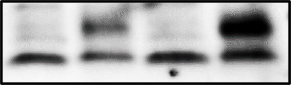
Detection of Human Endocan/ESM‑1 by Western Blot.
Western blot shows lysate of HUVEC human umbilical vein endothelial cells. PVDF membrane was probed with 1 µg/mL of Goat Anti-Human Endocan/ESM-1 Antigen Affinity-purified Polyclonal Antibody (Catalog # AF1810) followed by HRP-conjugated Anti-Goat IgG Secondary Antibody (Catalog # HAF017). A specific band was detected for Endocan/ESM-1 at approximately 20 kDa (as indicated). This experiment was conducted under reducing conditions and using Immunoblot Buffer Group 1.
Cell Adhesion Mediated by Endocan/ESM‑1 and Neutral-ization by Human Endocan/ESM‑1 Antibody.
Recombinant Human Endocan/ESM-1 (Catalog # 1810-EC), immobilized onto a microplate, supports the adhesion of the Jurkat human acute T cell leukemia cell line in a dose-dependent manner (orange line). Adhesion elicited by Recombinant Human Endocan/ESM-1 (20 µg/mL) is neutralized (green line) by increasing concentrations of Goat Anti-Human Endocan/ESM-1 Antigen Affinity-purified Polyclonal Antibody (Catalog # AF1810). The ND50 is typically 1-4 µg/mL.Applications for Human Endocan/ESM-1 Antibody
Application
Recommended Usage
Western Blot
1 µg/mL
Sample: HUVEC human umbilical vein endothelial cells
Sample: HUVEC human umbilical vein endothelial cells
Neutralization
Measured by its ability to neutralize Endocan/ESM‑1-mediated adhesion of the Jurkat human acute T cell leukemia cell line. The Neutralization Dose (ND50) is typically 1-4 µg/mL in the presence of 20 µg/mL Recombinant Human Endocan/ESM‑1.
Reviewed Applications
Read 1 review rated 4 using AF1810 in the following applications:
Formulation, Preparation, and Storage
Purification
Antigen Affinity-purified
Reconstitution
Reconstitute at 0.2 mg/mL in sterile PBS. For liquid material, refer to CoA for concentration.
Loading...
Formulation
Lyophilized from a 0.2 μm filtered solution in PBS with Trehalose. *Small pack size (SP) is supplied either lyophilized or as a 0.2 µm filtered solution in PBS.
Shipping
Lyophilized product is shipped at ambient temperature. Liquid small pack size (-SP) is shipped with polar packs. Upon receipt, store immediately at the temperature recommended below.
Stability & Storage
Use a manual defrost freezer and avoid repeated freeze-thaw cycles.
- 12 months from date of receipt, -20 to -70 °C as supplied.
- 1 month, 2 to 8 °C under sterile conditions after reconstitution.
- 6 months, -20 to -70 °C under sterile conditions after reconstitution.
Calculators
Background: Endocan/ESM-1
References
- Lassalle, P. et al. (1996) J. Biol. Chem. 271:20458.
- Bechard, D. et al. (2000) J. Vasc. Res. 37:417.
- Wellner, M. et al. (2003) Horm. Metab. Res. 35:217.
- Bechard, D. et al. (2001) J. Biol. Chem. 276:48341.
- Bechard, D. et al. (2001) J. Immunol. 167:3099
- Scherpereel, A. et al. (2003) Cancer Res. 63:6084.
Alternate Names
ESM-1, ESM1, IGFBP-rp6
Gene Symbol
ESM1
UniProt
Additional Endocan/ESM-1 Products
Product Documents for Human Endocan/ESM-1 Antibody
Certificate of Analysis
To download a Certificate of Analysis, please enter a lot or batch number in the search box below.
Note: Certificate of Analysis not available for kit components.
Product Specific Notices for Human Endocan/ESM-1 Antibody
For research use only
Related Research Areas
Citations for Human Endocan/ESM-1 Antibody
 Powered by Bioz
Customer Reviews for Human Endocan/ESM-1 Antibody (1)
4 out of 5
1 Customer Rating
Have you used Human Endocan/ESM-1 Antibody?
Submit a review and receive an Amazon gift card!
$25/€18/£15/$25CAN/¥2500 Yen for a review with an image
$10/€7/£6/$10CAN/¥1110 Yen for a review without an image
Submit a review
Customer Images
Showing
1
-
1 of
1 review
Showing All
Filter By:
-
Application: Western BlotSample Tested: Cell LysatesSpecies: HumanVerified Customer | Posted 06/09/2022

There are no reviews that match your criteria.
Protocols
Find general support by application which include: protocols, troubleshooting, illustrated assays, videos and webinars.
- Cellular Response to Hypoxia Protocols
- R&D Systems Quality Control Western Blot Protocol
- Troubleshooting Guide: Western Blot Figures
- Western Blot Conditions
- Western Blot Protocol
- Western Blot Protocol for Cell Lysates
- Western Blot Troubleshooting
- Western Blot Troubleshooting Guide
- View all Protocols, Troubleshooting, Illustrated assays and Webinars
Loading...