
FCRLB, also known as FCRL2, FCRY, and FREB2, is a 57-60 kDa protein with sequence homology to classical Fc receptors. There are at least six type 1 transmembrane FCRL proteins and two that lack transmembrane segments. Each family member contains between three and nine immunoglobulin-like domains. FCRL proteins are differentially expressed within the B cell lineage and can either promote or inhibit B cell proliferation and activation (1, 2). According to R&D Systems testing, FCRLB binds to purified human IgG with high affinity. Human FCRLB shares 82% and 85% amino acid sequence identity with mouse and rat FCRLB, respectively. It contains a putative signal peptide, three immunoglobulin-like domains, and a mucin-like stalk that is rich in Pro, Ser, and Thr residues (1 - 3). The stalk region also contains di-Leu motifs and an unpaired cysteine (1 - 3). Alternative splicing generates isoforms with deletions in the putative signal peptide and substitutions and/or truncations in the third Ig-like domain (3). When expressed in transfectants, FCRLB is not secreted but shows a diffuse intracellular localization (3, 4). FCRLB is expressed at a low level in placenta and in B lineage cells of the germinal center (3, 4). It is upregulated in B cells by BAFF and LPS (5). Among non-hematopoietic cells, FCRLB is expressed in fibroblasts, melanocytes, and melanoma (3, 5). It is preferentially expressed in nonproliferating cells and at the onset of apoptosis (4, 5).

Scientific images may be resized, cropped, or adjusted for brightness or contrast for presentation purposes. These updates are limited to presentation and do not affect the underlying scientific interpretation of the data. Where available, additional source data may be provided upon request.
Key Product Details
Species Reactivity
Applications
Label
Antibody Source
Product Specifications
Immunogen
Ala18-Ser426
Accession # Q6BAA4
Specificity
Clonality
Host
Isotype
Scientific Data Images for Human FCRLB/FCRY Antibody
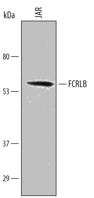
Detection of Human FCRLB/FCRY by Western Blot.
Western blot shows lysates of JAR human choriocarcinoma cell line. PVDF Membrane was probed with 1 µg/mL of Human FCRLB/FCRY Antigen Affinity-purified Polyclonal Antibody (Catalog # AF4705) followed by HRP-conjugated Anti-Sheep IgG Secondary Antibody (Catalog # HAF016). A specific band was detected for FCRLB/FCRY at approximately 57-60 kDa (as indicated). This experiment was conducted under reducing conditions and using Immunoblot Buffer Group 1.Applications for Human FCRLB/FCRY Antibody
Western Blot
Sample: JAR human choriocarcinoma cell line
Formulation, Preparation, and Storage
Purification
Reconstitution
Sterile PBS to a final concentration of 0.2 mg/mL. For liquid material, refer to CoA for concentration.
Formulation
Shipping
Stability & Storage
- 12 months from date of receipt, -20 to -70 °C as supplied.
- 1 month, 2 to 8 °C under sterile conditions after reconstitution.
- 6 months, -20 to -70 °C under sterile conditions after reconstitution.
Calculators
Background: FCRLB/FCRY
References
- Davis, R.S. (2007) Annu. Rev. Immunol. 25:525.
- Maltais, L.J. et al. (2006) Nat. Immunol. 7:431.
- Chikaev, N.A. et al. (2005) Genomics 85:264.
- Wilson, T.J. and M. Colonna (2005) Genes Immun. 6:341.
- Masuda, K. et al. (2005) Gene 363:32.
Long Name
Alternate Names
Gene Symbol
UniProt
Additional FCRLB/FCRY Products
Product Documents for Human FCRLB/FCRY Antibody
Certificate of Analysis
To download a Certificate of Analysis, please enter a lot or batch number in the search box below.
Note: Certificate of Analysis not available for kit components.
Product Specific Notices for Human FCRLB/FCRY Antibody
For research use only
Related Research Areas
Customer Reviews for Human FCRLB/FCRY Antibody
There are currently no reviews for this product. Be the first to review Human FCRLB/FCRY Antibody and earn rewards!
Have you used Human FCRLB/FCRY Antibody?
Submit a review and receive an Amazon gift card!
$25/€18/£15/$25CAN/¥2500 for a review with an image
$10/€7/£6/$10CAN/¥1110 for a review without an image
Submit a review
Protocols
Find general support by application which include: protocols, troubleshooting, illustrated assays, videos and webinars.
- Cellular Response to Hypoxia Protocols
- R&D Systems Quality Control Western Blot Protocol
- Troubleshooting Guide: Western Blot Figures
- Western Blot Conditions
- Western Blot Protocol
- Western Blot Protocol for Cell Lysates
- Western Blot Troubleshooting
- Western Blot Troubleshooting Guide
- View all Protocols, Troubleshooting, Illustrated assays and Webinars