
Discontinued Product
AF2606 has been discontinued.
View all GATA-4 products.
Key Product Details
Species Reactivity
Validated:
Human
Cited:
Human, Mouse
Applications
Validated:
Western Blot, Immunocytochemistry, Chromatin Immunoprecipitation (ChIP)
Cited:
Immunohistochemistry, Immunohistochemistry-Paraffin, Immunohistochemistry-Frozen, Western Blot, Neutralization, Flow Cytometry, Immunocytochemistry, Immunoprecipitation, Chromatin Immunoprecipitation (ChIP)
Label
Unconjugated
Antibody Source
Polyclonal Goat IgG
Loading...
Product Specifications
Immunogen
E. coli-derived recombinant human GATA-4
Met27-Phe211
Accession # P43694
Met27-Phe211
Accession # P43694
Specificity
Detects human GATA-4 in direct ELISAs and Western blots.
Clonality
Polyclonal
Host
Goat
Isotype
IgG
Scientific Data Images for Human GATA‑4 Antibody
Detection of Human GATA‑4 by Western Blot.
Western blot shows lysates of human heart tissue. PVDF membrane was probed with 1 µg/mL of Goat Anti-Human GATA-4 Antigen Affinity-purified Polyclonal Antibody (Catalog # AF2606) followed by HRP-conjugated Anti-Goat IgG Secondary Antibody (Catalog # HAF019). A specific band was detected for GATA-4 at approximately 46 kDa (as indicated). This experiment was conducted under reducing conditions and using Immunoblot Buffer Group 1.
Detection of GATA‑4-regulated Genes by Chromatin Immunoprecipitation.
KATO-III human gastric carcinoma cell line was fixed using formaldehyde, resuspended in lysis buffer, and sonicated to shear chromatin. GATA-4/DNA complexes were immunoprecipitated using 5 µg Goat Anti-Human GATA-4 Antigen Affinity-purified Polyclonal Antibody (Catalog # AF2606) or control antibody (Catalog # AB-108-C) for 15 minutes in an ultrasonic bath, followed by Biotinylated Anti-Goat IgG Secondary Antibody (Catalog # BAF109). Immunocomplexes were captured using 50 µL of MagCellect Streptavidin Ferrofluid (Catalog # MAG999) and DNA was purified using chelating resin solution. Themucin4promoter was detected by standard PCR.
GATA‑4 in Differentiated Human Embryonic Stem Cells.
GATA‑4 was detected in immersion fixed differentiated human embryonic stem cells using 10 µg/mL Goat Anti-Human GATA‑4 Antigen Affinity-purified Polyclonal Antibody (Catalog # AF2606) for 3 hours at room temperature. Cells were stained (green) and counterstained with DAPI (blue). View our protocol for Fluorescent ICC Staining of Cells on Coverslips.
GATA‑4 in Differentiated Human Embryonic Stem Cells.
GATA‑4 was detected in immersion fixed differentiated human embryonic stem cells using 10 µg/mL Goat Anti-Human GATA‑4 Antigen Affinity-purified Polyclonal Antibody (Catalog # AF2606) for 3 hours at room temperature. Cells were stained (green) and counterstained with DAPI (blue). View our protocol for Fluorescent ICC Staining of Cells on Coverslips.Applications for Human GATA‑4 Antibody
Application
Recommended Usage
Chromatin Immunoprecipitation (ChIP)
5 µg/5 x 106 cells
Sample: KATO‑III human gastric carcinoma cell line chromatin, mucin4 promoter detected by standard PCR.
Sample: KATO‑III human gastric carcinoma cell line chromatin, mucin4 promoter detected by standard PCR.
Immunocytochemistry
5-15 µg/mL
Sample: Immersion fixed differentiated human embryonic stem cells
Sample: Immersion fixed differentiated human embryonic stem cells
Western Blot
1 µg/mL
Sample: Human heart tissue
Sample: Human heart tissue
Reviewed Applications
Read 3 reviews rated 4.3 using AF2606 in the following applications:
Formulation, Preparation, and Storage
Purification
Antigen Affinity-purified
Reconstitution
Reconstitute at 0.2 mg/mL in sterile PBS. For liquid material, refer to CoA for concentration.
Formulation
Lyophilized from a 0.2 μm filtered solution in PBS with Trehalose. *Small pack size (SP) is supplied either lyophilized or as a 0.2 µm filtered solution in PBS.
Shipping
Lyophilized product is shipped at ambient temperature. Liquid small pack size (-SP) is shipped with polar packs. Upon receipt, store immediately at the temperature recommended below.
Stability & Storage
Use a manual defrost freezer and avoid repeated freeze-thaw cycles.
- 12 months from date of receipt, -20 to -70 °C as supplied.
- 1 month, 2 to 8 °C under sterile conditions after reconstitution.
- 6 months, -20 to -70 °C under sterile conditions after reconstitution.
Calculators
Background: GATA-4
Additional GATA-4 Products
Product Documents for Human GATA‑4 Antibody
Certificate of Analysis
To download a Certificate of Analysis, please enter a lot or batch number in the search box below.
Note: Certificate of Analysis not available for kit components.
Product Specific Notices for Human GATA‑4 Antibody
For research use only
Related Research Areas
Citations for Human GATA‑4 Antibody
 Powered by Bioz
Customer Reviews for Human GATA‑4 Antibody (3)
4.3 out of 5
3 Customer Ratings
Have you used Human GATA‑4 Antibody?
Submit a review and receive an Amazon gift card!
$25/€18/£15/$25CAN/¥2500 Yen for a review with an image
$10/€7/£6/$10CAN/¥1110 Yen for a review without an image
Submit a review
Customer Images
Showing
1
-
3 of
3 reviews
Showing All
Filter By:
-
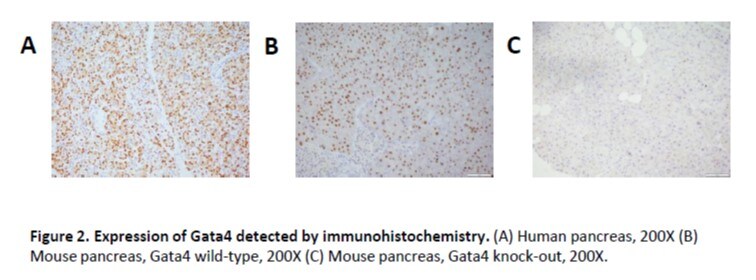
Application: ImmunohistochemistrySample Tested: Pancreas tissueSpecies: Human and MouseVerified Customer | Posted 03/31/2017(A) Human pancreas, 200X (B) Mouse pancreas, Gata4 wild-type, 200X (C) Mouse pancreas, Gata4 knock-out, 200X.

-
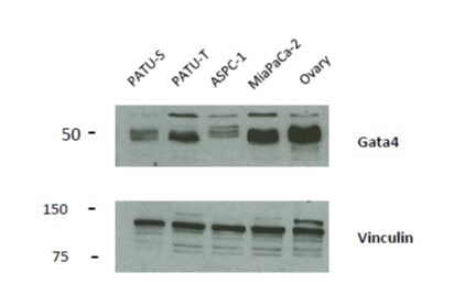
Application: Western BlotSample Tested: PDAC Cell Lines and Ovary tissueSpecies: MouseVerified Customer | Posted 03/31/201775ug of protein from PDAC cell lines and 50 ug of protein from mouse ovary were loaded and incubated overnight at 1:1000 dilution with Gata4 antibody (AF2606). HRP-conjugated rabbit Anti-Goat IgG Secondary Antibody Rabbit was used. Vinculin was used as a loading control.
-
Application: ImmunofluorescenceSample Tested: See PMID 23658023Species: HumanVerified Customer | Posted 01/07/2015
There are no reviews that match your criteria.
Protocols
Find general support by application which include: protocols, troubleshooting, illustrated assays, videos and webinars.
- Appropriate Fixation of IHC/ICC Samples
- Cellular Response to Hypoxia Protocols
- ChIP Protocol Video
- Chromatin Immunoprecipitation (ChIP) Protocol
- Chromatin Immunoprecipitation Protocol
- ClariTSA™ Fluorophore Kits
- Detection & Visualization of Antibody Binding
- ICC Cell Smear Protocol for Suspension Cells
- ICC Immunocytochemistry Protocol Videos
- ICC for Adherent Cells
- Immunocytochemistry (ICC) Protocol
- Immunocytochemistry Troubleshooting
- Immunofluorescence of Organoids Embedded in Cultrex Basement Membrane Extract
- Immunohistochemistry (IHC) and Immunocytochemistry (ICC) Protocols
- Preparing Samples for IHC/ICC Experiments
- Preventing Non-Specific Staining (Non-Specific Binding)
- Primary Antibody Selection & Optimization
- Protocol for VisUCyte™ HRP Polymer Detection Reagent
- Protocol for the Fluorescent ICC Staining of Cell Smears - Graphic
- Protocol for the Fluorescent ICC Staining of Cultured Cells on Coverslips - Graphic
- Protocol for the Preparation and Fluorescent ICC Staining of Cells on Coverslips
- Protocol for the Preparation and Fluorescent ICC Staining of Non-adherent Cells
- Protocol for the Preparation and Fluorescent ICC Staining of Stem Cells on Coverslips
- Protocol for the Preparation of a Cell Smear for Non-adherent Cell ICC - Graphic
- R&D Systems Quality Control Western Blot Protocol
- TUNEL and Active Caspase-3 Detection by IHC/ICC Protocol
- The Importance of IHC/ICC Controls
- Troubleshooting Guide: Western Blot Figures
- Western Blot Conditions
- Western Blot Protocol
- Western Blot Protocol for Cell Lysates
- Western Blot Troubleshooting
- Western Blot Troubleshooting Guide
- View all Protocols, Troubleshooting, Illustrated assays and Webinars

