
Key Product Details
Species Reactivity
Human
Applications
Immunohistochemistry, Western Blot
Label
Unconjugated
Antibody Source
Monoclonal Mouse IgG2B Clone # 714729
Loading...
Product Specifications
Immunogen
S. frugiperda insect ovarian cell line Sf 21-derived recombinant human Hexosaminidase A/HEXA
Met1-Thr529
Accession # P06865
Met1-Thr529
Accession # P06865
Specificity
Detects human Hexosaminidase A/HEXA in direct ELISAs and Western blots.
Clonality
Monoclonal
Host
Mouse
Isotype
IgG2B
Scientific Data Images for Human Hexosaminidase A/HEXA Antibody
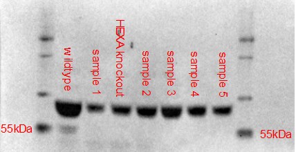
Detection of Human Hexosaminidase A/HEXA by Western Blot.
Western blot shows lysates of HepG2 human hepatocellular carcinoma cell line and human liver tissue. PVDF membrane was probed with 2 µg/mL of Mouse Anti-Human Hexosaminidase A/HEXA Monoclonal Antibody (Catalog # MAB6237) followed by HRP-conjugated Anti-Mouse IgG Secondary Antibody (Catalog # HAF007). A specific band was detected for Hexosaminidase A/HEXA at approximately 60 kDa (as indicated). This experiment was conducted under reducing conditions and using Immunoblot Buffer Group 1.
Hexosaminidase A/HEXA in Human Brain.
Hexosaminidase A/HEXA was detected in immersion fixed paraffin-embedded sections of human brain (hypothalamus) using Mouse Anti-Human Hexosaminidase A/HEXA Monoclonal Antibody (Catalog # MAB6237) at 15 µg/mL overnight at 4 °C. Before incubation with the primary antibody, tissue was subjected to heat-induced epitope retrieval using Antigen Retrieval Reagent-Basic (Catalog # CTS013). Tissue was stained using the Anti-Mouse HRP-DAB Cell & Tissue Staining Kit (brown; Catalog # CTS002) and counterstained with hematoxylin (blue). Specific staining was localized to the cytoplasm and lysosomes in neuronal cell bodies. View our protocol for Chromogenic IHC Staining of Paraffin-embedded Tissue Sections.Applications for Human Hexosaminidase A/HEXA Antibody
Application
Recommended Usage
Immunohistochemistry
8-25 µg/mL
Sample: Immersion fixed paraffin-embedded sections of human brain (hypothalamus)
Sample: Immersion fixed paraffin-embedded sections of human brain (hypothalamus)
Western Blot
2 µg/mL
Sample: HepG2 human hepatocellular carcinoma cell line and human liver tissue
Sample: HepG2 human hepatocellular carcinoma cell line and human liver tissue
Reviewed Applications
Read 1 review rated 3 using MAB6237 in the following applications:
Formulation, Preparation, and Storage
Purification
Protein A or G purified from hybridoma culture supernatant
Reconstitution
Sterile PBS to a final concentration of 0.5 mg/mL. For liquid material, refer to CoA for concentration.
Loading...
Formulation
Lyophilized from a 0.2 μm filtered solution in PBS with Trehalose. *Small pack size (SP) is supplied either lyophilized or as a 0.2 µm filtered solution in PBS.
Shipping
Lyophilized product is shipped at ambient temperature. Liquid small pack size (-SP) is shipped with polar packs. Upon receipt, store immediately at the temperature recommended below.
Stability & Storage
Use a manual defrost freezer and avoid repeated freeze-thaw cycles.
- 12 months from date of receipt, -20 to -70 °C as supplied.
- 1 month, 2 to 8 °C under sterile conditions after reconstitution.
- 6 months, -20 to -70 °C under sterile conditions after reconstitution.
Calculators
Background: Hexosaminidase A/HEXA
References
- Gilbert, F. et al. (1975) Proc. Natl. Acad. Sci. USA 72:263.
- Myerowitz, R. et al. (1985) Proc. Natl. Acad. Sci. USA 82:7830.
- Korneluk, R.G. et al. (1986) J. Biol. Chem. 261:8407.
- Mark, B.L. et al. (2003) J. Mol. Biol. 327:1093.
- Mahuran, D.J. et al. (1988) J. Biol. Chem. 263:4612.
- Hepbildikler, S.T. et al. (2002) J. Biol. Chem. 277:2562.
- Mahuran, D.J. (1991) Biochim. Biophys. Acta 1096:87.
- Mencarelli, S. et al. (2005) FEBS Lett. 579:5501.
- Neufeld, E.F. (1989) J. Biol. Chem. 264:10927.
- Ohno, K. et al. (2008) Mol. Genet. Metab. 94:462.
Alternate Names
HEXA, TSD
Entrez Gene IDs
3073 (Human)
Gene Symbol
HEXA
UniProt
Additional Hexosaminidase A/HEXA Products
Product Documents for Human Hexosaminidase A/HEXA Antibody
Certificate of Analysis
To download a Certificate of Analysis, please enter a lot or batch number in the search box below.
Note: Certificate of Analysis not available for kit components.
Product Specific Notices for Human Hexosaminidase A/HEXA Antibody
For research use only
Related Research Areas
Customer Reviews for Human Hexosaminidase A/HEXA Antibody (1)
3 out of 5
1 Customer Rating
Have you used Human Hexosaminidase A/HEXA Antibody?
Submit a review and receive an Amazon gift card!
$25/€18/£15/$25CAN/¥2500 Yen for a review with an image
$10/€7/£6/$10CAN/¥1110 Yen for a review without an image
Submit a review
Customer Images
Showing
1
-
1 of
1 review
Showing All
Filter By:
-
Application: Non-specific stainingSample Tested: HEK293T human embryonic kidney cell lineSpecies: HumanVerified Customer | Posted 01/30/2023Non-specific staining, expected HEXA band not detected unless membrane is covered/cut to remove molecular weight of non-specific bands.

There are no reviews that match your criteria.
Protocols
Find general support by application which include: protocols, troubleshooting, illustrated assays, videos and webinars.
- Antigen Retrieval Protocol (PIER)
- Antigen Retrieval for Frozen Sections Protocol
- Appropriate Fixation of IHC/ICC Samples
- Cellular Response to Hypoxia Protocols
- Chromogenic IHC Staining of Formalin-Fixed Paraffin-Embedded (FFPE) Tissue Protocol
- Chromogenic Immunohistochemistry Staining of Frozen Tissue
- ClariTSA™ Fluorophore Kits
- Detection & Visualization of Antibody Binding
- Fluorescent IHC Staining of Frozen Tissue Protocol
- Graphic Protocol for Heat-induced Epitope Retrieval
- Graphic Protocol for the Preparation and Fluorescent IHC Staining of Frozen Tissue Sections
- Graphic Protocol for the Preparation and Fluorescent IHC Staining of Paraffin-embedded Tissue Sections
- Graphic Protocol for the Preparation of Gelatin-coated Slides for Histological Tissue Sections
- IHC Sample Preparation (Frozen sections vs Paraffin)
- Immunofluorescent IHC Staining of Formalin-Fixed Paraffin-Embedded (FFPE) Tissue Protocol
- Immunohistochemistry (IHC) and Immunocytochemistry (ICC) Protocols
- Immunohistochemistry Frozen Troubleshooting
- Immunohistochemistry Paraffin Troubleshooting
- Preparing Samples for IHC/ICC Experiments
- Preventing Non-Specific Staining (Non-Specific Binding)
- Primary Antibody Selection & Optimization
- Protocol for Heat-Induced Epitope Retrieval (HIER)
- Protocol for Making a 4% Formaldehyde Solution in PBS
- Protocol for VisUCyte™ HRP Polymer Detection Reagent
- Protocol for the Preparation & Fixation of Cells on Coverslips
- Protocol for the Preparation and Chromogenic IHC Staining of Frozen Tissue Sections
- Protocol for the Preparation and Chromogenic IHC Staining of Frozen Tissue Sections - Graphic
- Protocol for the Preparation and Chromogenic IHC Staining of Paraffin-embedded Tissue Sections
- Protocol for the Preparation and Chromogenic IHC Staining of Paraffin-embedded Tissue Sections - Graphic
- Protocol for the Preparation and Fluorescent IHC Staining of Frozen Tissue Sections
- Protocol for the Preparation and Fluorescent IHC Staining of Paraffin-embedded Tissue Sections
- Protocol for the Preparation of Gelatin-coated Slides for Histological Tissue Sections
- R&D Systems Quality Control Western Blot Protocol
- TUNEL and Active Caspase-3 Detection by IHC/ICC Protocol
- The Importance of IHC/ICC Controls
- Troubleshooting Guide: Immunohistochemistry
- Troubleshooting Guide: Western Blot Figures
- Western Blot Conditions
- Western Blot Protocol
- Western Blot Protocol for Cell Lysates
- Western Blot Troubleshooting
- Western Blot Troubleshooting Guide
- View all Protocols, Troubleshooting, Illustrated assays and Webinars
Loading...