
The interleukin 17 (IL-17) family of cytokines, comprising six members (IL-17, IL-17B through IL-17F), are structurally related proteins with a conserved cysteine-knot structure. These proinflammatory cytokines can induce local cytokine productions and are involved in the regulation of the immune response. The cognate receptors activated by some of these cytokines have been identified (1). Interleukin-17 B Receptor (IL-17 RB), also known as IL-17Rh1, IL-17ER and EVI27, represents the second receptor of the IL-17 family of cytokines to be recognized (2-4). Human IL-17 RB cDNA encodes a 502 amino acid (aa) residue type I membrane protein with a putative 17 aa signal peptide, a 275 aa extracellular domain, a 21 aa transmembrane domain and a 189 aa cytoplasmic tail. By alternative splicing, a secreted variant of IL-17 RB has also been identified (4). Human and mouse IIL-17 RB share 76% aa sequence identity. The human IL-17 RB protein sequence is only 19.2% identical to the human IL-17 R sequence, but the two receptors share many conserved cysteine residues within their extracellular domains as well as additional conserved elements within their cytoplasmic domains. Three additional type I transmembrane receptors with homology to IL-17 R have been reported, increasing the number of the IL-17 R family members to five (5, 6). By Northern blot analysis, human IL-17 RB is highly expressed in kidneys and liver but is expressed at lower levels in testes, brain, small intestine and other endocrine tissues (2-4). The expression of IL-17 RB is significantly up-regulated under inflammatory conditions. IL-17 RB binds strongly to IL-17E and weakly to IL-17B. It does not bind IL-17, IL-17C, and IL-17F. Activation of IL-17 RB by its ligands results in the activation of nuclear factor kappa-B (2-4).
Human IL-17RB Antibody (938975)
R&D Systems | Catalog # MAB12072

Scientific images may be resized, cropped, or adjusted for brightness or contrast for presentation purposes. These updates are limited to presentation and do not affect the underlying scientific interpretation of the data. Where available, additional source data may be provided upon request.
Key Product Details
Species Reactivity
Validated:
Cited:
Applications
Validated:
Cited:
Label
Antibody Source
Product Specifications
Immunogen
Arg18-Gly289
Accession # Q9NRM6
Specificity
Clonality
Host
Isotype
Scientific Data Images for Human IL-17RB Antibody (938975)
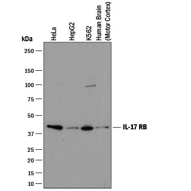
Detection of Human IL‑17 RB by Western Blot.
Western blot shows lysates of HeLa human cervical epithelial carcinoma cell line, HepG2 human hepatocellular carcinoma cell line, K562 human chronic myelogenous leukemia cell line, and human brain (motor cortex) tissue. PVDF membrane was probed with 2 µg/mL of Mouse Anti-Human IL-17 RB Monoclonal Antibody (Catalog # MAB12072) followed by HRP-conjugated Anti-Mouse IgG Secondary Antibody (Catalog # HAF018). A specific band was detected for IL-17 RB at approximately 46 kDa (as indicated). This experiment was conducted under reducing conditions and using Immunoblot Buffer Group 1.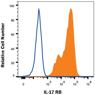
Detection of IL‑17 RB in HEK293 Human Cell Line Transfected with Human IL-17 RB by Flow Cytometry.
HEK293 human embryonic kidney cell line transfected with human IL-17 RB was stained with Mouse Anti-Human IL-17 RB Monoclonal Antibody (Catalog # MAB12072, filled histogram) or isotype control antibody (Catalog # MAB002, open histogram), followed by Phycoerythrin-conjugated Anti-Mouse IgG Secondary Antibody (Catalog # F0102B). View our protocol for Staining Membrane-associated Proteins.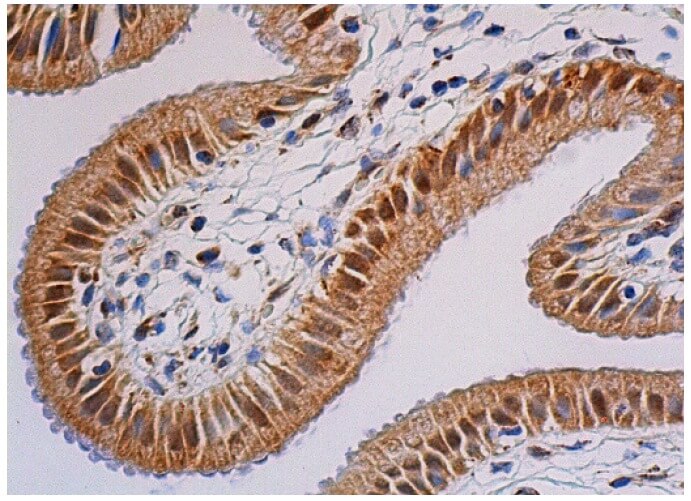
Detection of Human IL-17 RB antibody by Immunohistochemistry.
Immunoperoxidase staining of formalin-fixed, paraffin-embedded human gallbladder tissue showing nuclear and cytoplasmic staining of glandular cells.Applications for Human IL-17RB Antibody (938975)
Flow Cytometry
Sample: HEK293 human embryonic kidney cell line transfected with human IL-17 RB
Western Blot
Sample: HeLa human cervical epithelial carcinoma cell line, HepG2 human hepatocellular carcinoma cell line, K562 human chronic myelogenous leukemia cell line, and human brain (motor cortex) tissue
Flow Cytometry Panel Builder
Bio-Techne Knows Flow Cytometry
Save time and reduce costly mistakes by quickly finding compatible reagents using the Panel Builder Tool.
Advanced Features
- Spectra Viewer - Custom analysis of spectra from multiple fluorochromes
- Spillover Popups - Visualize the spectra of individual fluorochromes
- Antigen Density Selector - Match fluorochrome brightness with antigen density

Formulation, Preparation, and Storage
Purification
Reconstitution
Formulation
Shipping
Stability & Storage
- 12 months from date of receipt, -20 to -70 °C as supplied.
- 1 month, 2 to 8 °C under sterile conditions after reconstitution.
- 6 months, -20 to -70 °C under sterile conditions after reconstitution.
Calculators
Background: IL-17RB
References
- Aggarwal, S. and A.L. Gurney (2002) J. Leukoc. Biol. 71:1.
- Shi Y, et al. (2000) J. Biol. Chem. 275:19167.
- Lee, J, et al. (2001) J. Biol. Chem. 276:1660.
- Tian E, et al. (2000) Oncogene 19:2098.
- Haudenschild, D. et al. (2002) J. Biol. Chem. 277:4309.
- Hurst, S.D. et al. (2002) J. Immunol. 169:443.
Long Name
Alternate Names
Gene Symbol
UniProt
Additional IL-17RB Products
Product Documents for Human IL-17RB Antibody (938975)
Certificate of Analysis
To download a Certificate of Analysis, please enter a lot or batch number in the search box below.
Note: Certificate of Analysis not available for kit components.
Product Specific Notices for Human IL-17RB Antibody (938975)
For research use only
Related Research Areas
Citations for Human IL-17RB Antibody (938975)
 Powered by Bioz
Customer Reviews for Human IL-17RB Antibody (938975)
There are currently no reviews for this product. Be the first to review Human IL-17RB Antibody (938975) and earn rewards!
Have you used Human IL-17RB Antibody (938975)?
Submit a review and receive an Amazon gift card!
$25/€18/£15/$25CAN/¥2500 for a review with an image
$10/€7/£6/$10CAN/¥1110 for a review without an image
Submit a review
Protocols
Find general support by application which include: protocols, troubleshooting, illustrated assays, videos and webinars.
- 7-Amino Actinomycin D (7-AAD) Cell Viability Flow Cytometry Protocol
- Cellular Response to Hypoxia Protocols
- Extracellular Membrane Flow Cytometry Protocol
- Flow Cytometry Protocol for Cell Surface Markers
- Flow Cytometry Protocol for Staining Membrane Associated Proteins
- Flow Cytometry Staining Protocols
- Flow Cytometry Troubleshooting Guide
- Intracellular Flow Cytometry Protocol Using Alcohol (Methanol)
- Intracellular Flow Cytometry Protocol Using Detergents
- Intracellular Nuclear Staining Flow Cytometry Protocol Using Detergents
- Intracellular Staining Flow Cytometry Protocol Using Alcohol Permeabilization
- Intracellular Staining Flow Cytometry Protocol Using Detergents to Permeabilize Cells
- Propidium Iodide Cell Viability Flow Cytometry Protocol
- Protocol for Liperfluo
- Protocol for the Characterization of Human Th22 Cells
- Protocol for the Characterization of Human Th9 Cells
- Protocol: Annexin V and PI Staining by Flow Cytometry
- Protocol: Annexin V and PI Staining for Apoptosis by Flow Cytometry
- R&D Systems Quality Control Western Blot Protocol
- Troubleshooting Guide: Fluorokine Flow Cytometry Kits
- Troubleshooting Guide: Western Blot Figures
- Western Blot Conditions
- Western Blot Protocol
- Western Blot Protocol for Cell Lysates
- Western Blot Troubleshooting
- Western Blot Troubleshooting Guide
- View all Protocols, Troubleshooting, Illustrated assays and Webinars
Associated Pathways


