Delta-like protein 4 (DLL4) is a type I membrane protein belonging to the Delta/Serrate/Lag2 (DSL) family of Notch ligands (1). Notch signaling is an evolutionarily conserved pathway that controls cell fate and is required in multiple developmental processes including vascular development, hematopoiesis, somatogenesis, myogenesis, and neurogenesis (2-4). Dysregulation in the Notch pathway is associated with various human diseases. In mammals, four Notch homologs (Notch 1 to 4) and five ligands (DLL 1, 3 and 4, Jagged 1 and 2) have been identified. Notch ligands are transmembrane proteins with a DSL motif necessary for Notch binding, tandem EGF repeats, a transmembrane region and a short intracellular domain (ICD). Notch ligands are categorized into two subfamilies based on the presence of an extracellular cysteine-rich domain and insertions that interrupt some EGF repeats in the Jagged but not the Delta ligand family. Interactions of Notch receptors with their ligands results in reciprocal regulated intramembrane proteolysis (RIP) (4). RIP is a mechanism for transmembrane signal transduction that involves the sequential processing by a disintegrin metalloprotease (ADAM) and then by presenilin/ gamma secretase, resulting in shedding of the extracellular domains and the generation of the soluble ICD signaling fragments, respectively. The Notch ICD translocates to the nucleus and interacts with transcriptional coactivators, resulting in the transcription of target genes. The ICDs of the Notch ligands have also been shown to translocate to the nucleus where they may have a signaling function (5, 6). DLL4 is expressed highly and selectively within the arterial endothelium and has been shown to function as a ligand for Notch 1 and Notch 4. Human and mouse DLL4 share 86% amino acid sequence identity (1).

Key Product Details
Species Reactivity
Validated:
Human, Mouse
Cited:
Human, Mouse
Applications
Validated:
Immunohistochemistry, Western Blot
Cited:
Immunohistochemistry, Immunohistochemistry-Frozen, Western Blot, Immunocytochemistry
Label
Unconjugated
Antibody Source
Monoclonal Rat IgG2A Clone # 207822
Loading...
Product Specifications
Immunogen
Mouse myeloma cell line NS0-derived recombinant mouse DLL4
Ser28-Pro525
Accession # Q9JI71
Ser28-Pro525
Accession # Q9JI71
Specificity
Detects mouse DLL4 in direct ELISAs and Western blots. In direct ELISAs and Western blots, this antibody shows 100% cross-reactivity with recombinant human DLL4.
Clonality
Monoclonal
Host
Rat
Isotype
IgG2A
Scientific Data Images for DLL4 Antibody (207822)
DLL4 in Mouse Embryonic Heart.
DLL4 was detected in immersion fixed frozen sections of mouse embryonic heart using Rat Anti-Human/Mouse DLL4 Monoclonal Antibody (Catalog # MAB1389) at 8 µg/mL overnight at 4 °C. Tissue was stained using the Anti-Rat HRP-DAB Cell & Tissue Staining Kit (brown; Catalog # CTS017) and counterstained with hematoxylin (blue). Specific staining was localized to developing cardiomyocytes. View our protocol for Chromogenic IHC Staining of Frozen Tissue Sections.
Detection of DLL4 by Western Blot
mDia1is essential for CCN1-induced Cdc42 activation in tip cell formation.(A) HUVECs were treated with PBS (Control) or CCN1 (10 ng/mL) for 24 hr, and total RNA was used for the detection of DIAPH1 (mDia1) mRNA expression by qRT-PCR. *p<0.001 vs. Control. (B) After transfection with DIAPH1 WT full length and DIAPH1 double-negative mutant plasmids, HUVECs were treated with CCN1 and immunostained with anti-vinculin antibody (red) and phalloidin (green) to visualise filopodia. Scale bar = 10 µm. (C, D) HUVECs transfected with DIAPH1 WT full length and DIAPH1 double-negative mutant plasmids were treated with CCN1 (10 ng/mL) for 30 min, and active Cdc42 was assessed by western blotting analysis (C) and visualised by p-N-Wasp (D). Fold changes were noted under each protein band. (E) After HUVECs were transfected with DIAPH1 WT full length and DIAPH1 DN3 mutant plasmid, starved cells for 16 hr were treated with CCN1 10 ng/ml for 1 hr and detected YAP/TAZ by IF. Scale bar = 100 µm.10.7554/eLife.46012.012Figure 4—source data 1.Source data for Figure 4C.Source data for Figure 4C. Image collected and cropped by CiteAb from the following open publication (https://pubmed.ncbi.nlm.nih.gov/31429823), licensed under a CC-BY license. Not internally tested by R&D Systems.
Detection of DLL4 by Western Blot
CCN1 stimulates tip cell activity in ECs. (G) Western blotting analysis of VEGFR2, DLL4, and SOX17 protein levels in the same samples as used in F. beta -actin was used as an internal control. Fold increase was indicated as a number below each band. (H) Luciferase assays in HUVECs transfected with DLL4-Luc reporter vector and treated with CCN1 or VEGF (10 ng/mL) for 24 hr. Luciferase activity was normalised to Renilla luminescence. *p<0.001 vs. Control.10.7554/eLife.46012.005Figure 2—source data 1.Source data for Figure 2G.Source data for Figure 2G. Image collected and cropped by CiteAb from the following open publication (https://pubmed.ncbi.nlm.nih.gov/31429823), licensed under a CC-BY license. Not internally tested by R&D Systems.Applications for DLL4 Antibody (207822)
Application
Recommended Usage
Immunohistochemistry
5-25 µg/mL
Sample: Immersion fixed frozen sections of mouse embryonic heart
Sample: Immersion fixed frozen sections of mouse embryonic heart
Western Blot
1 µg/mL
Sample: Recombinant Mouse DLL4 (Catalog # 1389-D4)
Sample: Recombinant Mouse DLL4 (Catalog # 1389-D4)
Reviewed Applications
Read 3 reviews rated 4.3 using MAB1389 in the following applications:
Formulation, Preparation, and Storage
Purification
Protein A or G purified from hybridoma culture supernatant
Reconstitution
Reconstitute at 0.5 mg/mL in sterile PBS. For liquid material, refer to CoA for concentration.
Loading...
Formulation
Lyophilized from a 0.2 μm filtered solution in PBS with Trehalose. *Small pack size (SP) is supplied either lyophilized or as a 0.2 µm filtered solution in PBS.
Shipping
Lyophilized product is shipped at ambient temperature. Liquid small pack size (-SP) is shipped with polar packs. Upon receipt, store immediately at the temperature recommended below.
Stability & Storage
Use a manual defrost freezer and avoid repeated freeze-thaw cycles.
- 12 months from date of receipt, -20 to -70 °C as supplied.
- 1 month, 2 to 8 °C under sterile conditions after reconstitution.
- 6 months, -20 to -70 °C under sterile conditions after reconstitution.
Calculators
Background: DLL4
References
- Shutter, J.R. et al. (2000) Genes Dev. 14:1313.
- Iso, T. et al. (2002) Arterioscler. Thromb. Vasc. Biol. 23:543.
- Walker, L. et al. (2001) Stem Cells 19:543.
- Baron, M. (2002) Semin. Cell Dev. Biol. 14:113.
- Ikeuchi, T. and S.S. Sisodia (2003) J. Biol. Chem. 278:7751.
- Bland, C.E. et al. (2003) J. Biol. Chem. 278:13607.
Long Name
Delta-like 4
Alternate Names
delta 4, Delta 4 precursor, delta ligand 4, delta4, delta-like 4 (Drosophila), delta-like 4 homolog, delta-like 4 homolog (Drosophila), delta-like 4 protein, delta-like protein 4, Drosophila Delta homolog 4, hdelta2, MGC126344, notch ligand delta-2, notch ligand DLL4
Gene Symbol
DLL4
UniProt
Additional DLL4 Products
Product Documents for DLL4 Antibody (207822)
Certificate of Analysis
To download a Certificate of Analysis, please enter a lot or batch number in the search box below.
Note: Certificate of Analysis not available for kit components.
Product Specific Notices for DLL4 Antibody (207822)
For research use only
Related Research Areas
Citations for DLL4 Antibody (207822)
 Powered by Bioz
Customer Reviews for DLL4 Antibody (207822) (3)
4.3 out of 5
3 Customer Ratings
Have you used DLL4 Antibody (207822)?
Submit a review and receive an Amazon gift card!
$25/€18/£15/$25CAN/¥2500 Yen for a review with an image
$10/€7/£6/$10CAN/¥1110 Yen for a review without an image
Submit a review
Customer Images
Showing
1
-
3 of
3 reviews
Showing All
Filter By:
-
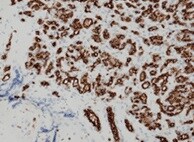
Application: ImmunohistochemistrySample Tested: Placental tissueSpecies: HumanVerified Customer | Posted 04/08/2022

-
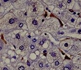
Application: ImmunohistochemistrySample Tested: Liver tissueSpecies: HumanVerified Customer | Posted 09/03/2021
-
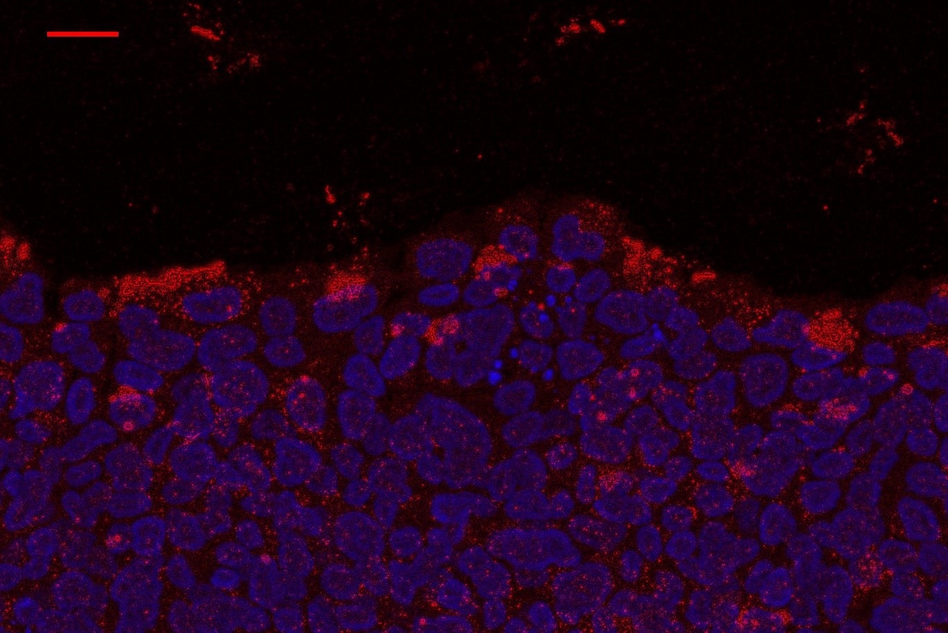
Application: Immunocytochemistry/ImmunofluorescenceSample Tested: Bladder cancer tissueSpecies: HumanVerified Customer | Posted 12/10/2017
There are no reviews that match your criteria.
Protocols
Find general support by application which include: protocols, troubleshooting, illustrated assays, videos and webinars.
- Antigen Retrieval Protocol (PIER)
- Antigen Retrieval for Frozen Sections Protocol
- Appropriate Fixation of IHC/ICC Samples
- Cellular Response to Hypoxia Protocols
- Chromogenic IHC Staining of Formalin-Fixed Paraffin-Embedded (FFPE) Tissue Protocol
- Chromogenic Immunohistochemistry Staining of Frozen Tissue
- ClariTSA™ Fluorophore Kits
- Detection & Visualization of Antibody Binding
- Fluorescent IHC Staining of Frozen Tissue Protocol
- Graphic Protocol for Heat-induced Epitope Retrieval
- Graphic Protocol for the Preparation and Fluorescent IHC Staining of Frozen Tissue Sections
- Graphic Protocol for the Preparation and Fluorescent IHC Staining of Paraffin-embedded Tissue Sections
- Graphic Protocol for the Preparation of Gelatin-coated Slides for Histological Tissue Sections
- IHC Sample Preparation (Frozen sections vs Paraffin)
- Immunofluorescent IHC Staining of Formalin-Fixed Paraffin-Embedded (FFPE) Tissue Protocol
- Immunohistochemistry (IHC) and Immunocytochemistry (ICC) Protocols
- Immunohistochemistry Frozen Troubleshooting
- Immunohistochemistry Paraffin Troubleshooting
- Preparing Samples for IHC/ICC Experiments
- Preventing Non-Specific Staining (Non-Specific Binding)
- Primary Antibody Selection & Optimization
- Protocol for Heat-Induced Epitope Retrieval (HIER)
- Protocol for Making a 4% Formaldehyde Solution in PBS
- Protocol for VisUCyte™ HRP Polymer Detection Reagent
- Protocol for the Preparation & Fixation of Cells on Coverslips
- Protocol for the Preparation and Chromogenic IHC Staining of Frozen Tissue Sections
- Protocol for the Preparation and Chromogenic IHC Staining of Frozen Tissue Sections - Graphic
- Protocol for the Preparation and Chromogenic IHC Staining of Paraffin-embedded Tissue Sections
- Protocol for the Preparation and Chromogenic IHC Staining of Paraffin-embedded Tissue Sections - Graphic
- Protocol for the Preparation and Fluorescent IHC Staining of Frozen Tissue Sections
- Protocol for the Preparation and Fluorescent IHC Staining of Paraffin-embedded Tissue Sections
- Protocol for the Preparation of Gelatin-coated Slides for Histological Tissue Sections
- R&D Systems Quality Control Western Blot Protocol
- TUNEL and Active Caspase-3 Detection by IHC/ICC Protocol
- The Importance of IHC/ICC Controls
- Troubleshooting Guide: Immunohistochemistry
- Troubleshooting Guide: Western Blot Figures
- Western Blot Conditions
- Western Blot Protocol
- Western Blot Protocol for Cell Lysates
- Western Blot Troubleshooting
- Western Blot Troubleshooting Guide
- View all Protocols, Troubleshooting, Illustrated assays and Webinars
Loading...
Associated Pathways
