Integrin alpha 11 (ITGA11) is a 150 kDa transmembrane glycoprotein that associates with the Integrin beta 1/CD29 chain to form a receptor for collagen I and IX. ITGA11 is most highly expressed in adult cardiac and uterine smooth muscle and developing myocytes. The extracellular region contains seven FG-GAP repeats and one VWF‑C domain. Within the extracellular domain, human and mouse ITGA11 share 90% aa sequence identity.
Integrin alpha 11 Antibody (396214)
R&D Systems | Catalog # MAB4235

Key Product Details
Validated by
Knockout/Knockdown
Species Reactivity
Validated:
Human, Mouse
Cited:
Human, Mouse, Rat
Applications
Validated:
Western Blot
Cited:
Western Blot, Flow Cytometry, Control
Label
Unconjugated
Antibody Source
Monoclonal Rat IgG1 Clone # 396214
Loading...
Product Specifications
Immunogen
Chinese hamster ovary cell line CHO-derived recombinant human Integrin alpha 11
Phe23-Pro1142 (Leu972Pro, Val1030 del)
Accession # EAW77820
Phe23-Pro1142 (Leu972Pro, Val1030 del)
Accession # EAW77820
Specificity
Detects human and mouse Integrin alpha 11 in direct ELISAs and Western blots. In direct ELISAs, does not cross-react with recombinant human Integrin alpha 2, alpha 10, alpha X, or alpha L.
Clonality
Monoclonal
Host
Rat
Isotype
IgG1
Scientific Data Images for Integrin alpha 11 Antibody (396214)
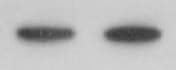
Detection of Human Integrin alpha 11 by Western Blot.
Western blot shows lysates of HeLa human cervical epithelial carcinoma cell line and human heart tissue. PVDF membrane was probed with 2 µg/mL of Human/Mouse Integrin a11 Monoclonal Antibody (Catalog # MAB4235) followed by HRP-conjugated Anti-Rat IgG Secondary Antibody (Catalog # HAF005). A specific band was detected for Integrin a11 at approximately 150 kDa (as indicated). This experiment was conducted under non-reducing conditions and using Immunoblot Buffer Group 1.
Detection of Mouse Integrin alpha 11 by Western Blot
YAP-1 and MYL9 mediate pro-fibrotic aspects of liver fibrosis in activated HSCs.(a,b) Quantified increase in total MYL9 and YAP-1 protein levels on activation of rat HSCs from n≥3 experiments (a) with representative immunoblot shown in b. (c–e) Total YAP-1 and MYL9 are diminished in activated mouse HSCs (‘Control') following integrin beta-1 loss (‘Itgb1-null'). Quantification from n=3 experiments shown in c with representative immunoblot in d. In the immunofluorescence in e, note the rounded inactivated appearance of the Itgb1-null cells. The remaining total YAP-1 signal is more cytoplasmic (see h,i). Scale bar, 50 μm. (f,g) Itga11 knockdown in activated rat HSCs using two different siRNA oligos. Data for each oligo are shown relative to its own scrambled control in either black or grey (n=3 for each). (f) Detection of Myl9 transcripts was diminished to an almost identical extent as for Itga11. (g) Protein detection of MYL9 and total YAP-1 was also diminished following Itga11 knockdown. (h,i) The proportion of phosphorylated YAP (PYAP, inactive form), is increased following integrin beta-1 loss (‘Itgb1-null') from activated mouse HSCs (‘Control'; n=3 experiments) and localises more predominantly to the cytoplasm (i). DAPI, blue nuclear counterstain, is shown. Scale bar, 50 μm. (j) Luciferase activity (in relative light units; RLU) following co-transfection of constructs containing the wild-type (MYL9-TEAD) or mutated (MYL9-delta TEAD) TEAD motif from the 3′-untranslated region of the MYL9 gene with empty vector (Control) or YAP expression vector. Results are normalized to a Renilla vector and expressed relative to the control MYL9 luciferase construct without YAP. (k) Transcript levels by qRT–PCR following inhibition of YAP-TEAD interaction using VP in activated rat HSCs expressed relative to DMSO control. Two-tailed unpaired t-test was used for statistical analysis. Data are shown as means±s.e.m. *P<0.05, **P<0.01, †P<0.005, ‡P<0.001. Image collected and cropped by CiteApplications for Integrin alpha 11 Antibody (396214)
Application
Recommended Usage
Western Blot
2 µg/mL
Sample: HeLa human cervical epithelial carcinoma cell line and human heart tissue under non-reducing conditions
Sample: HeLa human cervical epithelial carcinoma cell line and human heart tissue under non-reducing conditions
Reviewed Applications
Read 1 review rated 5 using MAB4235 in the following applications:
Formulation, Preparation, and Storage
Purification
Protein A or G purified from hybridoma culture supernatant
Reconstitution
Reconstitute at 0.5 mg/mL in sterile PBS. For liquid material, refer to CoA for concentration.
Loading...
Formulation
Lyophilized from a 0.2 μm filtered solution in PBS with Trehalose. *Small pack size (SP) is supplied either lyophilized or as a 0.2 µm filtered solution in PBS.
Shipping
Lyophilized product is shipped at ambient temperature. Liquid small pack size (-SP) is shipped with polar packs. Upon receipt, store immediately at the temperature recommended below.
Stability & Storage
Use a manual defrost freezer and avoid repeated freeze-thaw cycles.
- 12 months from date of receipt, -20 to -70 °C as supplied.
- 1 month, 2 to 8 °C under sterile conditions after reconstitution.
- 6 months, -20 to -70 °C under sterile conditions after reconstitution.
Calculators
Background: Integrin alpha 11
Alternate Names
ITGA11
Gene Symbol
ITGA11
UniProt
Additional Integrin alpha 11 Products
Product Documents for Integrin alpha 11 Antibody (396214)
Certificate of Analysis
To download a Certificate of Analysis, please enter a lot or batch number in the search box below.
Note: Certificate of Analysis not available for kit components.
Product Specific Notices for Integrin alpha 11 Antibody (396214)
For research use only
Related Research Areas
Citations for Integrin alpha 11 Antibody (396214)
 Powered by Bioz
Customer Reviews for Integrin alpha 11 Antibody (396214) (1)
5 out of 5
1 Customer Rating
Have you used Integrin alpha 11 Antibody (396214)?
Submit a review and receive an Amazon gift card!
$25/€18/£15/$25CAN/¥2500 Yen for a review with an image
$10/€7/£6/$10CAN/¥1110 Yen for a review without an image
Submit a review
Customer Images
Showing
1
-
1 of
1 review
Showing All
Filter By:
-
Application: Western BlotSample Tested: Heart tissueSpecies: MouseVerified Customer | Posted 12/06/2021Heart tissue lysates

There are no reviews that match your criteria.
Protocols
Find general support by application which include: protocols, troubleshooting, illustrated assays, videos and webinars.
- Cellular Response to Hypoxia Protocols
- R&D Systems Quality Control Western Blot Protocol
- Troubleshooting Guide: Western Blot Figures
- Western Blot Conditions
- Western Blot Protocol
- Western Blot Protocol for Cell Lysates
- Western Blot Troubleshooting
- Western Blot Troubleshooting Guide
- View all Protocols, Troubleshooting, Illustrated assays and Webinars
Loading...