
Key Product Details
Validated by
Biological Validation
Species Reactivity
Validated:
Human, Mouse
Cited:
Human, Mouse
Applications
Validated:
Western Blot
Cited:
Western Blot, Immunoprecipitation
Label
Unconjugated
Antibody Source
Monoclonal Mouse IgG2A Clone # 604421
Loading...
Product Specifications
Immunogen
E. coli-derived recombinant human MCPIP1
Asp426-Glu599
Accession # Q5D1E8
Asp426-Glu599
Accession # Q5D1E8
Specificity
Detects human MCPIP1 in direct ELISAs and human and mouse MCPIP1 in Western blots. In direct ELISAs, no cross-reactivity with recombinant human MCPIP3 is observed.
Clonality
Monoclonal
Host
Mouse
Isotype
IgG2A
Scientific Data Images for MCPIP1 Antibody (604421)
Detection of Human and Mouse MCPIP1 by Western Blot.
Western blot shows lysates of untreated (-) HeLa human cervical epithelial carcinoma cell line and RAW 264.7 mouse monocyte/macrophage cell line and THP-1 human acute monocytic leukemia cell line untreated (-) or treated (+) with 1 µg/mL LPS for 24 hours or 10 µg/mL LPS for 3 hours, respectively. PVDF membrane was probed with 1 µg/mL of Mouse Anti-Human MCPIP1 Monoclonal Antibody (Catalog # MAB7875) followed by HRP-conjugated Anti-Mouse IgG Secondary Antibody (Catalog # HAF018). A specific band was detected for MCPIP1 at approximately 70 kDa (as indicated). This experiment was conducted under reducing conditions and using Immunoblot Buffer Group 1.
Detection of Mouse MCPIP1 by Western Blot
Effects of single or combined TRAF6 KO and MALT1 paracaspase mutation on signaling in CD4+ T cells. (A) Schematic overview of the four conditional mouse strains used in the analyses. (B) Western blots showing MALT1 substrate cleavage in unstimulated purified CD4+ T cells from Wthet, T6-delta T;M1 PD-T, Traf6-delta T and Malt1 PD-T mice (two independent mice each). Asterisks indicate unspecific signals. (C) Analyses of NF-kappa B activation (EMSA), p65 phosphorylation and MALT1 substrate cleavage (Western blot, WB) in PMA/Ionomycin (P/I) stimulated purified CD4+ T cells of mice as depicted in (A). (D) Expression of ICOS on CD4+ and CD8+ T cells by flow cytometric analysis of spleen of mice as depicted in (A). Bars show the means ± SEM, and P values were calculated by one-way ANOVA with Tukey’s multiple comparison test. All analyses were performed with mice 9-11 weeks of age. Each dot represents one mouse. Ct, C-terminus; l.e., long exposure. Image collected and cropped by CiteAb from the following open publication (https://pubmed.ncbi.nlm.nih.gov/36761777), licensed under a CC-BY license. Not internally tested by R&D Systems.
Detection of Mouse MCPIP1 by Western Blot
Effects of single or combined TRAF6 KO and MALT1 paracaspase mutation on signaling in CD4+ T cells. (A) Schematic overview of the four conditional mouse strains used in the analyses. (B) Western blots showing MALT1 substrate cleavage in unstimulated purified CD4+ T cells from Wthet, T6-delta T;M1 PD-T, Traf6-delta T and Malt1 PD-T mice (two independent mice each). Asterisks indicate unspecific signals. (C) Analyses of NF-kappa B activation (EMSA), p65 phosphorylation and MALT1 substrate cleavage (Western blot, WB) in PMA/Ionomycin (P/I) stimulated purified CD4+ T cells of mice as depicted in (A). (D) Expression of ICOS on CD4+ and CD8+ T cells by flow cytometric analysis of spleen of mice as depicted in (A). Bars show the means ± SEM, and P values were calculated by one-way ANOVA with Tukey’s multiple comparison test. All analyses were performed with mice 9-11 weeks of age. Each dot represents one mouse. Ct, C-terminus; l.e., long exposure. Image collected and cropped by CiteAb from the following open publication (https://pubmed.ncbi.nlm.nih.gov/36761777), licensed under a CC-BY license. Not internally tested by R&D Systems.Applications for MCPIP1 Antibody (604421)
Application
Recommended Usage
Western Blot
1 µg/mL
Sample: Untreated HeLa human cervical epithelial carcinoma cell line and RAW 264.7 mouse monocyte/macrophage cell line and THP‑1 human acute monocytic leukemia cell line, both treated with LPS
Sample: Untreated HeLa human cervical epithelial carcinoma cell line and RAW 264.7 mouse monocyte/macrophage cell line and THP‑1 human acute monocytic leukemia cell line, both treated with LPS
Reviewed Applications
Read 2 reviews rated 5 using MAB7875 in the following applications:
Formulation, Preparation, and Storage
Purification
Protein A or G purified from hybridoma culture supernatant
Reconstitution
Sterile PBS to a final concentration of 0.5 mg/mL. For liquid material, refer to CoA for concentration.
Loading...
Formulation
Lyophilized from a 0.2 μm filtered solution in PBS with Trehalose. *Small pack size (SP) is supplied either lyophilized or as a 0.2 µm filtered solution in PBS.
Shipping
Lyophilized product is shipped at ambient temperature. Liquid small pack size (-SP) is shipped with polar packs. Upon receipt, store immediately at the temperature recommended below.
Stability & Storage
Use a manual defrost freezer and avoid repeated freeze-thaw cycles.
- 12 months from date of receipt, -20 to -70 °C as supplied.
- 1 month, 2 to 8 °C under sterile conditions after reconstitution.
- 6 months, -20 to -70 °C under sterile conditions after reconstitution.
Calculators
Background: MCPIP1
Long Name
MCP-1-induced Protein 1
Alternate Names
ZC3H12A
Gene Symbol
ZC3H12A
UniProt
Additional MCPIP1 Products
Product Documents for MCPIP1 Antibody (604421)
Certificate of Analysis
To download a Certificate of Analysis, please enter a lot or batch number in the search box below.
Note: Certificate of Analysis not available for kit components.
Product Specific Notices for MCPIP1 Antibody (604421)
For research use only
Related Research Areas
Citations for MCPIP1 Antibody (604421)
 Powered by Bioz
Customer Reviews for MCPIP1 Antibody (604421) (2)
5 out of 5
2 Customer Ratings
Have you used MCPIP1 Antibody (604421)?
Submit a review and receive an Amazon gift card!
$25/€18/£15/$25CAN/¥2500 Yen for a review with an image
$10/€7/£6/$10CAN/¥1110 Yen for a review without an image
Submit a review
Customer Images
Showing
1
-
2 of
2 reviews
Showing All
Filter By:
-
Application: Western BlotSample Tested: B lymphocytesSpecies: MouseVerified Customer | Posted 11/11/2021

-
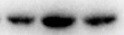
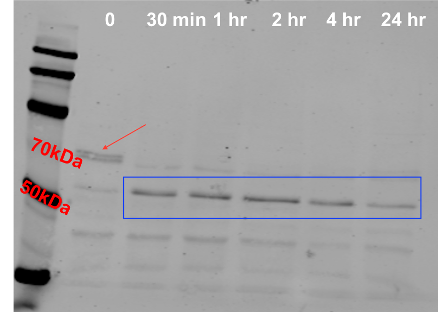
Application: Western BlotSample Tested: T cellsSpecies: HumanVerified Customer | Posted 07/12/2018Primary T cells were activated with PMA/ionomycin for the time points as shown. Cell lysates were harvested in a NP40-based lysis buffer. 8% SDS-PAGE, transferred to a nitrocellulose membrane by semi-dry transfer machine. Blocked by 5% milk for 30 mins. Antibody dilution was used at 1:500. 680nm Donkey anti-mouse was used as secondary antibody (1:30,000). Image captured by Licor Odyssey. The arrow shows the full length Regnase-1, and the rectangle shows the cleaved product upon TCR stimulation.
There are no reviews that match your criteria.
Protocols
Find general support by application which include: protocols, troubleshooting, illustrated assays, videos and webinars.
- Cellular Response to Hypoxia Protocols
- R&D Systems Quality Control Western Blot Protocol
- Troubleshooting Guide: Western Blot Figures
- Western Blot Conditions
- Western Blot Protocol
- Western Blot Protocol for Cell Lysates
- Western Blot Troubleshooting
- Western Blot Troubleshooting Guide
- View all Protocols, Troubleshooting, Illustrated assays and Webinars
Loading...