Neogenin (NEO) is a type I transmembrane protein that is crucial for axonal guidance and neuronal migration. It is also involved in regulating differentiation programs in many embryonic and adult tissues (1). Mouse NEO is widely expressed in adult tissues and is expressed throughout the mid to late stages of gestation, in both neuronal and non-neuronal tissues. It is a member of the immunoglobulin (Ig) superfamily and is closely related to deleted in colorectal cancer (DCC). Mouse NEO cDNA encodes a 1493 amino acid residue (aa) precursor with a putative 36 aa signal peptide, a 1100 aa extracellular domain with six Ig-like C2 type domains and three fibronectin type III domains, a 21 aa transmembrane domain, and a 345 aa cytoplasmic domain. At least five isoforms are produced in mice by alternative splicing. Mouse NEO shares 96%, 93%, and 86% aa sequence identity with rat, human, and chicken NEO, respectively. It also has 46% and 29% sequence homology with mouse DCC and C. elegans UNC40, a homolog of DCC. NEO and DCC, together with the UNC5 family of type I transmembrane proteins, are receptors for the netrin/UNC6 family of laminin-related bifunctional guidance molecules that both attract some axons and repel others (2, 3). In mouse, at least five netrins (netrin‑1,
‑3, -4, G1, and G2) have been identified (3-5). Mouse netrin-1 and netrin-3 have been shown to be ligands for mouse NEO.

Key Product Details
Species Reactivity
Validated:
Cited:
Applications
Validated:
Cited:
Label
Antibody Source
Product Specifications
Immunogen
Ala42-Ile1033 (Asp442-Leu461 del)
Accession # NP_032710
Specificity
Clonality
Host
Isotype
Endotoxin Level
Scientific Data Images for Neogenin Antibody
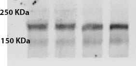
Detection of Human and Mouse Neogenin by Western Blot.
Western blot shows lysates of 3T3-L1 mouse embryonic fibroblast adipose-like cell line, Jurkat human acute T cell leukemia cell line, and Hepa 1-6 mouse hepatoma cell line. PVDF membrane was probed with 0.5 µg/mL of Goat Anti-Human/Mouse Neogenin Antigen Affinity-purified Polyclonal Antibody (Catalog # AF1079) followed by HRP-conjugated Anti-Goat IgG Secondary Antibody (HAF017). A specific band was detected for Neogenin at approximately 230 kDa (as indicated). This experiment was conducted under reducing conditions and using Immunoblot Buffer Group 1.
Neogenin in Mouse Embryo.
Neogenin was detected in perfusion fixed frozen sections of mouse embryo (15 d.p.c.) using Goat Anti-Human/Mouse Neogenin Antigen Affinity-purified Polyclonal Antibody (Catalog # AF1079) at 15 µg/mL overnight at 4 °C. Tissue was stained using the Anti-Goat HRP-DAB Cell & Tissue Staining Kit (brown; CTS008) and counterstained with hematoxylin (blue). Specific staining was localized to developing neurons. View our protocol for Chromogenic IHC Staining of Frozen Tissue Sections.
Detection of Mouse Neogenin by Simple WesternTM.
Simple Western lane view shows lysates of beta TC-6 mouse beta cell insulinoma cell line, loaded at 0.2 mg/mL. A specific band was detected for Neogenin at approximately 207 kDa (as indicated) using 10 µg/mL of Goat Anti-Human/Mouse Neogenin Antigen Affinity-purified Polyclonal Antibody (Catalog # AF1079) followed by 1:50 dilution of HRP-conjugated Anti-Goat IgG Secondary Antibody (HAF109). This experiment was conducted under reducing conditions and using the 12-230 kDa separation system.
Detection of Neogenin by Immunohistochemistry
Subregion-specific expression of RGMs and Neogenin in the hippocampus.In situ hybridization (A–C, J–L) and immunohistochemistry (D–I, M–O) on coronal mouse brain sections at E16.5 (A–I) and P5 (J–O). Sections in D–I and M–O are counterstained in blue with fluorescent Nissl. (A–F) RGMa mRNA and protein are expressed in the ventricular zone (VZ), dentate gyrus (DG) and cornu ammonis (CA) region. Strong expression of RGMb mRNA and protein is detected in the pial surface lining the hippocampal fissure (HF). Neogenin transcripts and protein are widely expressed in the developing hippocampus (Hip). (G–I) Immunostaining with isotype matched controls. (J–L) In situ hybridization at P5 shows strong but differential expression patterns of RGMa, RGMb and Neogenin in the CA pyramidal cell layers (Pyr). In addition, strong expression of Neogenin is detected in the granular layer (GC) of the DG. (M–O) Immunohistochemistry reveals expression of RGMa and weak expression of RGMb in the stratum lacunosum moleculare (SLM) and fimbria (FIM). Neogenin strongly labels different hippocampal layers. CX, cortex; Hb, habenula; HC, hippocampal commissure; PO, polymorph layer; SO, stratum oriens; SR, stratum radiatum; Th, thalamus. Scale bar A–C: 400 µm, D–F: 300 µm, G–I: 300 µm, J–L: 500 µm and M–O: 400 µm. Image collected and cropped by CiteAb from the following open publication (https://pubmed.ncbi.nlm.nih.gov/23457482), licensed under a CC-BY license. Not internally tested by R&D Systems.
Detection of Neogenin by Immunohistochemistry
RGM and Neogenin expression in the mouse olfactory system.In situ hybridization on coronal mouse brain sections at E16.5 (A–C′) and P5 (L–N′). Panels A′–C′ and L′–N′ show higher magnifications of boxed areas in A–C and L–N, respectively. Immunohistochemistry (D–F, H–J) and RGMa-AP section binding (G, K) on E16.5 coronal mouse brain sections. Sections in D–F and H–J are counterstained in blue with fluorescent Nissl. (A–C′) In situ hybridization shows differential expression patterns of RGMa, RGMb and Neogenin in the olfactory bulb and olfactory epithelium (OE). In line with this, immunohistochemistry reveals that axons of olfactory sensory neurons in the OE stain strongly for RGMb and weakly for RGMa and Neogenin. Furthermore, RGMa, RGMb and Neogenin are expressed on olfactory bulb axon projections such as the lateral olfactory tract (LOT). a, apical; ACa, anterior commissure pars anterior; AOB, accessory olfactory bulb; b, basal; CP, cortical plate; CRP, cribriform plate; EPL, external plexiform layer; GL, glomerular layer; GR, granule cell layer; IPL, internal plexiform layer; IZ, intermediate zone; LV, lateral ventricle; MCL, mitral cell layer; ONL, olfactory nerve layer; OVZ, olfactory ventricular zone; S, septum; STR, striatum; VN, vomeronasal nerve. Scale bar A–C 200 µm, A′–C′ 100 µm, D–F 300 µm, G 500 µm, H–J 400 µm, K 500 µm, L–N 400 µm and L′–N′ 200 µm. Image collected and cropped by CiteAb from the following open publication (https://pubmed.ncbi.nlm.nih.gov/23457482), licensed under a CC-BY license. Not internally tested by R&D Systems.Applications for Neogenin Antibody
Blockade of Receptor-ligand Interaction
Immunohistochemistry
Sample: Perfusion fixed frozen sections of mouse embryo (15 d.p.c.)
Simple Western
Sample: beta TC‑6 mouse beta cell insulinoma cell line
Western Blot
Sample: 3T3‑L1 mouse embryonic fibroblast adipose-like cell line, Jurkat human acute T cell leukemia cell line, and Hepa 1‑6 mouse hepatoma cell line
Reviewed Applications
Read 1 review rated 4 using AF1079 in the following applications:
Formulation, Preparation, and Storage
Purification
Reconstitution
Reconstitute at 0.2 mg/mL in sterile PBS. For liquid material, refer to CoA for concentration.
Formulation
*Small pack size (-SP) is supplied either lyophilized or as a 0.2 µm filtered solution in PBS.
Shipping
Stability & Storage
- 12 months from date of receipt, -20 to -70 °C as supplied.
- 1 month, 2 to 8 °C under sterile conditions after reconstitution.
- 6 months, -20 to -70 °C under sterile conditions after reconstitution.
Calculators
Background: Neogenin
References
- Keeling, S.L. et al. (1997) Oncogene 15:691.
- Hong, K. et al. (1999) Cell 97:927.
- Livesey, F.J. (1999) Cell Mol. Life Sci. 56:62.
- Nakashiba, T. et al. (2000) J. Neurosci. 20:6540.
- Nakashiba, T. et al. (2002) Mech. Dev. 111:47.
Alternate Names
Gene Symbol
UniProt
Additional Neogenin Products
Product Documents for Neogenin Antibody
Certificate of Analysis
To download a Certificate of Analysis, please enter a lot or batch number in the search box below.
Note: Certificate of Analysis not available for kit components.
Product Specific Notices for Neogenin Antibody
This product or the use of this product is covered by U.S. Patents owned by The Regents of the University of California. This product is for research use only and is not to be used for commercial purposes. Use of this product to produce products for sale or for diagnostic, therapeutic or drug discovery purposes is prohibited. In order to obtain a license to use this product for such purposes, contact The Regents of the University of California.
U.S. Patent # 5,939,271, 6,277,585, and other U.S. and international patents pending.
For research use only
Citations for Neogenin Antibody
 Powered by Bioz
Customer Reviews for Neogenin Antibody (1)
Have you used Neogenin Antibody?
Submit a review and receive an Amazon gift card!
$25/€18/£15/$25CAN/¥2500 Yen for a review with an image
$10/€7/£6/$10CAN/¥1110 Yen for a review without an image
Submit a review
Customer Images
-
Application: Western BlotSample Tested: Brain tissueSpecies: MouseVerified Customer | Posted 01/25/2019Dilution used for the western is 1:250

There are no reviews that match your criteria.
Protocols
Find general support by application which include: protocols, troubleshooting, illustrated assays, videos and webinars.
- Antigen Retrieval Protocol (PIER)
- Antigen Retrieval for Frozen Sections Protocol
- Appropriate Fixation of IHC/ICC Samples
- Cellular Response to Hypoxia Protocols
- Chromogenic IHC Staining of Formalin-Fixed Paraffin-Embedded (FFPE) Tissue Protocol
- Chromogenic Immunohistochemistry Staining of Frozen Tissue
- ClariTSA™ Fluorophore Kits
- Detection & Visualization of Antibody Binding
- Fluorescent IHC Staining of Frozen Tissue Protocol
- Graphic Protocol for Heat-induced Epitope Retrieval
- Graphic Protocol for the Preparation and Fluorescent IHC Staining of Frozen Tissue Sections
- Graphic Protocol for the Preparation and Fluorescent IHC Staining of Paraffin-embedded Tissue Sections
- Graphic Protocol for the Preparation of Gelatin-coated Slides for Histological Tissue Sections
- IHC Sample Preparation (Frozen sections vs Paraffin)
- Immunofluorescent IHC Staining of Formalin-Fixed Paraffin-Embedded (FFPE) Tissue Protocol
- Immunohistochemistry (IHC) and Immunocytochemistry (ICC) Protocols
- Immunohistochemistry Frozen Troubleshooting
- Immunohistochemistry Paraffin Troubleshooting
- Preparing Samples for IHC/ICC Experiments
- Preventing Non-Specific Staining (Non-Specific Binding)
- Primary Antibody Selection & Optimization
- Protocol for Heat-Induced Epitope Retrieval (HIER)
- Protocol for Making a 4% Formaldehyde Solution in PBS
- Protocol for VisUCyte™ HRP Polymer Detection Reagent
- Protocol for the Preparation & Fixation of Cells on Coverslips
- Protocol for the Preparation and Chromogenic IHC Staining of Frozen Tissue Sections
- Protocol for the Preparation and Chromogenic IHC Staining of Frozen Tissue Sections - Graphic
- Protocol for the Preparation and Chromogenic IHC Staining of Paraffin-embedded Tissue Sections
- Protocol for the Preparation and Chromogenic IHC Staining of Paraffin-embedded Tissue Sections - Graphic
- Protocol for the Preparation and Fluorescent IHC Staining of Frozen Tissue Sections
- Protocol for the Preparation and Fluorescent IHC Staining of Paraffin-embedded Tissue Sections
- Protocol for the Preparation of Gelatin-coated Slides for Histological Tissue Sections
- R&D Systems Quality Control Western Blot Protocol
- TUNEL and Active Caspase-3 Detection by IHC/ICC Protocol
- The Importance of IHC/ICC Controls
- Troubleshooting Guide: Immunohistochemistry
- Troubleshooting Guide: Western Blot Figures
- Western Blot Conditions
- Western Blot Protocol
- Western Blot Protocol for Cell Lysates
- Western Blot Troubleshooting
- Western Blot Troubleshooting Guide
- View all Protocols, Troubleshooting, Illustrated assays and Webinars