
Endophilin A1 (also SH3GL2, SH3P4, EEN-B1, CNS-A2, GL2 and SH3D2A) is a 39-42 kDa member of the A subfamily, endophilin (attracted to endocytic protein) family of molecules. It is a neuronally-expressed cytosolic protein that is concentrated in presynaptic areas. Endophilin A1 is a key player in clathrin-associated endocytosis, participating in invagination plus vesicle fission and recycling. It is able to impact membrane curvature through electroststic differences via its N‑terminus, and interact with multiple synaptic region proteins such as dynamin and synaptojanin through an SH3 homology domain at its C-terminus. Although Endophilin A1 is a monomer in the cytosol, it will homodimerize and heterodimerize with endophilin-2 when active at the synapse. Endophilin A1 is also associated with endocytic trafficking of growth factor receptors. In concert with CIN85, Cbl and ataxin-2, Endophilin A1 extend the half-life of EGFR at the plasma membrane. Human Endophilin A1 is 352 amino acids (aa) in length. It contains an aforementioned N-terminal BAR domain (aa 18-249) plus an SH3 domain that binds Pro‑rich aa sequences (aa 290-349). There is one potential alternative start site at Met36. Endophilin A1 is one of three groupA endophilin isoforms, all of which are the product of separate genes. Although the -A2 and -A3 isoforms share approximately 70% aa overall sequence identity with -A1, over aa 217-293, -A2 and -A3 share less than 50% aa sequence identity with Endophilin A1. Over this same aa sequence, human and mouse Endophilin A1 share more that 97% aa sequence identity.

Scientific images may be resized, cropped, or adjusted for brightness or contrast for presentation purposes. These updates are limited to presentation and do not affect the underlying scientific interpretation of the data. Where available, additional source data may be provided upon request.
Key Product Details
Species Reactivity
Applications
Label
Antibody Source
Product Specifications
Immunogen
Ala217-Pro293
Accession # Q99962
Specificity
Clonality
Host
Isotype
Scientific Data Images for Endophilin A1/SH3GL2 Antibody
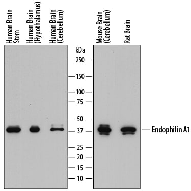
Detection of Human, Mouse, and Rat Endophilin A1/SH3GL2 by Western Blot.
Western blot shows lysates of human brain stem tissue, human brain (hypothalamus) tissue, human brain (cerebellum) tissue, mouse brain (cerebellum) tissue, and rat brain tissue. PVDF membrane was probed with 1 µg/mL of Sheep Anti-Human Endophilin A1/SH3GL2 Antigen Affinity-purified Polyclonal Antibody (Catalog # AF8006) followed by HRP-conjugated Anti-Sheep IgG Secondary Antibody (Catalog # HAF016). A specific band was detected for Endophilin A1/SH3GL2 at approximately 40 kDa (as indicated). This experiment was conducted under reducing conditions and using Immunoblot Buffer Group 1.Applications for Endophilin A1/SH3GL2 Antibody
Western Blot
Sample: Human brain stem tissue, human brain (hypothalamus) tissue, human brain (cerebellum) tissue, mouse brain (cerebellum) tissue, and rat brain tissue
Formulation, Preparation, and Storage
Purification
Reconstitution
Sterile PBS to a final concentration of 0.2 mg/mL. For liquid material, refer to CoA for concentration.
Formulation
Shipping
Stability & Storage
- 12 months from date of receipt, -20 to -70 °C as supplied.
- 1 month, 2 to 8 °C under sterile conditions after reconstitution.
- 6 months, -20 to -70 °C under sterile conditions after reconstitution.
Calculators
Background: Endophilin A1/SH3GL2
Long Name
Alternate Names
Gene Symbol
UniProt
Additional Endophilin A1/SH3GL2 Products
Product Documents for Endophilin A1/SH3GL2 Antibody
Certificate of Analysis
To download a Certificate of Analysis, please enter a lot or batch number in the search box below.
Note: Certificate of Analysis not available for kit components.
Product Specific Notices for Endophilin A1/SH3GL2 Antibody
For research use only
Customer Reviews for Endophilin A1/SH3GL2 Antibody
There are currently no reviews for this product. Be the first to review Endophilin A1/SH3GL2 Antibody and earn rewards!
Have you used Endophilin A1/SH3GL2 Antibody?
Submit a review and receive an Amazon gift card!
$25/€18/£15/$25CAN/¥2500 for a review with an image
$10/€7/£6/$10CAN/¥1110 for a review without an image
Submit a review
Protocols
Find general support by application which include: protocols, troubleshooting, illustrated assays, videos and webinars.
- Cellular Response to Hypoxia Protocols
- R&D Systems Quality Control Western Blot Protocol
- Troubleshooting Guide: Western Blot Figures
- Western Blot Conditions
- Western Blot Protocol
- Western Blot Protocol for Cell Lysates
- Western Blot Troubleshooting
- Western Blot Troubleshooting Guide
- View all Protocols, Troubleshooting, Illustrated assays and Webinars