Human/Mouse/Rat Phospho-Ribosomal Protein S6/RPS6 (S235/S236) Antibody
Human/Mouse/Rat Phospho-Ribosomal Protein S6/RPS6 (S235/S236) Antibody Summary
Applications
Please Note: Optimal dilutions should be determined by each laboratory for each application. General Protocols are available in the Technical Information section on our website.
Scientific Data
 View Larger
Ribosomal Protein S6 in Human Colon Cancer Tissue. Ribosomal Protein S6 was detected in immersion fixed paraffin-embedded sections of human colon cancer tissue using 5 µg/mL Rabbit Anti-Human/Mouse/Rat Phospho-Ribosomal Protein S6 (S235/S236) Antigen Affinity-purified Polyclonal Antibody (Catalog # AF3918) overnight at 4 °C. Tissue was stained with the Anti-Rabbit HRP-DAB Cell & Tissue Staining Kit (brown; Catalog # CTS005) and counterstained with hematoxylin (blue). Specific labeling was localized to vascular endothelial cell within glomeruli. View our protocol for Chromogenic IHC Staining of Paraffin-embedded Tissue Sections.
 View Larger
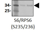
Detection of Human and Mouse Phospho-Ribosomal Protein S6 (S235/S236) by Western Blot. Western blot shows lysates of MCF-7 human breast cancer cell line untreated (-) or treated (+) with 100 ng/mL Recombinant Human IGF-1 (Catalog # 291-G1) for 20 minutes and NIH-3T3 mouse embryonic fibroblast cell line untreated or treated with 10 ng/mL Human PDGF (Catalog # 120-HD) for 20 minutes. PVDF membrane was probed with 0.5 µg/mL of Rabbit Anti-Human/Mouse/Rat Phospho-Ribosomal Protein S6 (S235/S236) Antigen Affinity-purified Polyclonal Antibody (Catalog # AF3918), followed by HRP-conjugated Anti-Rabbit IgG Secondary Antibody (Catalog # HAF008). A specific band was detected for Phospho-Ribosomal Protein S6 (S235/S236) at approximately 32 kDa (as indicated). This experiment was conducted under reducing conditions and using Immunoblot Buffer Group 1.
 View Larger
Detection of Human Phospho-Ribosomal Protein S6/RPS6 (S235/S236) by Simple WesternTM. Simple Western lane view shows lysates of MCF-7 human breast cancer cell line untreated (-) or treated (+) with 100 ng/mL Recombinant Human IGF-I (Catalog # 291-G1) for 20 minutes, loaded at 0.5 mg/mL. A specific band was detected for Phospho-Ribosomal Protein S6/RPS6 (S235/S236) at approximately 40 kDa (as indicated) using 5 µg/mL of Rabbit Anti-Human/Mouse/Rat Phospho-Ribosomal Protein S6/RPS6 (S235/S236) Antigen Affinity-purified Polyclonal Antibody (Catalog # AF3918). This experiment was conducted under reducing conditions and using the 12-230 kDa separation system.
 View Larger
Detection of Human Ribosomal Protein S6/RPS6 by Western Blot ALM inhibits HIF-1 alpha translation by down-regulating mTOR pathway. (A) Hep3B and PC3 cells were exposed to vehicle or the hypoxia mimics dimethyloxalylglycine (DMOG), cobalt chloride (CoCl2), desferrioxamine (DFX) or MG132 in the presence of ALM at indicated concentrations or vehicle for 24 h and whole cell lysates were subjected to Western blot. (B) Hep3B cells were pretreated for 4 h with vehicle (Vhc), 25 nM rapamycin (Rapa), 20 μg/mL cycloheximide (Chx), or 200 nM ALM, [35S] methionine/cysteine was added for 1 h followed by cell lysis, HIF-1 alpha immunoprecipitation (IP), SDS/PAGE, and autoradiography. (C–E) PC3 cells were cultured at 20% or 1% O2 for 24 h in the presence of ALM (at the indicated concentrations) and whole cell lysates were subjected to for: (C) p-P70, p-Akt, p-mTOR (2448/2481), mTOR, (D) p-4E-BP1, (E) p-RPS6, HIF-1 alpha, or beta -actin. Image collected and cropped by CiteAb from the following publication (https://pubmed.ncbi.nlm.nih.gov/31083403), licensed under a CC-BY license. Not internally tested by R&D Systems.
Reconstitution Calculator
Preparation and Storage
- 12 months from date of receipt, -20 to -70 °C as supplied.
- 1 month, 2 to 8 °C under sterile conditions after reconstitution.
- 6 months, -20 to -70 °C under sterile conditions after reconstitution.
Background: Ribosomal Protein S6/RPS6
40S ribosomal protein S6 is the major substrate of protein kinases, particularly p70 S6 kinase, in eukaryotic ribosomes. S6 phosphorylation at S235, S236, S240, and S244 upregulates the translation of mRNAs containing an oligopyrimidine tract at their transcriptional start sites. This phosphorylation is stimulated by growth factors, tumor promoting agents, and other mitogens.
Product Datasheets
Citations for Human/Mouse/Rat Phospho-Ribosomal Protein S6/RPS6 (S235/S236) Antibody
R&D Systems personnel manually curate a database that contains references using R&D Systems products. The data collected includes not only links to publications in PubMed, but also provides information about sample types, species, and experimental conditions.
6
Citations: Showing 1 - 6
Filter your results:
Filter by:
-
Ancient familial Mediterranean fever mutations in human pyrin and resistance to Yersinia pestis
Authors: YH Park, EF Remmers, W Lee, AK Ombrello, LK Chung, Z Shilei, DL Stone, MI Ivanov, NA Loeven, KS Barron, P Hoffmann, M Nehrebecky, YZ Akkaya-Ulu, E Sag, B Balci-Peyn, I Aksentijev, A Gül, CN Rotimi, H Chen, JB Bliska, S Ozen, DL Kastner, D Shriner, JJ Chae
Nat. Immunol., 2020-06-29;0(0):.
-
2-Oxonanonoidal Antibiotic Actinolactomycin Inhibits Cancer Progression by Suppressing HIF-1 alpha.
Authors: Cheng Jiadong, Hu Lan, Yang Zheng et al.
Cells
-
Multiscale networks in multiple sclerosis
Authors: Kennedy, KE;Kerlero de Rosbo, N;Uccelli, A;Cellerino, M;Ivaldi, F;Contini, P;De Palma, R;Harbo, HF;Berge, T;Bos, SD;Høgestøl, EA;Brune-Ingebretsen, S;de Rodez Benavent, SA;Paul, F;Brandt, AU;Bäcker-Koduah, P;Behrens, J;Kuchling, J;Asseyer, S;Scheel, M;Chien, C;Zimmermann, H;Motamedi, S;Kauer-Bonin, J;Saez-Rodriguez, J;Rinas, M;Alexopoulos, LG;Andorra, M;Llufriu, S;Saiz, A;Blanco, Y;Martinez-Heras, E;Solana, E;Pulido-Valdeolivas, I;Martinez-Lapiscina, EH;Garcia-Ojalvo, J;Villoslada, P;
PLoS computational biology
Species: Human
Sample Types: Whole Cells
Applications: Flow Cytometry -
Branched-chain amino acid catabolism breaks glutamine addiction to sustain hepatocellular carcinoma progression
Authors: D Yang, H Liu, Y Cai, K Lu, X Zhong, S Xing, W Song, Y Zhang, L Ye, X Zhu, T Wang, P Zhang, ST Li, J Feng, W Jia, H Zhang, P Gao
Cell Reports, 2022-11-22;41(8):111691.
Species: Xenograft
Sample Types: Cell Lysates
Applications: Western Blot -
PAPP-A proteolytic activity enhances IGF bioactivity in ascites from women with ovarian carcinoma
Authors: Jacob Thomsen, Rikke Hjortebjerg, Ulrick Espelund, Gitte Ørtoft, Poul Vestergaard, Nils E. Magnusson et al.
Oncotarget
Species: Human
Sample Types: Ascites Fluid
Applications: Western Blot -
The protein-tyrosine phosphatase, SRC homology-2 domain containing protein tyrosine phosphatase-2, is a crucial mediator of exogenous insulin-like growth factor signaling to human trophoblast.
Authors: Forbes K, West G, Garside R, Aplin JD, Westwood M
Endocrinology, 2009-07-09;150(10):4744-54.
Species: Human
Sample Types: Whole Tissue
Applications: IHC-P
FAQs
No product specific FAQs exist for this product, however you may
View all Antibody FAQsReviews for Human/Mouse/Rat Phospho-Ribosomal Protein S6/RPS6 (S235/S236) Antibody
Average Rating: 4.5 (Based on 2 Reviews)
Have you used Human/Mouse/Rat Phospho-Ribosomal Protein S6/RPS6 (S235/S236) Antibody?
Submit a review and receive an Amazon gift card.
$25/€18/£15/$25CAN/¥75 Yuan/¥2500 Yen for a review with an image
$10/€7/£6/$10 CAD/¥70 Yuan/¥1110 Yen for a review without an image
Filter by:
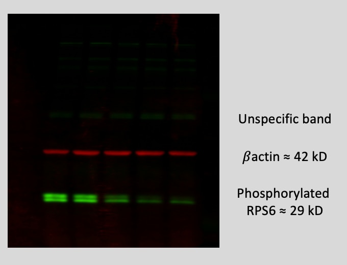
Mouse bone marrow stromal cells were lysed with NP-40 buffer for protein extraction. 10 ug of proteins were loaded in a 10% Acrylamide gel.
Western Blot: Phospho S6/RPS6 [AF3918] - Total protein from mouse skeletal muscle tissue, separated on a 4-12% gel by SDS-PAGE, transferred to nitrocellulose membrane and blocked in 5% non-fat milk for 1h at room temperature. The membrane was probed with anti-phospho S6/RPS6 1:400 in non-fat milk.


