The cAMP-dependent protein kinase (PKA) holoenzyme is composed of two regulatory and two catalytic subunits, designated PKA R and PKA C, respectively. PKA R subunits exist as two classes, RI and RII, with each class containing two isoforms, alpha and beta. Upon PKA R subunit binding to the second messenger cAMP, active PKA C subunits are released, initiating a phosphorylation cascade that regulates such cellular functions as metabolism, ion transport, and gene transcription.

Key Product Details
Species Reactivity
Validated:
Human, Mouse, Rat
Cited:
Human, Mouse
Applications
Validated:
Western Blot, Simple Western
Cited:
Immunohistochemistry-Frozen, Western Blot
Label
Unconjugated
Antibody Source
Polyclonal Sheep IgG
Loading...
Product Specifications
Immunogen
E. coli-derived recombinant human PKA RI beta
Met1-Val381
Accession # P31321
Met1-Val381
Accession # P31321
Specificity
Detects human, mouse, and rat PKA RI beta in Western blots. In Western blots, less than 1% cross-reactivity with recombinant human (rh) PKA RI alpha or rhPKA RII beta is observed; cross-reactivity with PKA RII alpha is unknown.
Clonality
Polyclonal
Host
Sheep
Isotype
IgG
Scientific Data Images for PKA RI beta Antibody
Detection of Human PKA RI beta by Western Blot.
Western blot shows lysates of MDA-MB-468 human breast cancer cell line, Jurkat human acute T cell leukemia cell line, SW480 human colorectal adenocarcinoma cell line, Raji human Burkitt's lymphoma cell line, and Ramos human Burkitt's lymphoma cell line. PVDF membrane was probed with 1 µg/mL Sheep Anti-Human/Mouse/Rat PKA RI beta Antigen Affinity-purified Polyclonal Antibody (Catalog # AF4177) followed by HRP-conjugated Anti-Sheep IgG Secondary Antibody (Catalog # HAF016). A specific band for PKA RI beta was detected at approximately 48 kDa (as indicated). This experiment was conducted under reducing conditions and using Immunoblot Buffer Group 1.
Detection of Human PKA RI beta by Western Blot.
Western blot shows recombinant human PKA RIa, PKA RI beta, and PKA RII beta (5 ng/lane). PVDF membrane was probed with 1 µg/mL Sheep Anti-Human/ Mouse/Rat PKA RI beta Antigen Affinity-purified Polyclonal Antibody (Catalog # AF4177) followed by HRP-conjugated Anti-Sheep IgG Secondary Antibody (Catalog # HAF016). A specific band for PKA RI beta was detected at approximately 48 kDa (as indicated). This experiment was conducted under reducing conditions and using Immunoblot Buffer Group 1.
Detection of Human PKA RI beta by Simple WesternTM.
Simple Western lane view shows lysates of Jurkat human acute T cell leukemia cell line, loaded at 0.2 mg/mL. A specific band was detected for PKA RI beta at approximately 56 kDa (as indicated) using 10 µg/mL of Sheep Anti-Human/Mouse/Rat PKA RI beta Antigen Affinity-purified Polyclonal Antibody (Catalog # AF4177) followed by 1:50 dilution of HRP-conjugated Anti-Sheep IgG Secondary Antibody (Catalog # HAF016). This experiment was conducted under reducing conditions and using the 12-230 kDa separation system.
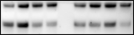
Detection of PKA RI beta by Western Blot
ARHGAP36 downregulates PKAC levels.(a) MDCK cells transfected with YFP-ARHGAP36, or a YFP control, were fixed after 8 or 24 h and subjected to immunofluorescence using antibodies against GFP and PKAC. Images were collected by confocal microscopy. Scale bars, 10 μm. (b) HEK293T cells were transfected with YFP-ARHGAP36 or YFP-Cherry control for 24 h. Lysates were immunoblotted with the indicated antibodies. (c) HEK293T cells were transfected with YFP-ARHGAP36 or YFP control. Lysates were immunoblotted with antibodies against RI alpha, RI beta RII alpha, RII beta, GAPDH and GFP. (d) MDCK cells transfected with CFP-PKI were fixed after 24 h and subjected to immunofluorescence as in a. Scale bar, 10 μm. (e) MDCK cells transfected with YFP-delta N (195–516), YFP-N (1–194), YFP-N2 (118–195), YFP-RRV or Cherry-36i were fixed after 24 h and subjected to immunofluorescence as in a. 36i signal was not amplified using antibodies. Scale bars, 10 μm. Image collected and cropped by CiteAb from the following open publication (https://pubmed.ncbi.nlm.nih.gov/27713425), licensed under a CC-BY license. Not internally tested by R&D Systems.Applications for PKA RI beta Antibody
Application
Recommended Usage
Simple Western
10 µg/mL
Sample: Jurkat human acute T cell leukemia cell line
Sample: Jurkat human acute T cell leukemia cell line
Western Blot
1 µg/mL
Sample: MDA-MB-468 human breast cancer cell line, Jurkat human acute T cell leukemia cell line, SW480 human colorectal adenocarcinoma cell line, Raji human Burkitt's lymphoma cell line, and Ramos human Burkitt's lymphoma cell line
Sample: MDA-MB-468 human breast cancer cell line, Jurkat human acute T cell leukemia cell line, SW480 human colorectal adenocarcinoma cell line, Raji human Burkitt's lymphoma cell line, and Ramos human Burkitt's lymphoma cell line
Reviewed Applications
Read 1 review rated 5 using AF4177 in the following applications:
Formulation, Preparation, and Storage
Purification
Antigen Affinity-purified
Reconstitution
Reconstitute at 0.2 mg/mL in sterile PBS. For liquid material, refer to CoA for concentration.
Loading...
Formulation
Lyophilized from a 0.2 μm filtered solution in PBS with Trehalose. *Small pack size (SP) is supplied either lyophilized or as a 0.2 µm filtered solution in PBS.
Shipping
Lyophilized product is shipped at ambient temperature. Liquid small pack size (-SP) is shipped with polar packs. Upon receipt, store immediately at the temperature recommended below.
Stability & Storage
Use a manual defrost freezer and avoid repeated freeze-thaw cycles.
- 12 months from date of receipt, -20 to -70 °C as supplied.
- 1 month, 2 to 8 °C under sterile conditions after reconstitution.
- 6 months, -20 to -70 °C under sterile conditions after reconstitution.
Calculators
Background: PKA RI beta
Long Name
cAMP-dependent Protein Kinase Regulatory Type I beta
Alternate Names
PRKAR1B, RIbeta
Gene Symbol
PRKAR1B
UniProt
Additional PKA RI beta Products
Product Documents for PKA RI beta Antibody
Certificate of Analysis
To download a Certificate of Analysis, please enter a lot or batch number in the search box below.
Note: Certificate of Analysis not available for kit components.
Product Specific Notices for PKA RI beta Antibody
For research use only
Related Research Areas
Citations for PKA RI beta Antibody
 Powered by Bioz
Customer Reviews for PKA RI beta Antibody (1)
5 out of 5
1 Customer Rating
Have you used PKA RI beta Antibody?
Submit a review and receive an Amazon gift card!
$25/€18/£15/$25CAN/¥2500 Yen for a review with an image
$10/€7/£6/$10CAN/¥1110 Yen for a review without an image
Submit a review
Customer Images
Showing
1
-
1 of
1 review
Showing All
Filter By:
-
Application: Western BlotSample Tested: Adult lungSpecies: MouseVerified Customer | Posted 11/04/2019

There are no reviews that match your criteria.
Protocols
Find general support by application which include: protocols, troubleshooting, illustrated assays, videos and webinars.
- Cellular Response to Hypoxia Protocols
- R&D Systems Quality Control Western Blot Protocol
- Troubleshooting Guide: Western Blot Figures
- Western Blot Conditions
- Western Blot Protocol
- Western Blot Protocol for Cell Lysates
- Western Blot Troubleshooting
- Western Blot Troubleshooting Guide
- View all Protocols, Troubleshooting, Illustrated assays and Webinars
Loading...