Smads are a family of intracellular proteins that transmit transforming growth factor beta (TGF-beta ) superfamily signals from the cell surface to the nucleus. The Smad family is divided into three subclasses: receptor regulated Smads, (Smads 1, 2, 3, 5 and 8); the common partner, (Smad4) that functions via its interaction to the various Smads; and the inhibitory Smads, (Smads 6 and 7). Smad7, also known as Mothers Against Decapentaplegic homolog 7 (MADH7), inhibits selected pathways by binding directly to cell-surface receptors and preventing the activation-induced phosphorylation of other Smad subunits.

Key Product Details
Validated by
Knockout/Knockdown, Biological Validation
Species Reactivity
Validated:
Human, Mouse, Rat
Cited:
Human, Mouse, Rat
Applications
Validated:
Immunohistochemistry, Western Blot, Simple Western
Cited:
Immunohistochemistry, Immunohistochemistry-Paraffin, Western Blot, Immunofluorescence, Immunocytochemistry, Simple Western, Immunoprecipitation, Proximity Ligation Assay, IF/ICC
Label
Unconjugated
Antibody Source
Monoclonal Mouse IgG2B Clone # 293039
Loading...
Product Specifications
Immunogen
E. coli-derived recombinant human Smad7
Gly320-Ser398
Accession # O15105
Gly320-Ser398
Accession # O15105
Specificity
Detects human, mouse, and rat Smad7.
Clonality
Monoclonal
Host
Mouse
Isotype
IgG2B
Scientific Data Images for Smad7 Antibody (293039)
Smad7 in Human Liver Cancer Tissue.
Smad7 was detected in immersion fixed paraffin-embedded sections of human liver cancer tissue using 25 µg/mL Human/Mouse/Rat Smad7 Monoclonal Antibody (Catalog # MAB2029) overnight at 4 °C. Tissue was stained with the Anti-Mouse HRP-DAB Cell & Tissue Staining Kit (brown; Catalog # CTS002) and counterstained with hematoxylin (blue). View our protocol for Chromogenic IHC Staining of Paraffin-embedded Tissue Sections.
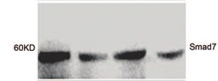
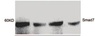
Detection of Human/Mouse/Rat Smad7 by Western Blot.
Western blot shows lysates of H9 human cutaneous T lymphoma cell line, PT18 mouse mast/basophil cell line, and Rat-2 rat embryonic fibroblast cell line. PVDF membrane was probed with 1 µg/mL of Human/Mouse/Rat Smad7 Monoclonal Antibody (Catalog # MAB2029) followed by HRP-conjugated Anti-Mouse IgG Secondary Antibody (Catalog # HAF007). A specific band was detected for Smad7 at approximately 50 kDa (as indicated). This experiment was conducted under reducing conditions and using Immunoblot Buffer Group 2.
Smad7 in Human Liver.
Smad7 was detected in immersion fixed paraffin-embedded sections of human liver array using Human/Mouse/Rat Smad7 Monoclonal Antibody (Catalog # MAB2029) at 25 µg/mL overnight at 4 °C. Tissue was stained using the Anti-Mouse HRP-DAB Cell & Tissue Staining Kit (brown; Catalog # CTS002) and counterstained with hematoxylin (blue). Lower panel shows a lack of labeling if primary antibodies are omitted and tissue is stained only with secondary antibody followed by incubation with detection reagents. View our protocol for Chromogenic IHC Staining of Paraffin-embedded Tissue Sections.
Detection of Human Smad7 by Simple WesternTM.
Simple Western lane view shows lysates ofSf21S. frugiperdainsect ovarian cell line transfected with human Smad7, loaded at 0.2 mg/mL. A specific band was detected for Smad7 at approximately 56 kDa (as indicated) using 10 µg/mL of Mouse Anti-Human/Mouse/Rat Smad7 Monoclonal Antibody (Catalog # MAB2029). This experiment was conducted under reducing conditions and using the 12-230 kDa separation system.
Detection of Human Smad7 by Western Blot
Adenovirus-mediated SMAD7 expression inhibits STAT3 phosphorylation in HuH-7. (a) Ectopic SMAD7 expression inhibits both endogenous and IL-6-induced STAT3 phosphorylation in HuH-7 cells. (b) Ectopic SMAD7 expression inhibits IL-6-induced MCL-1 expression in HuH-7 cells. Quantification of immunoblot analysis was performed of two independent experiments. One result is shown representatively. (c) IL-6 expression levels were investigated by RT–PCR upon ectopic SMAD7 expression. (d) Theoretical pathway connections between SMAD7 and STAT3 in HCC analyzed by cBioPortal analysis (https://www.cbioportal.org/). Image collected and cropped by CiteAb from the following publication (https://www.nature.com/articles/oncsis201685), licensed under a CC-BY license. Not internally tested by R&D Systems.
Detection of Human Smad7 by Western Blot
USP11 interacts with ALK5. (a) HEK293 cells were transiently transfected with FLAG-ALK5, HA-USP11 and/or HA-SMAD7, as indicated. Extracts or FLAG IPs were resolved by SDS–PAGE and immunoblotted with antibodies against HA-USP11, HA-SMAD7 and ALK5. (b) HEK293 cells were transiently transfected with 3XFLAG-ALK5, and HA-SMAD7, as indicated. Extracts or FLAG IPs were resolved by SDS–PAGE and immunoblotted with antibodies against endogenous USP11, HA-SMAD7 and 3XFLAG-ALK5. All immunoblots are representative of at least three biological replicates. (c) Lysates from HEK293 cells treated with vehicle or TGF beta (50 pM 45 min) were immunoprecipitated using pre-immune IgG or an ALK5 antibody covalently bound to Dynabeads. IPs, flow-through extracts and lysate inputs were immunoblotted with endogenous USP11, ALK5 and phospho-SMAD2 antibodies. The arrowhead denotes the native molecular weight ALK5. Image collected and cropped by CiteAb from the following publication (https://pubmed.ncbi.nlm.nih.gov/22773947), licensed under a CC-BY license. Not internally tested by R&D Systems.
Detection of Mouse Smad7 by Immunohistochemistry
DEN-dependent HCC in Tamoxifen (TAM)-inducible hepatocyte-specific SMAD7 Tg and SMAD7 KO mice. (a) Plasmid constructs used for generating Albumin-SMAD7 Tg and SMAD7fl/fl mice. (b) Experimental design of the DEN-induced HCC in TTR-Cre × SMAD7 Tg or KO mice. (c) Smad7 levels were analyzed by RT–PCR and (d) immunohistochemistry (positive staining is indicated by brown color). Image collected and cropped by CiteAb from the following publication (https://www.nature.com/articles/oncsis201685), licensed under a CC-BY license. Not internally tested by R&D Systems.
Detection of Mouse Smad7 by Western Blot
TGF‐ beta 1 signaling in MSCs is regulated by environmental TGF‐ beta 1 concentrationAAT‐MSCs were cultured in the labeling media containing [35S]‐Met/Cys and increasing concentrations of r.h. TGF‐ beta 1 at 0, 0.1, 0.3, 1, 3, and 10 ng/ml for overnight. Supernatants were harvested and incubated with TGF‐ beta 1 capture antibody. After washing, the captured TGF‐ beta 1 was transferred to glass filter membrane, and the radioactivity was counted with a scintillation counter (black bars). The supernatants were incubated with new TGF‐ beta 1 capture antibody, and the radioactivity of captured TGF‐ beta 1 was counted again (white bars). The data are given as mean ± SD of five repeated measurements. This experiment was performed four times with comparable results. CPM, count per minute.B, CAT‐MSCs (B) or HDFs (C) were cultured in media containing increasing concentrations of r.h. TGF‐ beta 1 at 0, 0.1, 0.3, 1, 3, and 10 ng/ml for 24 h, before protein lysates or total RNAs were harvested. Western blot analysis of Smad7 was performed with protein lysates and semi‐quantitatively analyzed with densitometry. beta ‐actin served as loading control. Data are given as mean ± SEM of three independent blots. **P < 0.01, by one‐way ANOVA with Tukey's test. AU, arbitrary unit.DFrom total RNA, the mature miR‐21 and endogenous control RNU6B were converted to cDNA and quantified by qPCR‐based TaqMan microRNA assays. The expression of miR‐21 was normalized on RNU6B. Data are given as mean ± SEM of three independent experiments.Source data are available online for this figure. Image collected and cropped by CiteAb from the following publication (https://pubmed.ncbi.nlm.nih.gov/32080965), licensed under a CC-BY license. Not internally tested by R&D Systems.
Detection of Human Smad7 by Western Blot
SMAD protein expression data from healthy controls, celiac disease, and environmental enteropathy.A) Representative western blot example investigating SMAD7; p-SMAD2,3 expression (using ERK1/2 as a normalizing protein) from controls (Ctl), celiac disease (CD), and environmental enteropathy (EE) patients. Note that for p-SMAD2,3, patients 3 and 4 have a double band that controls lacked. This band was likely an artifact and consequently was not considered in the densitometric analyses. B) Elevated SMAD7 densitometry levels in EE compared to controls (Ctl mean±SD = 0.47±0.20 a.u., EE = 1.13±0.25 a.u., p-value<0.05). Corresponds to WB5 (see S3 Table). C) Elevated p-SMAD2,3 densitometry levels in EE compared to controls (Ctl mean±SD = 0.38±0.14 a.u., EE = 0.60±0.10 a.u., p-value<0.05). Corresponds to WB2 (see S3 Table). D) Elevated p-SMAD2,3 densitometry levels in EE and CD compared to controls, with significant differences only between EE and controls (p-value<0.05) (Ctl mean±SD = 0.34±0.12 a.u., CD = 0.87±0.36 a.u., EE = 0.97±0.11 a.u.). Corresponds to WB1 (see S3 Table). Image collected and cropped by CiteAb from the following publication (https://dx.plos.org/10.1371/journal.pntd.0006224), licensed under a CC-BY license. Not internally tested by R&D Systems.
Detection of Human Smad7 by Knockdown Validated
SMAD7 is involved in the regulation of IL-1R1 expression in PSCs. a Gene expression of SMAD7 and IL-1R1 from PSCs established from different patients (n = 19) were analysed by qPCR. PSCs were transfected with non-signaling siRNA (control) or SMAD7 siRNA (100nM) for 3 days. Correlation was determined using Pearson correlation coefficient. *p < 0.05. b Pancreatic stellate cells were incubated in the presence of increasing concentrations of recombinant TGF beta (30 pg/ml – 8 ng/ml) to assess the optimal concentration required to down-regulate the gene expression of IL-1R1 and to investigate the impact of TGF beta on PSC expression of SMAD7. c SMAD7 protein expression was visualized by western blotting after 3 days of culture and (d) IL-1R1 was visualized after additional 2 days in serum free medium Image collected and cropped by CiteAb from the following publication (https://jeccr.biomedcentral.com/articles/10.1186/s13046-016-0400-5), licensed under a CC-BY license. Not internally tested by R&D Systems.
Detection of Human Human/Mouse/Rat Smad7 Antibody by Western Blot
SMAD protein expression data from healthy controls, celiac disease, and environmental enteropathy.A) Representative western blot example investigating SMAD7; p-SMAD2,3 expression (using ERK1/2 as a normalizing protein) from controls (Ctl), celiac disease (CD), and environmental enteropathy (EE) patients. Note that for p-SMAD2,3, patients 3 and 4 have a double band that controls lacked. This band was likely an artifact and consequently was not considered in the densitometric analyses. B) Elevated SMAD7 densitometry levels in EE compared to controls (Ctl mean±SD = 0.47±0.20 a.u., EE = 1.13±0.25 a.u., p-value<0.05). Corresponds to WB5 (see S3 Table). C) Elevated p-SMAD2,3 densitometry levels in EE compared to controls (Ctl mean±SD = 0.38±0.14 a.u., EE = 0.60±0.10 a.u., p-value<0.05). Corresponds to WB2 (see S3 Table). D) Elevated p-SMAD2,3 densitometry levels in EE and CD compared to controls, with significant differences only between EE and controls (p-value<0.05) (Ctl mean±SD = 0.34±0.12 a.u., CD = 0.87±0.36 a.u., EE = 0.97±0.11 a.u.). Corresponds to WB1 (see S3 Table). Image collected and cropped by CiteAb from the following publication (https://pubmed.ncbi.nlm.nih.gov/29415065), licensed under a CC-BY license. Not internally tested by R&D Systems.
Detection of Human Human/Mouse/Rat Smad7 Antibody by Western Blot
Adenovirus-mediated SMAD7 expression inhibits STAT3 phosphorylation in HuH-7. (a) Ectopic SMAD7 expression inhibits both endogenous and IL-6-induced STAT3 phosphorylation in HuH-7 cells. (b) Ectopic SMAD7 expression inhibits IL-6-induced MCL-1 expression in HuH-7 cells. Quantification of immunoblot analysis was performed of two independent experiments. One result is shown representatively. (c) IL-6 expression levels were investigated by RT–PCR upon ectopic SMAD7 expression. (d) Theoretical pathway connections between SMAD7 and STAT3 in HCC analyzed by cBioPortal analysis (http://www.cbioportal.org/). Image collected and cropped by CiteAb from the following publication (https://pubmed.ncbi.nlm.nih.gov/28134936), licensed under a CC-BY license. Not internally tested by R&D Systems.
Detection of Smad7 by Western Blot
IUGR alters expression and phosphorylation of Smad proteins in rats.A: Representative immunoblots illustrating the expression of TGF-beta -specific Smad2, Smad3, the co-Smad, Smad4, and the inhibitory Smad, Smad7, in lungs extracted at days P1 and P70 from rats with and without IUGR. beta -actin served as loading control. Immunoblot data were quantified for Smad4 and Smad7 for both days P1 and P70 (Co as black bar, and IUGR as white bar); n = 4–6 for each bar. The significance for each bar is indicated by p values, IUGR vs. CO; two-tailed Mann-Whitney test. B: The expression of active TGF-beta signaling components in lung homogenates of rats with and without IUGR was analyzed by immunoblotting of phosphorylated (p) and total Smad2 and Smad3. beta -actin served as loading control. Immunoblot data were quantified for pSmad2 and pSmad3 for both days P1 and P70 (Co as black bar, and IUGR as white bar); n = 4–6 for each bar. The significance for each bar is indicated by p values, IUGR vs. CO; two-tailed Mann-Whitney test. C: Immunhistochemical localization and expression pattern of pSmad2 and pSmad3 in lungs of rats with IUGR (right column) and without IUGR (left column). A–F: representative fields illustrating the expression and localization of pSmad2 in bronchi (A–D) and in the alveoli (E–F) of lungs extracted on day P70. G–L: representative fields illustrating the expression and localization of pSmad3 in bronchi (G–J) and in the alveoli (K–L) of lungs extracted on day P70. M–N: negative control. Image collected and cropped by CiteAb from the following open publication (https://pubmed.ncbi.nlm.nih.gov/22028866), licensed under a CC-BY license. Not internally tested by R&D Systems.
Detection of Human Smad7 by Western Blot
EZH2 increases pS465/467-Smad2 and pY397-FAK levels in response to TGF beta stimulation.a Western blotting of the expression of the indicated proteins in MDA-MB-231, 231.KO#1, and 231.KO#2 cells treated with a vehicle or TGF beta (5 ng/mL) for 2 h. b Western blotting of the expression of the indicated proteins in 231.KO#1.vector, 231.KO#1.EZH2, and 231.KO#1.H689A cells treated with a vehicle or TGF beta (5 ng/mL) for 2 h. c RPPA analysis of MDA-MB-231, 231.sgCtrl, 231.KO#1, and 231.KO#2 cells treated with a vehicle or TGF beta (5 ng/mL) for 2 h. The heatmap shows the top downregulated proteins in 231.KO#1 and 231.KO#2 cells compared with MDA-MB-231 and 231.sgCtrl cells. d Western blotting of the expression of the indicated proteins in MDA-MB-231, 231.sgCtrl, 231.KO#1, and 231.KO#2 cells treated with a vehicle or TGF beta (5 ng/mL, 2 h). e Western blotting of the expression of the indicated proteins in MDA-MB-231 cells treated with the FAKi VS-4718 at different concentrations (0–10 μM) and with TGF beta (5 ng/mL) for 2 h. f Western blotting of the expression of the indicated proteins in 231.shScr, 231.shFAK#2, and 231.shFAK#3 cells treated with TGF beta (5 ng/mL) for 2 h. Image collected and cropped by CiteAb from the following open publication (https://pubmed.ncbi.nlm.nih.gov/35538070), licensed under a CC-BY license. Not internally tested by R&D Systems.
Detection of Human Smad7 by Western Blot
EZH2 increases pS465/467-Smad2 and pY397-FAK levels in response to TGF beta stimulation.a Western blotting of the expression of the indicated proteins in MDA-MB-231, 231.KO#1, and 231.KO#2 cells treated with a vehicle or TGF beta (5 ng/mL) for 2 h. b Western blotting of the expression of the indicated proteins in 231.KO#1.vector, 231.KO#1.EZH2, and 231.KO#1.H689A cells treated with a vehicle or TGF beta (5 ng/mL) for 2 h. c RPPA analysis of MDA-MB-231, 231.sgCtrl, 231.KO#1, and 231.KO#2 cells treated with a vehicle or TGF beta (5 ng/mL) for 2 h. The heatmap shows the top downregulated proteins in 231.KO#1 and 231.KO#2 cells compared with MDA-MB-231 and 231.sgCtrl cells. d Western blotting of the expression of the indicated proteins in MDA-MB-231, 231.sgCtrl, 231.KO#1, and 231.KO#2 cells treated with a vehicle or TGF beta (5 ng/mL, 2 h). e Western blotting of the expression of the indicated proteins in MDA-MB-231 cells treated with the FAKi VS-4718 at different concentrations (0–10 μM) and with TGF beta (5 ng/mL) for 2 h. f Western blotting of the expression of the indicated proteins in 231.shScr, 231.shFAK#2, and 231.shFAK#3 cells treated with TGF beta (5 ng/mL) for 2 h. Image collected and cropped by CiteAb from the following open publication (https://pubmed.ncbi.nlm.nih.gov/35538070), licensed under a CC-BY license. Not internally tested by R&D Systems.Applications for Smad7 Antibody (293039)
Application
Recommended Usage
Immunohistochemistry
8-25 µg/mL
Sample: Immersion fixed paraffin-embedded sections of human liver and human liver cancer tissue
Sample: Immersion fixed paraffin-embedded sections of human liver and human liver cancer tissue
Simple Western
10 µg/mL
Sample: Sf 21 S. frugiperda insect ovarian cell line transfected with human Smad7
Sample: Sf 21 S. frugiperda insect ovarian cell line transfected with human Smad7
Western Blot
1 µg/mL
Sample: H9 human cutaneous T lymphoma cell line, PT18 mouse mast/basophil cell line, and Rat-2 rat embryonic fibroblast cell line
Sample: H9 human cutaneous T lymphoma cell line, PT18 mouse mast/basophil cell line, and Rat-2 rat embryonic fibroblast cell line
Reviewed Applications
Read 3 reviews rated 4.3 using MAB2029 in the following applications:
Formulation, Preparation, and Storage
Purification
Protein A or G purified from hybridoma culture supernatant
Reconstitution
Reconstitute at 0.5 mg/mL in sterile PBS. For liquid material, refer to CoA for concentration.
Loading...
Formulation
Lyophilized from a 0.2 μm filtered solution in PBS with Trehalose. *Small pack size (SP) is supplied either lyophilized or as a 0.2 µm filtered solution in PBS.
Shipping
Lyophilized product is shipped at ambient temperature. Liquid small pack size (-SP) is shipped with polar packs. Upon receipt, store immediately at the temperature recommended below.
Stability & Storage
Use a manual defrost freezer and avoid repeated freeze-thaw cycles.
- 12 months from date of receipt, -20 to -70 °C as supplied.
- 1 month, 2 to 8 °C under sterile conditions after reconstitution.
- 6 months, -20 to -70 °C under sterile conditions after reconstitution.
Calculators
Background: Smad7
Long Name
Mothers Against DPP Homolog 7
Alternate Names
MADH7, MADH8
Entrez Gene IDs
4092 (Human)
Gene Symbol
SMAD7
UniProt
Additional Smad7 Products
Product Documents for Smad7 Antibody (293039)
Certificate of Analysis
To download a Certificate of Analysis, please enter a lot or batch number in the search box below.
Note: Certificate of Analysis not available for kit components.
Product Specific Notices for Smad7 Antibody (293039)
For research use only
Related Research Areas
Citations for Smad7 Antibody (293039)
 Powered by Bioz
Customer Reviews for Smad7 Antibody (293039) (3)
4.3 out of 5
3 Customer Ratings
Have you used Smad7 Antibody (293039)?
Submit a review and receive an Amazon gift card!
$25/€18/£15/$25CAN/¥2500 Yen for a review with an image
$10/€7/£6/$10CAN/¥1110 Yen for a review without an image
Submit a review
Customer Images
Showing
1
-
3 of
3 reviews
Showing All
Filter By:
-
Application: Western BlotSample Tested: Liver tissueSpecies: MouseVerified Customer | Posted 07/19/2021

-
Application: Western BlotSample Tested: Spinal cordSpecies: MouseVerified Customer | Posted 05/04/2021Antibody worked very well.
-
Application: Western BlotSample Tested: See PMID 22433854Species: MouseVerified Customer | Posted 02/16/2015
There are no reviews that match your criteria.
Protocols
Find general support by application which include: protocols, troubleshooting, illustrated assays, videos and webinars.
- Antigen Retrieval Protocol (PIER)
- Antigen Retrieval for Frozen Sections Protocol
- Appropriate Fixation of IHC/ICC Samples
- Cellular Response to Hypoxia Protocols
- Chromogenic IHC Staining of Formalin-Fixed Paraffin-Embedded (FFPE) Tissue Protocol
- Chromogenic Immunohistochemistry Staining of Frozen Tissue
- ClariTSA™ Fluorophore Kits
- Detection & Visualization of Antibody Binding
- Fluorescent IHC Staining of Frozen Tissue Protocol
- Graphic Protocol for Heat-induced Epitope Retrieval
- Graphic Protocol for the Preparation and Fluorescent IHC Staining of Frozen Tissue Sections
- Graphic Protocol for the Preparation and Fluorescent IHC Staining of Paraffin-embedded Tissue Sections
- Graphic Protocol for the Preparation of Gelatin-coated Slides for Histological Tissue Sections
- IHC Sample Preparation (Frozen sections vs Paraffin)
- Immunofluorescent IHC Staining of Formalin-Fixed Paraffin-Embedded (FFPE) Tissue Protocol
- Immunohistochemistry (IHC) and Immunocytochemistry (ICC) Protocols
- Immunohistochemistry Frozen Troubleshooting
- Immunohistochemistry Paraffin Troubleshooting
- Preparing Samples for IHC/ICC Experiments
- Preventing Non-Specific Staining (Non-Specific Binding)
- Primary Antibody Selection & Optimization
- Protocol for Heat-Induced Epitope Retrieval (HIER)
- Protocol for Making a 4% Formaldehyde Solution in PBS
- Protocol for VisUCyte™ HRP Polymer Detection Reagent
- Protocol for the Preparation & Fixation of Cells on Coverslips
- Protocol for the Preparation and Chromogenic IHC Staining of Frozen Tissue Sections
- Protocol for the Preparation and Chromogenic IHC Staining of Frozen Tissue Sections - Graphic
- Protocol for the Preparation and Chromogenic IHC Staining of Paraffin-embedded Tissue Sections
- Protocol for the Preparation and Chromogenic IHC Staining of Paraffin-embedded Tissue Sections - Graphic
- Protocol for the Preparation and Fluorescent IHC Staining of Frozen Tissue Sections
- Protocol for the Preparation and Fluorescent IHC Staining of Paraffin-embedded Tissue Sections
- Protocol for the Preparation of Gelatin-coated Slides for Histological Tissue Sections
- R&D Systems Quality Control Western Blot Protocol
- TUNEL and Active Caspase-3 Detection by IHC/ICC Protocol
- The Importance of IHC/ICC Controls
- Troubleshooting Guide: Immunohistochemistry
- Troubleshooting Guide: Western Blot Figures
- Western Blot Conditions
- Western Blot Protocol
- Western Blot Protocol for Cell Lysates
- Western Blot Troubleshooting
- Western Blot Troubleshooting Guide
- View all Protocols, Troubleshooting, Illustrated assays and Webinars
Loading...
Associated Pathways
