Tenascin C, also known as hexabrachion, cytotactin, neuronectin, GMEM, JI, myotendinous antigen, glioma-associated-extracellular matrix antigen, and GP 150‑225, is a member of the Tenascin family of extracellular matrix proteins. It is secreted as a disulfide-linked homohexamer whose subunits can vary in size from approximately 200 kDa to over 300 kDa due to differences in glycosylation (1). Rotary-shadowed electron micrographs of the purified molecule show six strands joined to one another at one end in a globular domain with each arm terminating in a knob-like structure (2, 3). The human Tenascin C monomer is synthesized as a precursor with a 22 amino acid (aa) signal sequence and a 2179 aa mature chain. The mature chain consists of a coiled-coil region (aa 118‑145), followed by
15 EGF‑like domains, 15 fibronectin type-III domains, and a fibrinogen C-terminal domain. In addition, there are 23 potential sites of N‑linked glycosylation. Alternative splicing within the fibronectin type-III repeats produces six isoforms for human Tenascin C. Mature human Tenascin C (isoform 1) shares 84% aa sequence identity with mature mouse Tenascin C. In the developing embryo, Tenascin C is expressed during neural, skeletal, and vascular morphogenesis (1, 2). In the adult, it virtually disappears with continued basal expression detectable only in tendon-associated tissues (1, 2). However, great up-regulation in expression occurs in tissues undergoing remodeling processes seen during wound repair and neovascularization or in pathological states such as inflammation or tumorigenesis (1, 4, 5). Biologically, Tenascin C functions as an adhesion-modulatory extracellular matrix protein (1, 4‑8). Specifically, it antagonizes the adhesive effects of fibronectin, and impacts the ability of fibroblasts to deposit and contract the matrix by affecting the morphology and signaling pathways of adherent cells (5‑7). Tenascin C acts by blocking syndecan-4 binding at the edges of the wound and by suppressing fibronectin-mediated activation of RhoA and focal adhesion kinase (FAK) (4‑8). Tenascin C thus promotes epidermal cell migration and proliferation during wound repair.

Key Product Details
Species Reactivity
Validated:
Human, Mouse
Cited:
Human, Mouse, Transgenic Mouse
Applications
Validated:
Western Blot, Neutralization, Immunocytochemistry
Cited:
Immunohistochemistry, Immunohistochemistry-Paraffin, Immunohistochemistry-Frozen, Western Blot, Neutralization, Flow Cytometry, Immunocytochemistry, Functional Assay
Label
Unconjugated
Antibody Source
Monoclonal Rat IgG2A Clone # 578
Loading...
Product Specifications
Immunogen
Mouse immature astrocyte-derived Tenascin C
Specificity
Detect human and mouse Tenascin C in Western blots.
Clonality
Monoclonal
Host
Rat
Isotype
IgG2A
Endotoxin Level
<0.10 EU per 1 μg of the antibody by the LAL method.
Scientific Data Images for Tenascin C Antibody (578)
Tenascin C in U‑118 MG Human Cell Line.
Tenascin C was detected in immersion fixed U-118 MG human glioblastoma/astrocytoma cell line using Rat Anti-Human/Mouse Tenascin C Monoclonal Antibody (Catalog # MAB2138) at 10 µg/mL for 3 hours at room temperature. Cells were stained using the NorthernLights™ 557-conjugated Anti-Rat IgG Secondary Antibody (yellow; Catalog # NL013) and counterstained with DAPI (blue). View our protocol for Fluorescent ICC Staining of Cells on Coverslips.
Detection of Tenascin C by Immunocytochemistry/ Immunofluorescence
P62 mediates selective autophagic degradation of TNC.a Immunoblot of MDA-MB-231-WT or MDA-MB-231-Atg5KO#4 cells treated with EBSS for the indicated time points. b HEK293T cells were transfected with Flag-tagged p62, NDP52, NBR1, TAX1BP1, Tollip, OPTN or BNIP3L, followed by immunoprecipitation with anti-Flag beads and immunoblot analysis with anti-TNC. c Coimmunoprecipitation and immunoassay of extracts of HEK293T cells transfected with FLAG-tagged wild-type p62 or its UBA domain deletion mutant, together with HA-tagged TNC. d Immunoprecipitation and immunoassay of extracts HEK293T cells transfected with Flag-p62, HA-TNC, and treated with EBSS for different hours. e Confocal microscopy of MDA-MB-231 cells treated with EBSS for 3 h or exposed to hypoxia for 12 h in the presence of BafA1. Scar bar, 10 µm. Line-scan analysis for each image is also shown. Green, TNC; Red, P62. f HEK293T cells were transiently transfected with p62 siRNA for 12 h, then co-transfected with HA-tagged TNC for another 48 h. Then the cells were treated with EBSS for different hours. g Construct deletion mutants of Flag-TNC according to the conserved domains of TNC. h HEK293T cells were transfected with Flag-tagged TNC or deletion mutants. Endogenous p62 was immunoprecipitated and the bound Flag-TNC proteins examined by immunoblot. All data are representative of three independent experiments. Image collected and cropped by CiteAb from the following open publication (https://pubmed.ncbi.nlm.nih.gov/32732922), licensed under a CC-BY license. Not internally tested by R&D Systems.
Detection of Tenascin C by Western Blot
TNC is overexpressed in autophagy-deficient TNBC cells and inhibits T-cell priming. A The Top10 KEGG pathways enriched for commonly upregulated proteins in MDA-MB-231-Atg5KO#4 cells and MEF-Atg5−/− cells compared to control cells. b Immunoassay of extracts of the indicated MDA-MB-231 cells and MEF cells. c The indicated MDA-MB-231 cells were co-cultured with CD3/CD28- activated human T-lymphocyte cells. Upper, representative dot plots of the cleavage of caspase-3 in tumour cells measured by flow cytometry. Bottom, percentage of the cleaved caspase-3 in tumour cells (n = 3 biological independent samples). d The effect of TNC knockout in 4T1-Atg5KO cells using CRISPR-Cas9 technology. e Tumour growth of indicated mouse 4T1-Atg5KO#1 cells in BALB/c mice (n = 5 mice per group). Tumour volumes were calculated (left), and tumour weights from experiment on autopsy on day 27 (right). f FACS analysis of CD45+CD4+, CD45+CD8+, and IFN gamma + in CD45+CD4+T and CD45+CD8+T-cell populations from the isolated TILs in (e) (n = 5 mice per group, right). Representative dot plots from a representative mouse for each group (left). Error bars represent mean ± SEM. The P value in c was determined by one-way ANOVA with Tukey’s multiple comparisons test, no adjustments were made for multiple comparisons. The P value in e, f was determined by a two-tailed unpaired Student’s t test. NS no significance. All data are representative of three independent experiments. Image collected and cropped by CiteAb from the following open publication (https://pubmed.ncbi.nlm.nih.gov/32732922), licensed under a CC-BY license. Not internally tested by R&D Systems.
Detection of Tenascin C by Flow Cytometry
Blockade of TNC sensitizes checkpoint blockade immunotherapy in vitro.a MDA-MB-231-Atg5KO#4 cells were pre-treated with anti-TNC (10 µg per ml) or anti-PD-L1 (10 µg/ml) for 2 h, then co-cultured with CD3/CD28-activated human T cells. Left, representative dot plots of the cleavage of caspase-3 in tumour cells measured by flow cytometry. Right, percentage of cleaved caspase-3+ tumour cells. b MDA-MB-231-Atg5KO#4 cells were pre-treated with anti-TNC (10 µg/ml) for 2 h, then co-cultured with CD3/CD28-activated human T cells in the presence of nivolumab (10 µg/ml). Left, representative dot plots of the cleavage of caspase-3 in tumour cells measured by flow cytometry. Right, percentage of cleaved caspase-3+ tumour cells. c MDA-MB-231-Atg7KO#5 cells were pre-treated with anti-TNC (10 µg/ml) or anti-PD-L1 (10 µg/ml) for 2 h, then co-cultured with P53 antigen-specific activated human T cells. Upper, representative dot plots of the cleaved caspase-3 in tumour cells measured by flow cytometry. Bottom, percentage of cleaved caspase-3+ tumour cells. d MDA-MB-231-WT cells were pre-treated with 50 µM Resveratrol for 24 h, then co-cultured with CD3/CD28-activated human T cells. Upper, representative dot plots of the cleavage of caspase-3 in tumour cells measured by flow cytometry. Bottom, percentage of cleaved caspase-3+ tumour cells. Error bars represent mean ± SEM, n = 3 biological independent samples. The P value was determined by one-way ANOVA with the Dunnett’s multiple comparisons test, no adjustments were made for multiple comparisons. NS no significance. All data are representative of three independent experiments. Image collected and cropped by CiteAb from the following open publication (https://pubmed.ncbi.nlm.nih.gov/32732922), licensed under a CC-BY license. Not internally tested by R&D Systems.Applications for Tenascin C Antibody (578)
Application
Recommended Usage
Immunocytochemistry
8-25 µg/mL
Sample: Immersion fixed U-87 MG human glioblastoma/astrocytoma cell line and U-118 MG human glioblastoma/astrocytoma cell line
Sample: Immersion fixed U-87 MG human glioblastoma/astrocytoma cell line and U-118 MG human glioblastoma/astrocytoma cell line
Western Blot
Morganti, M. et al. (1990) Exp. Neurol. 109:98.
Neutralization
Husmann, K. et al. (1992) J. Cell Biol. 116:1475.
Reviewed Applications
Read 6 reviews rated 4.3 using MAB2138 in the following applications:
Formulation, Preparation, and Storage
Purification
Protein A or G purified from hybridoma culture supernatant
Reconstitution
Reconstitute at 0.5 mg/mL in sterile PBS. For liquid material, refer to CoA for concentration.
Loading...
Formulation
Lyophilized from a 0.2 μm filtered solution in PBS with Trehalose. *Small pack size (SP) is supplied either lyophilized or as a 0.2 µm filtered solution in PBS.
Shipping
Lyophilized product is shipped at ambient temperature. Liquid small pack size (-SP) is shipped with polar packs. Upon receipt, store immediately at the temperature recommended below.
Stability & Storage
Use a manual defrost freezer and avoid repeated freeze-thaw cycles.
- 12 months from date of receipt, -20 to -70 °C as supplied.
- 1 month, 2 to 8 °C under sterile conditions after reconstitution.
- 6 months, -20 to -70 °C under sterile conditions after reconstitution.
Calculators
Background: Tenascin C
References
- Hsia, H.C. and J.E. Schwarzbauer (2005) J. Biol. Chem. 280:26641.
- Nies, D.E. et al. (1991) J. Biol. Chem. 266:2818.
- Erickson, H.P and J.L. Iglesias (1984) Nature 311:267.
- Orend, G. et al. (2003) Oncogene 22:3917.
- Wenk, M.B. et al. (2000) J. Cell Biol. 150:913.
- Midwood, K.S. et al. (2004) Mol. Biol. Cell 15:5670.
- Midwood, K.S. and J. E. Schwarzbauer (2002) Mol. Biol. Cell 13:3601.
- Hsia, H.C. and J.E. Schwarzbauer (2006) J. Surg. Res. 136:92.
Alternate Names
Cytotactin, HXB, Tenascin J1, TNC
Gene Symbol
TNC
Additional Tenascin C Products
Product Documents for Tenascin C Antibody (578)
Certificate of Analysis
To download a Certificate of Analysis, please enter a lot or batch number in the search box below.
Note: Certificate of Analysis not available for kit components.
Product Specific Notices for Tenascin C Antibody (578)
For research use only
Related Research Areas
Citations for Tenascin C Antibody (578)
 Powered by Bioz
Customer Reviews for Tenascin C Antibody (578) (6)
4.3 out of 5
6 Customer Ratings
Have you used Tenascin C Antibody (578)?
Submit a review and receive an Amazon gift card!
$25/€18/£15/$25CAN/¥2500 Yen for a review with an image
$10/€7/£6/$10CAN/¥1110 Yen for a review without an image
Submit a review
Customer Images
Showing
1
-
5 of
6 reviews
Showing All
Filter By:
-
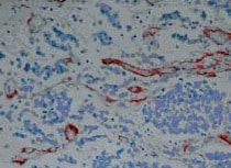
Application: Immunohistochemistry-ParaffinSample Tested: tumor tissue and Skin tumorSpecies: HumanVerified Customer | Posted 09/25/2021Paraffin-embedded sections from human skin tumor stained with tenascin c antibody (1:200).
 Bio-Techne ResponseThis review was submitted through the legacy Novus Innovators Program, reflecting a new species or application tested on a primary antibody.
 -
Application: Immunocytochemistry/ImmunofluorescenceSample Tested: Melanoma tissueSpecies: HumanVerified Customer | Posted 11/23/2020
-
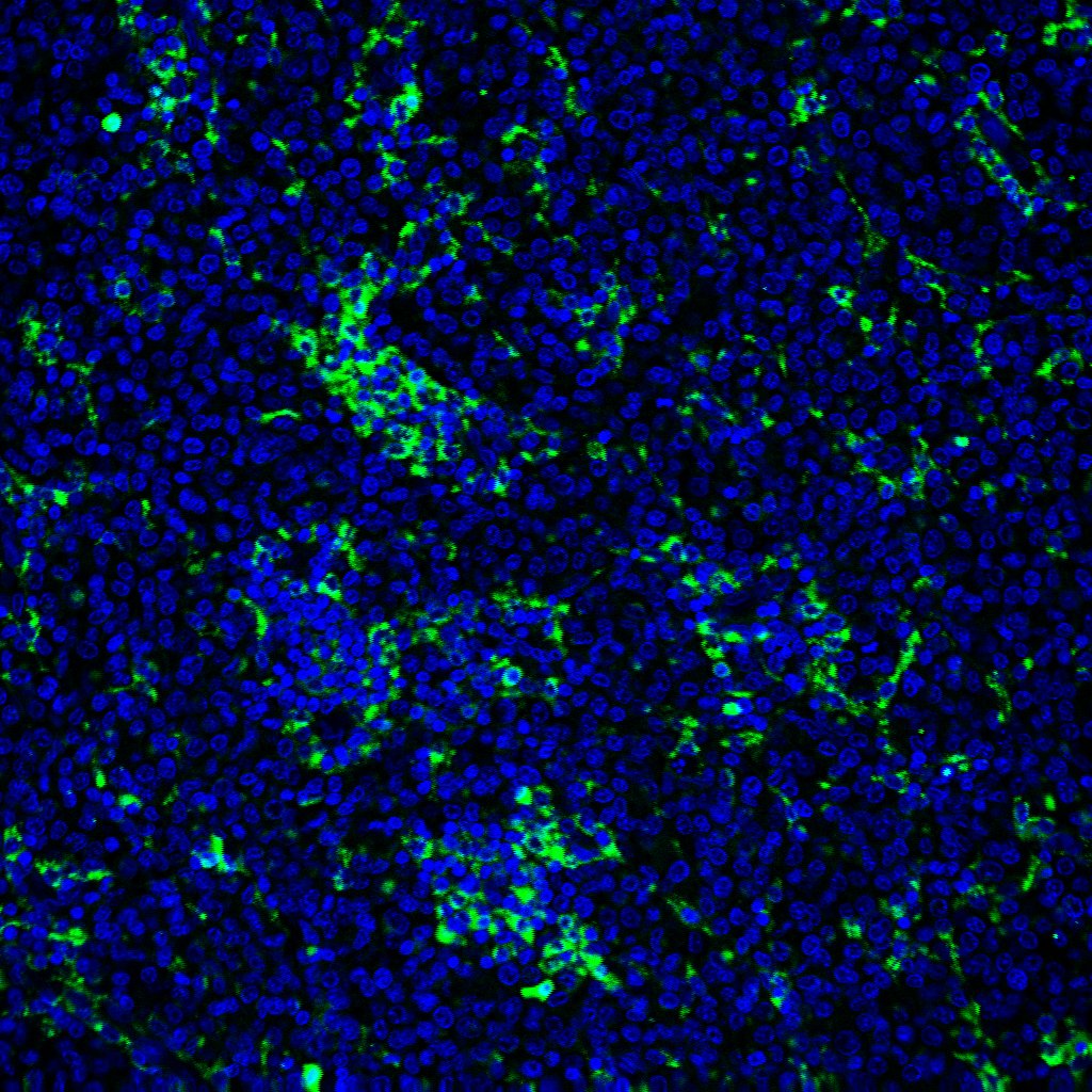
Application: Immunocytochemistry/ImmunofluorescenceSample Tested: Skin tissueSpecies: MouseVerified Customer | Posted 11/11/2020Skin tissue derived from a mouse model of epidermolysis bullosa. The skin displays a splitting in the dermal-epidermal junction, and an exacerbated tenascin C deposition in the dermis.
-
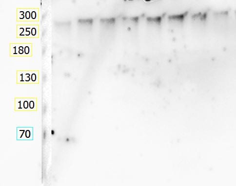
Application: Western BlotSample Tested: SVG-A immortalized astrocytesSpecies: HumanVerified Customer | Posted 06/17/2019SVGA immortalized astrocytes were stimulated with different cytokines for 24 h (unstimulated control: right lane); SDS-page was performed using 15 µg protein/lane, and anti-tenascin 1:500. Band at predicted molecular weight, no additional bands.
-
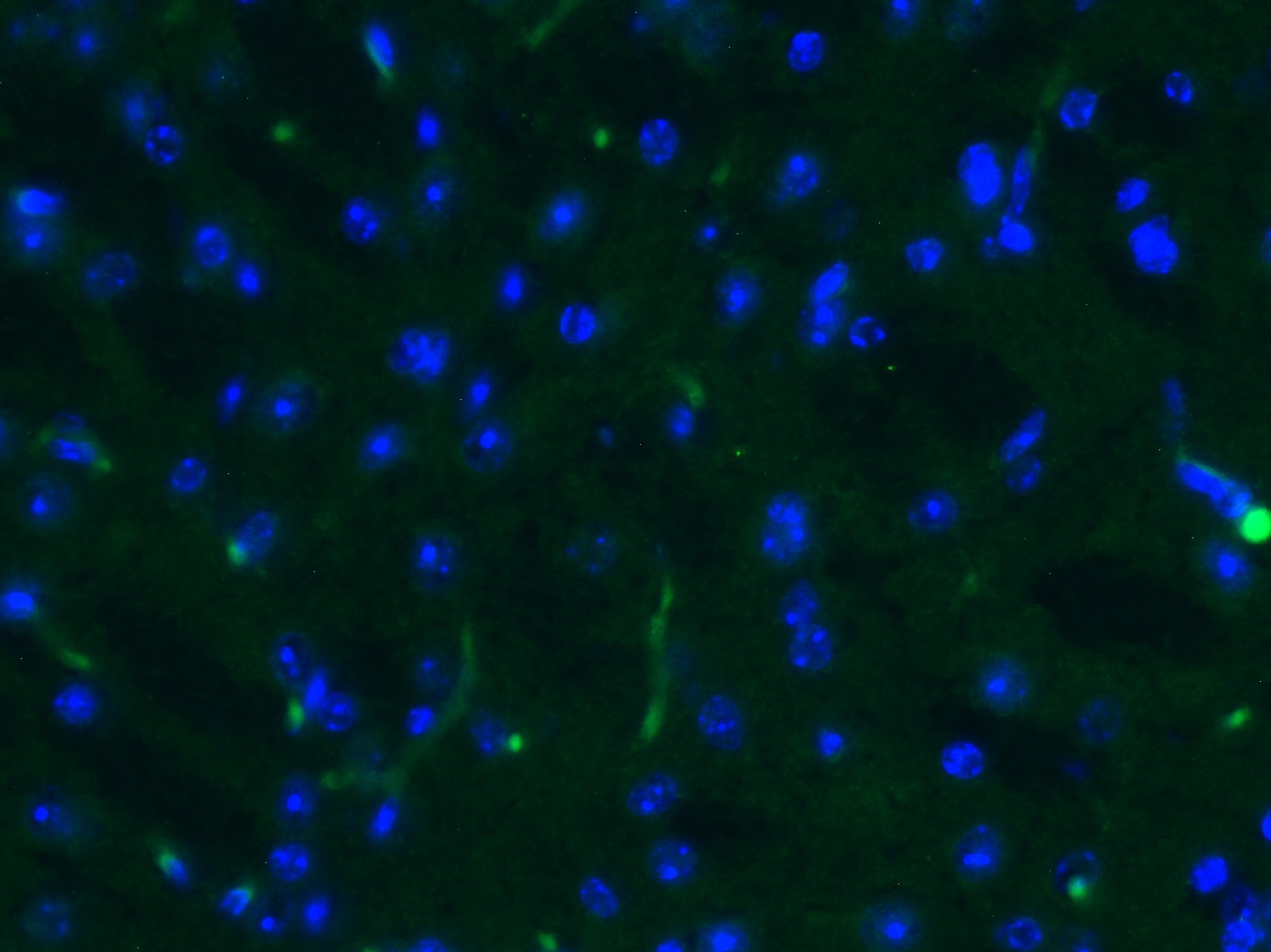
Application: Immunohistochemistry-FrozenSample Tested: Mouse brainSpecies: MouseVerified Customer | Posted 03/11/201910 µm Cryosections from adult mouse brain stained with tenascin c antibody (1:200) and Alexa Fluor 488 donkey anti rat IgG. Staining is quite faint (it's adult brain), but looks rather specific.
-
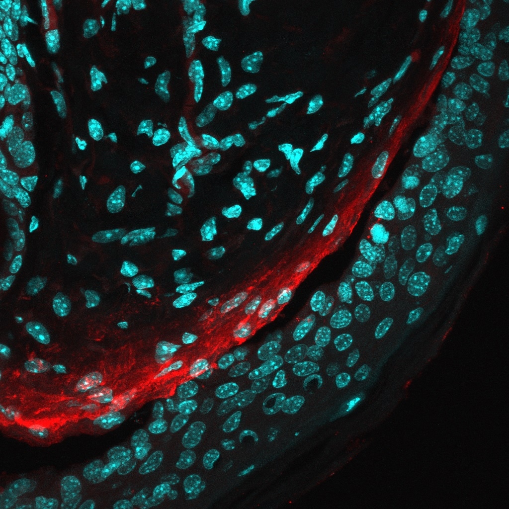
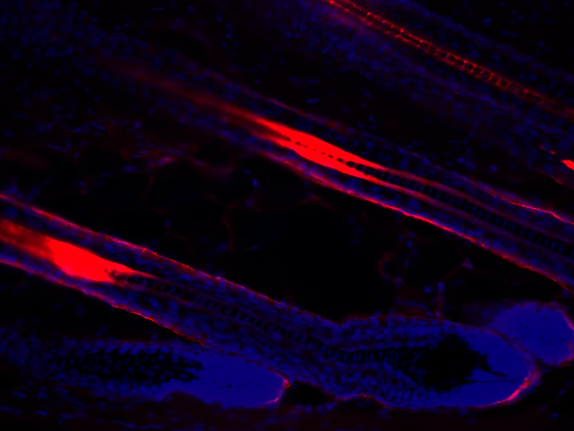
Application: ImmunofluorescenceSample Tested: Mouse skin (hair follicle)Species: MouseVerified Customer | Posted 12/11/2015Tenascin C (red) found outside the hair follicle outer root sheath
There are no reviews that match your criteria.
Protocols
Find general support by application which include: protocols, troubleshooting, illustrated assays, videos and webinars.
- Appropriate Fixation of IHC/ICC Samples
- Cellular Response to Hypoxia Protocols
- ClariTSA™ Fluorophore Kits
- Detection & Visualization of Antibody Binding
- ICC Cell Smear Protocol for Suspension Cells
- ICC Immunocytochemistry Protocol Videos
- ICC for Adherent Cells
- Immunocytochemistry (ICC) Protocol
- Immunocytochemistry Troubleshooting
- Immunofluorescence of Organoids Embedded in Cultrex Basement Membrane Extract
- Immunohistochemistry (IHC) and Immunocytochemistry (ICC) Protocols
- Preparing Samples for IHC/ICC Experiments
- Preventing Non-Specific Staining (Non-Specific Binding)
- Primary Antibody Selection & Optimization
- Protocol for VisUCyte™ HRP Polymer Detection Reagent
- Protocol for the Fluorescent ICC Staining of Cell Smears - Graphic
- Protocol for the Fluorescent ICC Staining of Cultured Cells on Coverslips - Graphic
- Protocol for the Preparation and Fluorescent ICC Staining of Cells on Coverslips
- Protocol for the Preparation and Fluorescent ICC Staining of Non-adherent Cells
- Protocol for the Preparation and Fluorescent ICC Staining of Stem Cells on Coverslips
- Protocol for the Preparation of a Cell Smear for Non-adherent Cell ICC - Graphic
- R&D Systems Quality Control Western Blot Protocol
- TUNEL and Active Caspase-3 Detection by IHC/ICC Protocol
- The Importance of IHC/ICC Controls
- Troubleshooting Guide: Western Blot Figures
- Western Blot Conditions
- Western Blot Protocol
- Western Blot Protocol for Cell Lysates
- Western Blot Troubleshooting
- Western Blot Troubleshooting Guide
- View all Protocols, Troubleshooting, Illustrated assays and Webinars
Loading...