Human phospho-Chk2 (T68) Antibody
R&D Systems | Catalog # AF1626
 by Western Blot.")
Discontinued Product
AF1626 has been discontinued.
View all Chk2 products.
Key Product Details
Validated by
Biological Validation
Species Reactivity
Validated:
Human
Cited:
Human, Mouse
Applications
Validated:
Western Blot, Immunocytochemistry
Cited:
Western Blot, Immunocytochemistry
Label
Unconjugated
Antibody Source
Polyclonal Rabbit IgG
Loading...
Product Specifications
Immunogen
Phosphopeptide containing human Chk2 T68 site
Specificity
Detects human Chk2 when phosphorylated at T68.
Clonality
Polyclonal
Host
Rabbit
Isotype
IgG
Scientific Data Images for Human phospho-Chk2 (T68) Antibody
Detection of Human Phospho-Chk2 (T68) by Western Blot.
Western blot shows lysates of HeLa human cervical epithelial carcinoma cell line untreated (-) or treated (+) with 1 µM camptothecin (CPT) for 1 hour. PVDF membrane was probed with 0.5 µg/mL Rabbit Anti-Human Phospho-Chk2 (T68) Antigen Affinity-purified Polyclonal Antibody (Catalog # AF1626) followed by HRP-conjugated Anti-Rabbit IgG Secondary Antibody (Catalog # HAF008). A specific band for Phospho-Chk2 (T68) was detected at approximately 64 kDa (as indicated). The phospho-specificity of this antibody was supported by decreased labeling following treatment with 600 U lambda-phosphatase (lambda-PPase) for 60 minutes. This experiment was conducted under reducing conditions and using Immunoblot Buffer Group 1. in HepG2 Human Cell Line.")
Phospho-Chk2 (T68) in HepG2 Human Cell Line.
Phopsho-Chk2 (T68) was detected in immersion fixed HepG2 human hepatocellular carcinoma cell line treated with 1 µM camptothecin using Rabbit Anti-Human Phospho-Chk2 (T68) Antigen Affinity-purified Polyclonal Antibody (Catalog # AF1626) at 1 µg/mL for 3 hours at room temperature. Cells were stained using the NorthernLights™ 557-conjugated Anti-Rabbit IgG Secondary Antibody (red; Catalog # NL004) and counterstained with DAPI (blue). Specific staining was localized to nuclei in treated cells. View our protocol for Fluorescent ICC Staining of Cells on Coverslips.Applications for Human phospho-Chk2 (T68) Antibody
Application
Recommended Usage
Immunocytochemistry
5-15 µg/mL
Sample: Immersion fixed HeLa human cervical epithelial carcinoma cell line treated with camptothecin, immersion fixed HepG2 human hepatocellular carcinoma cell line treated with camptothecin, and immersion fixed equine peripheral blood mononuclear cells (PBMCs) treated with calcium ionomycin and PMA
Sample: Immersion fixed HeLa human cervical epithelial carcinoma cell line treated with camptothecin, immersion fixed HepG2 human hepatocellular carcinoma cell line treated with camptothecin, and immersion fixed equine peripheral blood mononuclear cells (PBMCs) treated with calcium ionomycin and PMA
Western Blot
0.5 µg/mL
Sample: HeLa human cervical epithelial carcinoma cell line treated with camptothecin
Sample: HeLa human cervical epithelial carcinoma cell line treated with camptothecin
Reviewed Applications
Read 2 reviews rated 4.5 using AF1626 in the following applications:
Formulation, Preparation, and Storage
Purification
Antigen Affinity-purified
Reconstitution
Reconstitute at 0.2 mg/mL in sterile PBS. For liquid material, refer to CoA for concentration.
Formulation
Lyophilized from a 0.2 μm filtered solution in PBS with Trehalose. *Small pack size (SP) is supplied either lyophilized or as a 0.2 µm filtered solution in PBS.
Shipping
Lyophilized product is shipped at ambient temperature. Liquid small pack size (-SP) is shipped with polar packs. Upon receipt, store immediately at the temperature recommended below.
Stability & Storage
Use a manual defrost freezer and avoid repeated freeze-thaw cycles.
- 12 months from date of receipt, -20 to -70 °C as supplied.
- 1 month, 2 to 8 °C under sterile conditions after reconstitution.
- 6 months, -20 to -70 °C under sterile conditions after reconstitution.
Calculators
Background: Chk2
Long Name
Checkpoint Kinase 2
Alternate Names
CDS1, CHEK2, HuCds1, LFS2, PP1425, Rad53
Gene Symbol
CHEK2
Additional Chk2 Products
Product Documents for Human phospho-Chk2 (T68) Antibody
Certificate of Analysis
To download a Certificate of Analysis, please enter a lot or batch number in the search box below.
Note: Certificate of Analysis not available for kit components.
Product Specific Notices for Human phospho-Chk2 (T68) Antibody
For research use only
Citations for Human phospho-Chk2 (T68) Antibody
 Powered by Bioz
Customer Reviews for Human phospho-Chk2 (T68) Antibody (2)
4.5 out of 5
2 Customer Ratings
Have you used Human phospho-Chk2 (T68) Antibody?
Submit a review and receive an Amazon gift card!
$25/€18/£15/$25CAN/¥2500 Yen for a review with an image
$10/€7/£6/$10CAN/¥1110 Yen for a review without an image
Submit a review
Customer Images

Showing
1
-
2 of
2 reviews
Showing All
Filter By:
-
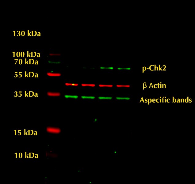
Application: Western BlotSample Tested: HepG2 human hepatocellular carcinoma cell lineSpecies: HumanVerified Customer | Posted 01/17/2023Ab concentration 0.5 ug/mL

-
Application: Western BlotSample Tested: U2OS human osteosarcoma cell lineSpecies: HumanVerified Customer | Posted 09/23/2021Excellent antibody, it worked extremely well for western blotting.
There are no reviews that match your criteria.
Protocols
Find general support by application which include: protocols, troubleshooting, illustrated assays, videos and webinars.
- Appropriate Fixation of IHC/ICC Samples
- Cellular Response to Hypoxia Protocols
- ClariTSA™ Fluorophore Kits
- Detection & Visualization of Antibody Binding
- ICC Cell Smear Protocol for Suspension Cells
- ICC Immunocytochemistry Protocol Videos
- ICC for Adherent Cells
- Immunocytochemistry (ICC) Protocol
- Immunocytochemistry Troubleshooting
- Immunofluorescence of Organoids Embedded in Cultrex Basement Membrane Extract
- Immunohistochemistry (IHC) and Immunocytochemistry (ICC) Protocols
- Preparing Samples for IHC/ICC Experiments
- Preventing Non-Specific Staining (Non-Specific Binding)
- Primary Antibody Selection & Optimization
- Protocol for VisUCyte™ HRP Polymer Detection Reagent
- Protocol for the Fluorescent ICC Staining of Cell Smears - Graphic
- Protocol for the Fluorescent ICC Staining of Cultured Cells on Coverslips - Graphic
- Protocol for the Preparation and Fluorescent ICC Staining of Cells on Coverslips
- Protocol for the Preparation and Fluorescent ICC Staining of Non-adherent Cells
- Protocol for the Preparation and Fluorescent ICC Staining of Stem Cells on Coverslips
- Protocol for the Preparation of a Cell Smear for Non-adherent Cell ICC - Graphic
- R&D Systems Quality Control Western Blot Protocol
- TUNEL and Active Caspase-3 Detection by IHC/ICC Protocol
- The Importance of IHC/ICC Controls
- Troubleshooting Guide: Western Blot Figures
- Western Blot Conditions
- Western Blot Protocol
- Western Blot Protocol for Cell Lysates
- Western Blot Troubleshooting
- Western Blot Troubleshooting Guide
- View all Protocols, Troubleshooting, Illustrated assays and Webinars
Loading...