
Siglecs are sialic acid specific I-type lectins that belong to the immunoglobulin superfamily. Structurally, they are transmembrane proteins with an N-terminal Ig-like V‑set domain followed by varying numbers of Ig-like C2-set domains (1, 2). Human Siglec-1, also known as sialoadhesin and CD169, is a 175‑185 kDa glycoprotein. It contains a 1622 amino acid (aa) extracellular domain (ECD) with one Ig-like V‑set domain and 16 Ig-like C2-set domains, a 21 aa transmembrane segment, and a 44 aa cytoplasmic domain (3). Within the ECD, human Siglec-1 shares approximately 70% aa sequence identity with mouse and rat Siglec-1. Alternate splicing generates a potentially soluble form of the ECD, and a second isoform with a substituted cytoplasmic domain. Siglec-1 expression is restricted to lymph node and splenic macrophages, plus some tissue macrophages (3). The adhesive function of Siglec-1 is supported by the N-terminal Ig-like domain which shows a selectivity for alpha 2,3‑linked sialic acid residues (3‑5). Siglec-1 binds a number of sialylated molecules including the mannose receptor, MGL1, MUC1, PSGL-1, and different glycoforms of CD43 (6‑9). Its binding capacity can be masked by endogenous sialylated molecules (10, 11). The sialylated and sulfated N-linked carbohydrates that modify Siglec-1 itself are required for ligand binding (6, 7). Siglec-1 is expressed on dendritic cells following rhinovirus exposure, and these DC promote T cell anergy (12). It is also induced on circulating monocytes during systemic sclerosis and HIV-1 infection (13‑15). Siglec-1 can trap HIV-1 particles for trans infection of permissive cells (14).
Human Siglec-1/CD169 Antibody
R&D Systems | Catalog # AF5197

Scientific images may be resized, cropped, or adjusted for brightness or contrast for presentation purposes. These updates are limited to presentation and do not affect the underlying scientific interpretation of the data. Where available, additional source data may be provided upon request.
Key Product Details
Species Reactivity
Validated:
Cited:
Applications
Validated:
Cited:
Label
Antibody Source
Product Specifications
Immunogen
Ser20-Gln1641
Accession # Q9BZZ2
Specificity
Clonality
Host
Isotype
Endotoxin Level
Scientific Data Images for Human Siglec-1/CD169 Antibody
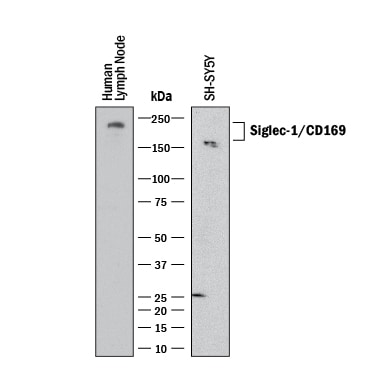
Detection of Human Siglec‑1/CD169 by Western Blot.
Western blot shows lysates of SH‑SY5Y human neuroblastoma cell line and human lymph node. PVDF membrane was probed with 0.5 µg/mL of Sheep Anti-Human Siglec‑1/CD169 Antigen Affinity-purified Polyclonal Antibody (Catalog # AF5197) followed by HRP-conjugated Anti-Sheep IgG Secondary Antibody (HAF016). A specific band was detected for Siglec‑1/CD169 at approximately 180-200 kDa (as indicated). This experiment was conducted under reducing conditions and using Western Blot Buffer Group 1.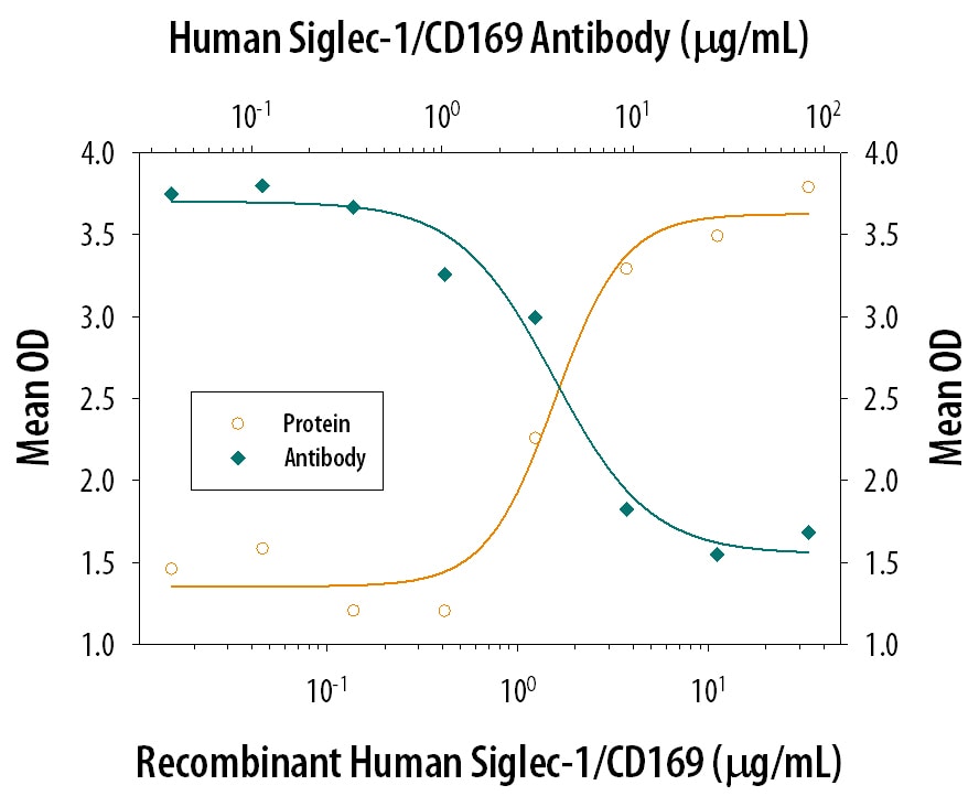
Cell Adhesion Mediated by Siglec‑1/CD169 and Neutralization by Human Siglec‑1/CD169 Antibody.
Recombinant Human Siglec-1/CD169 Fc Chimera (Catalog # 5197-SL), immobilized onto a microplate, supports the adhesion of human red blood cells in a dose-dependent manner (orange line). Adhesion elicited by Recombinant Human Siglec-1/CD169 Fc Chimera (5 µg/mL) is neutralized (green line) by increasing concentrations of Sheep Anti-Human Siglec-1/CD169 Antigen Affinity-purified Polyclonal Antibody (Catalog # AF5197). The ND50 is typically 1.5-7.5 µg/mL.Applications for Human Siglec-1/CD169 Antibody
Western Blot
Sample: SH‑SY5Y human neuroblastoma cell line and human lymph node
Neutralization
Formulation, Preparation, and Storage
Purification
Reconstitution
Reconstitute at 0.2 mg/mL in sterile PBS. For liquid material, refer to CoA for concentration.
Formulation
Shipping
Stability & Storage
- 12 months from date of receipt, -20 to -70 °C as supplied.
- 1 month, 2 to 8 °C under sterile conditions after reconstitution.
- 6 months, -20 to -70 °C under sterile conditions after reconstitution.
Calculators
Background: Siglec-1/CD169
References
- Varki, A. and T. Angata (2006) Glycobiology 16:1R.
- Crocker, P.R. et al. (2007) Nat. Rev. Immunol. 7:255.
- Hartnell, A. et al. (2001) Blood 97:288.
- Nath, D. et al. (1995) J. Biol. Chem. 270:26184.
- Crocker, P.R. et al. (1991) EMBO J. 10:1661.
- Martinez-Pomares, L. et al. (1999) J. Biol. Chem. 274:35211.
- Kumamoto, Y. et al. (2004) J. Biol. Chem. 279:49274.
- Nath, D. et al. (1999) Immunology 98:213.
- van den Berg, T.K. et al. (2001) J. Immunol. 166:3637.
- Nakamura, K. et al. (2002) Glycobiology 12:209.
- Barnes, Y.C. et al. (1999) Blood 93:1245.
- Kirchberger, S. et al. (2005) J. Immunol. 175:1145.
- York, M.R. et al. (2007) Arthritis Rheum. 56:1010.
- Rempel, H. et al. (2008) PloS ONE 3:e1967.
- van der Kuyl, A.C. et al. (2007) Plos ONE 2:e257.
Long Name
Alternate Names
Gene Symbol
UniProt
Additional Siglec-1/CD169 Products
Product Documents for Human Siglec-1/CD169 Antibody
Certificate of Analysis
To download a Certificate of Analysis, please enter a lot or batch number in the search box below.
Note: Certificate of Analysis not available for kit components.
Product Specific Notices for Human Siglec-1/CD169 Antibody
For research use only
Related Research Areas
Citations for Human Siglec-1/CD169 Antibody
 Powered by Bioz
Customer Reviews for Human Siglec-1/CD169 Antibody
There are currently no reviews for this product. Be the first to review Human Siglec-1/CD169 Antibody and earn rewards!
Have you used Human Siglec-1/CD169 Antibody?
Submit a review and receive an Amazon gift card!
$25/€18/£15/$25CAN/¥2500 for a review with an image
$10/€7/£6/$10CAN/¥1110 for a review without an image
Submit a review
Protocols
Find general support by application which include: protocols, troubleshooting, illustrated assays, videos and webinars.
- Cellular Response to Hypoxia Protocols
- R&D Systems Quality Control Western Blot Protocol
- Troubleshooting Guide: Western Blot Figures
- Western Blot Conditions
- Western Blot Protocol
- Western Blot Protocol for Cell Lysates
- Western Blot Troubleshooting
- Western Blot Troubleshooting Guide
- View all Protocols, Troubleshooting, Illustrated assays and Webinars