SPARCL1 (Secreted Protein, Acidic and Rich in Cysteines-like 1), also known as hevin, SC1 or MAST9, is a member of the SPARC family of extracellular glycoproteins (1, 2). SPARCL1 is an anti-adhesive protein that is widely expressed in tissues such as brain, heart, lung, muscle and kidney, but not liver (3, 4). Human SPARCL1 contains a 16 amino acid (aa) signal sequence and a 648 aa mature region with four domains: a 416 aa N-terminal acidic region, a 23 aa follistatin-like domain, a 55 aa kazal-like segment and a 48 aa EF-hand/calcium-binding domain (3, 4). SPARCL1 is predicted at 75 kDa, but migrates at ~130 kDa, which has been explained either by disulfide-linked homodimerization or by glycosylation and high acidity (3-5). Some truncated forms have been reported. In mouse, a 55 kDa C-terminal fragment is the only form in kidney and represents a portion of SPARCL1 in other tissues (6). In humans, a 25 kDa form is increased in liver tumors that are encapsulated, while the full-length form is downregulated in many epithelial cell-derived tumors (7, 8). SPARCL1 inhibits adhesion and spreading on a variety of substrates (5, 9). It is thought to cause antiadhesive signaling that terminates neuronal migration, consistent with production by glial and neuronal cells during development or in response to trauma (10). In tonsillar high endothelial venules (HEV), SPARCL1 may induce endothelial cell dissociation, promoting extravasation (3). SPARCL1 binds collagen; in mice, deletion causes dermal collagen fibrils that are smaller in diameter and deficient in decorin (6, 11). Human mature SPARCL1 shares 67%, 69%, 78%, 76%, 72%, and 72% aa identity with mouse, rat, equine, canine, porcine and bovine SPARCL1, respectively. The follistatin-like, kazal-like and calcium-binding domains of SPARCL1 show 61% aa identity with corresponding regions of SPARC.
Human SPARC-like 1/SPARCL1 Antibody
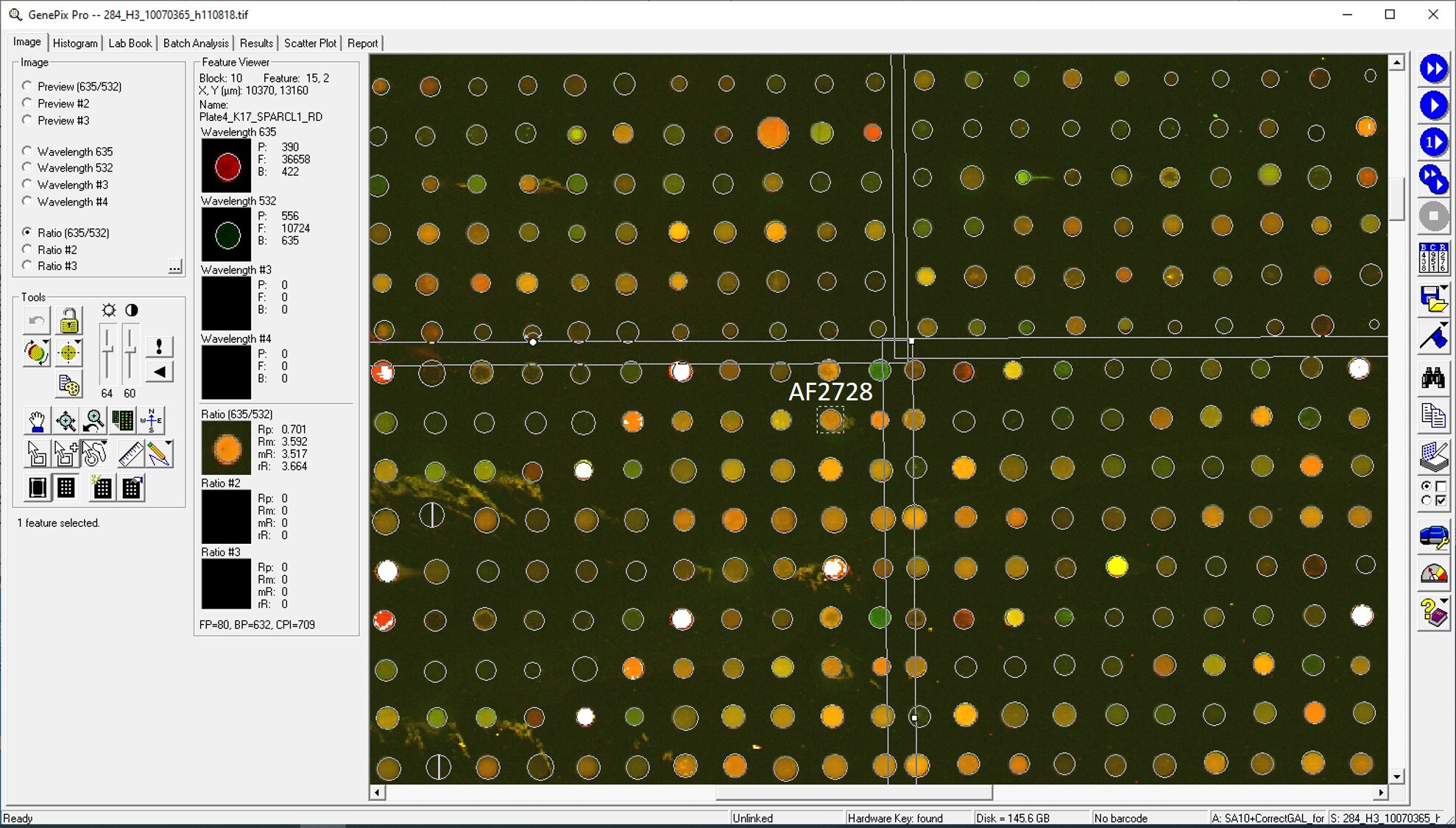
R&D Systems | Catalog # AF2728

Discontinued Product
AF2728 has been discontinued.
View all SPARC-like 1/SPARCL1 products.
Key Product Details
Species Reactivity
Validated:
Human
Cited:
Human, Mouse, Xenograft
Applications
Validated:
Immunohistochemistry, Western Blot, Intracellular Staining by Flow Cytometry, CyTOF-ready
Cited:
Immunohistochemistry, Immunohistochemistry-Paraffin, Western Blot, Neutralization, Immunocytochemistry, FACS
Label
Unconjugated
Antibody Source
Polyclonal Goat IgG
Loading...
Product Specifications
Immunogen
Mouse myeloma cell line NS0-derived recombinant human SPARC-like 1/SPARCL1 (R&D Systems, Catalog # 2728-SL)
Ile17-Phe664
Accession # Q8N4S1
Ile17-Phe664
Accession # Q8N4S1
Specificity
Detects human SPARC-like 1/SPARCL1 in direct ELISAs and Western blots. In direct ELISAs, less than 15% cross-reactivity with recombinant mouse (rm) SPARC-like 1/SPARCL1 is observed and less than 1% cross-reactivity with recombinant human SPARC is observed.
Clonality
Polyclonal
Host
Goat
Isotype
IgG
Scientific Data Images for Human SPARC-like 1/SPARCL1 Antibody
Detection of Human SPARC‑like 1/SPARCL1 by Western Blot.
Western blot shows lysates of human lung tissue. PVDF membrane was probed with 1 µg/mL of Goat Anti-Human SPARC-like 1/ SPARCL1 Antigen Affinity-purified Polyclonal Antibody (Catalog # AF2728) followed by HRP-conjugated Anti-Goat IgG Secondary Antibody (Catalog # HAF017). A specific band was detected for SPARC-like 1/SPARCL1 at approximately 130 kDa (as indicated). This experiment was conducted under reducing conditions and using Immunoblot Buffer Group 1.
SPARC‑like 1/SPARCL1 in Human Brain.
SPARC-like 1/SPARCL1 was detected in immersion fixed paraffin-embedded sections of human brain using Goat Anti-Human SPARC-like 1/SPARCL1 Antigen Affinity-purified Polyclonal Antibody (Catalog # AF2728) at 15 µg/mL overnight at 4 °C. Before incubation with the primary antibody, tissue was subjected to heat-induced epitope retrieval using Antigen Retrieval Reagent-Basic (Catalog # CTS013). Tissue was stained using the Anti-Goat HRP-DAB Cell & Tissue Staining Kit (brown; Catalog # CTS008) and counterstained with hematoxylin (blue). Specific staining was localized to neurons. View our protocol for Chromogenic IHC Staining of Paraffin-embedded Tissue Sections.
Detection of SPARC‑like 1/SPARCL1 in HL‑60 Human Cell Line by Flow Cytometry.
HL-60 human acute promyelocytic leukemia cell line was stained with Goat Anti-Human SPARC-like 1/SPARCL1 Antigen Affinity-purified Polyclonal Antibody (Catalog # AF2728, filled histogram) or control antibody (Catalog # AB-108-C, open histogram), followed by Phycoerythrin-conjugated Anti-Goat IgG Secondary Antibody (Catalog # F0107). To facilitate intracellular staining, cells were fixed with paraformaldehyde and permeabilized with saponin.Applications for Human SPARC-like 1/SPARCL1 Antibody
Application
Recommended Usage
CyTOF-ready
Ready to be labeled using established conjugation methods. No BSA or other carrier proteins that could interfere with conjugation.
Immunohistochemistry
5-15 µg/mL
Sample: Immersion fixed paraffin-embedded sections of human brain
Sample: Immersion fixed paraffin-embedded sections of human brain
Intracellular Staining by Flow Cytometry
2.5 µg/106 cells
Sample:
Sample:
HL‑60 human acute promyelocytic leukemia cell line fixed with paraformaldehyde and permeabilized with saponin.
Western Blot
1 µg/mL
Sample: Human lung tissue
Sample: Human lung tissue
Reviewed Applications
Read 4 reviews rated 4.3 using AF2728 in the following applications:
Flow Cytometry Panel Builder
Bio-Techne Knows Flow Cytometry
Save time and reduce costly mistakes by quickly finding compatible reagents using the Panel Builder Tool.
Advanced Features
- Spectra Viewer - Custom analysis of spectra from multiple fluorochromes
- Spillover Popups - Visualize the spectra of individual fluorochromes
- Antigen Density Selector - Match fluorochrome brightness with antigen density

Formulation, Preparation, and Storage
Purification
Antigen Affinity-purified
Reconstitution
Reconstitute at 0.2 mg/mL in sterile PBS. For liquid material, refer to CoA for concentration.
Formulation
Lyophilized from a 0.2 μm filtered solution in PBS with Trehalose. *Small pack size (SP) is supplied either lyophilized or as a 0.2 µm filtered solution in PBS.
Shipping
Lyophilized product is shipped at ambient temperature. Liquid small pack size (-SP) is shipped with polar packs. Upon receipt, store immediately at the temperature recommended below.
Stability & Storage
Use a manual defrost freezer and avoid repeated freeze-thaw cycles.
- 12 months from date of receipt, -20 to -70 °C as supplied.
- 1 month, 2 to 8 °C under sterile conditions after reconstitution.
- 6 months, -20 to -70 °C under sterile conditions after reconstitution.
Calculators
Background: SPARC-like 1/SPARCL1
References
- Framson, P.E. and E.H. Sage (2004) J. Cell. Biochem. 92:679.
- Sullivan, M.M. and E.H. Sage (2004) Int. J. Biochem. Cell Biol. 36:991.
- Girard, J.P. and T.A. Springer (1995) Immunity 2:113.
- Bendik, I. et al. (1998) Cancer Res. 58:626.
- Brekken, R.A. et al. (2004) J. Histochem. Cytochem. 52:735.
- Hambrock, H.O. et al. (2003) J. Biol. Chem. 278:11351.
- Lau, C.P. et al. (2006) J. Pathol. 210:469.
- Isler, S.G. et al. (2001) Int. J. Oncol. 18:521.
- Girard, J.P. and T.A. Springer (1996) J. Biol. Chem. 271:4511.
- Gongidi, V. et al. (2004) Neuron 41:57.
- Sullivan, M.M. et al. (2006) J. Biol. Chem. 281:27621.
Alternate Names
Hevin, MAST9, SC1, SPARC like 1, SPARCL1
Gene Symbol
SPARCL1
UniProt
Additional SPARC-like 1/SPARCL1 Products
Product Documents for Human SPARC-like 1/SPARCL1 Antibody
Certificate of Analysis
To download a Certificate of Analysis, please enter a lot or batch number in the search box below.
Note: Certificate of Analysis not available for kit components.
Product Specific Notices for Human SPARC-like 1/SPARCL1 Antibody
For research use only
Related Research Areas
Citations for Human SPARC-like 1/SPARCL1 Antibody
 Powered by Bioz
Customer Reviews for Human SPARC-like 1/SPARCL1 Antibody (4)
4.3 out of 5
4 Customer Ratings
Have you used Human SPARC-like 1/SPARCL1 Antibody?
Submit a review and receive an Amazon gift card!
$25/€18/£15/$25CAN/¥2500 Yen for a review with an image
$10/€7/£6/$10CAN/¥1110 Yen for a review without an image
Submit a review
Customer Images
Showing
1
-
4 of
4 reviews
Showing All
Filter By:
-
Application: MicroarraysSample Tested: EDTA PlasmaSpecies: HumanVerified Customer | Posted 01/14/2021

-
Application: MicroarraysSample Tested: EDTA PlasmaSpecies: HumanVerified Customer | Posted 11/14/2018
-
Application: MicroarraySample Tested: EDTA PlasmaSpecies: HumanVerified Customer | Posted 10/09/2018
-
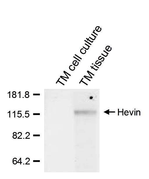
Application: Western BlotSample Tested: Trabecular meshwork tissueSpecies: HumanVerified Customer | Posted 10/05/2017
There are no reviews that match your criteria.
Protocols
Find general support by application which include: protocols, troubleshooting, illustrated assays, videos and webinars.
- 7-Amino Actinomycin D (7-AAD) Cell Viability Flow Cytometry Protocol
- Antigen Retrieval Protocol (PIER)
- Antigen Retrieval for Frozen Sections Protocol
- Appropriate Fixation of IHC/ICC Samples
- Cellular Response to Hypoxia Protocols
- Chromogenic IHC Staining of Formalin-Fixed Paraffin-Embedded (FFPE) Tissue Protocol
- Chromogenic Immunohistochemistry Staining of Frozen Tissue
- ClariTSA™ Fluorophore Kits
- Detection & Visualization of Antibody Binding
- Extracellular Membrane Flow Cytometry Protocol
- Flow Cytometry Protocol for Cell Surface Markers
- Flow Cytometry Protocol for Staining Membrane Associated Proteins
- Flow Cytometry Staining Protocols
- Flow Cytometry Troubleshooting Guide
- Fluorescent IHC Staining of Frozen Tissue Protocol
- Graphic Protocol for Heat-induced Epitope Retrieval
- Graphic Protocol for the Preparation and Fluorescent IHC Staining of Frozen Tissue Sections
- Graphic Protocol for the Preparation and Fluorescent IHC Staining of Paraffin-embedded Tissue Sections
- Graphic Protocol for the Preparation of Gelatin-coated Slides for Histological Tissue Sections
- IHC Sample Preparation (Frozen sections vs Paraffin)
- Immunofluorescent IHC Staining of Formalin-Fixed Paraffin-Embedded (FFPE) Tissue Protocol
- Immunohistochemistry (IHC) and Immunocytochemistry (ICC) Protocols
- Immunohistochemistry Frozen Troubleshooting
- Immunohistochemistry Paraffin Troubleshooting
- Intracellular Flow Cytometry Protocol Using Alcohol (Methanol)
- Intracellular Flow Cytometry Protocol Using Detergents
- Intracellular Nuclear Staining Flow Cytometry Protocol Using Detergents
- Intracellular Staining Flow Cytometry Protocol Using Alcohol Permeabilization
- Intracellular Staining Flow Cytometry Protocol Using Detergents to Permeabilize Cells
- Preparing Samples for IHC/ICC Experiments
- Preventing Non-Specific Staining (Non-Specific Binding)
- Primary Antibody Selection & Optimization
- Propidium Iodide Cell Viability Flow Cytometry Protocol
- Protocol for Heat-Induced Epitope Retrieval (HIER)
- Protocol for Liperfluo
- Protocol for Making a 4% Formaldehyde Solution in PBS
- Protocol for VisUCyte™ HRP Polymer Detection Reagent
- Protocol for the Characterization of Human Th22 Cells
- Protocol for the Characterization of Human Th9 Cells
- Protocol for the Preparation & Fixation of Cells on Coverslips
- Protocol for the Preparation and Chromogenic IHC Staining of Frozen Tissue Sections
- Protocol for the Preparation and Chromogenic IHC Staining of Frozen Tissue Sections - Graphic
- Protocol for the Preparation and Chromogenic IHC Staining of Paraffin-embedded Tissue Sections
- Protocol for the Preparation and Chromogenic IHC Staining of Paraffin-embedded Tissue Sections - Graphic
- Protocol for the Preparation and Fluorescent IHC Staining of Frozen Tissue Sections
- Protocol for the Preparation and Fluorescent IHC Staining of Paraffin-embedded Tissue Sections
- Protocol for the Preparation of Gelatin-coated Slides for Histological Tissue Sections
- Protocol: Annexin V and PI Staining by Flow Cytometry
- Protocol: Annexin V and PI Staining for Apoptosis by Flow Cytometry
- R&D Systems Quality Control Western Blot Protocol
- TUNEL and Active Caspase-3 Detection by IHC/ICC Protocol
- The Importance of IHC/ICC Controls
- Troubleshooting Guide: Fluorokine Flow Cytometry Kits
- Troubleshooting Guide: Immunohistochemistry
- Troubleshooting Guide: Western Blot Figures
- Western Blot Conditions
- Western Blot Protocol
- Western Blot Protocol for Cell Lysates
- Western Blot Troubleshooting
- Western Blot Troubleshooting Guide
- View all Protocols, Troubleshooting, Illustrated assays and Webinars
Loading...