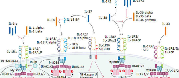
ST2, also known as IL-1 R4 and T1, is an Interleukin-1 receptor family glycoprotein that contributes to Th2 immune responses (1, 2). Human ST2 consists of a 310 amino acid (aa) extracellular domain (ECD) with three Ig-like domains, a 21 aa transmembrane segment, and a 207 aa cytoplasmic domain with an intracellular TIR domain (3, 4). Alternate splicing of the 120 kDa human ST2 generates a soluble 60 kDa isoform that lacks the transmembrane and cytoplasmic regions as well as an isoform that additionally lacks the third Ig‑like domain (4). Within the ECD, human ST2 shares 68% and 64% aa sequence identity with mouse and rat ST2, respectively. ST2 is expressed on the surface of mast cells, activated Th2 cells, macrophages, and cardiac myocytes (5-8). It binds IL-33, a cytokine that is upregulated by inflammation or mechanical strain in smooth muscle cells, airway epithelia, keratinocytes, and cardiac fibroblasts (5, 9). IL-33 binding induces the association of ST2 with IL-1R AcP, a shared signaling subunit that also associates with IL-1 RI and IL-1 R rp2 (1, 10, 11). In macrophages, ST2 interferes with signaling from IL-1 RI and TLR4 by sequestering the adaptor proteins MyD88 and Mal (7). In addition to its role in promoting mast cell and Th2 dependent inflammation, ST2 activation enhances antigen induced hypernociception and protects from atherosclerosis and cardiac hypertrophy (5, 12-14). The soluble ST2 isoform is released by activated Th2 cells and strained cardiac myocytes and is elevated in the serum in allergic asthma (6, 8, 15). Soluble ST2 functions as a decoy receptor that blocks IL‑33’s ability to signal through transmembrane ST2 (10, 13‑15).
Human ST2/IL-33R LlaMABodyTM Bivalent VHH HuIgG2 Fusion Antibody (L000.2.694H)
R&D Systems | Catalog # LMAB108591

Scientific images may be resized, cropped, or adjusted for brightness or contrast for presentation purposes. These updates are limited to presentation and do not affect the underlying scientific interpretation of the data. Where available, additional source data may be provided upon request.
Key Product Details
Species Reactivity
Applications
Label
Antibody Source
Product Specifications
Immunogen
Lys19-Phe328
Accession # BAA02233
Specificity
Clonality
Host
Isotype
Scientific Data Images
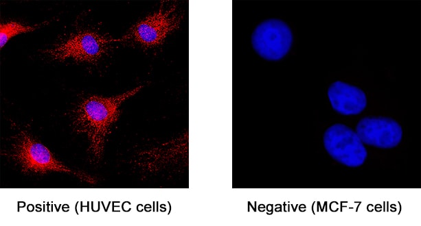
ST2/IL-33R in HUVEC Human Cells.
ST2/IL-33R was detected in immersion fixed HUVEC human umbilical vein endothelial cells (positive staining) and MCF‑7 human breast cancer cell line (negative staining) using Llama Anti-Human ST2/IL-33R LlamabodyTM Bivalent VHH HuIgG2 Fusion Monoclonal Antibody (Catalog # LMAB108591) at 8 µg/mL for 3 hours at room temperature. Cells were stained using an Anti-alpaca Alexa Fluor 594 (red) and counterstained with DAPI (blue). Specific staining was localized to cytoplasm. Staining was performed using our protocol for Fluorescent ICC Staining of Non-adherent Cells.Applications
Immunocytochemistry
Sample: Immersion fixed HUVEC human umbilical vein endothelial cells
Formulation, Preparation, and Storage
Purification
Reconstitution
Reconstitute at 0.5 mg/mL in sterile PBS. For liquid material, refer to CoA for concentration.
Formulation
Shipping
Stability & Storage
- 12 months from date of receipt, -20 to -70 °C as supplied.
- 1 month, 2 to 8 °C under sterile conditions after reconstitution.
- 6 months, -20 to -70 °C under sterile conditions after reconstitution.
Calculators
Background: ST2/IL-33R
References
-
Barksby, H.E. et al. (2007) Clin. Exp. Immunol. 149:217.
-
Gadina, M. and C.A. Jefferies (2007) Science STKE 2007:pe31.
-
Tominaga, S. et al. (1992) Biochim. Biophys. Acta 1171:215.
-
Li, H. et al. (2000) Genomics 67:284.
-
Schmitz, J. et al. (2005) Immunity 23:479.
-
Lecart, S. et al. (2002) Eur. J. Immunol. 32:2979.
-
Brint, E.K. et al. (2004) Nat. Immunol. 5:373.
-
Weinberg, E.O. et al. (2002) Circulation 106:2961.
-
Sanada S. et al. (2007) J. Clin. Invest. 117:1538.
-
Palmer, G. et al. (2008) Cytokine 42:358.
-
Chackerian, A.A. et al. (2007) J. Immunol. 179:2551.
-
Allakhverdi, Z. et al. (2007) J. Immunol. 179:2051.
-
Verri Jr., W.A. et al. (2008) Proc. Natl. Acad. Sci. 105:2723.
-
Miller, A.M. et al. (2008) J. Exp. Med. 205:339.
-
Hayakawa, H. et al. (2007) J. Biol. Chem. 282:26369.
Long Name
Alternate Names
Gene Symbol
UniProt
Additional ST2/IL-33R Products
Product Documents
Certificate of Analysis
To download a Certificate of Analysis, please enter a lot or batch number in the search box below.
Note: Certificate of Analysis not available for kit components.
Product Specific Notices
For research use only
Related Research Areas
Customer Reviews
There are currently no reviews for this product. Be the first to review Human ST2/IL-33R LlaMABodyTM Bivalent VHH HuIgG2 Fusion Antibody (L000.2.694H) and earn rewards!
Have you used Human ST2/IL-33R LlaMABodyTM Bivalent VHH HuIgG2 Fusion Antibody (L000.2.694H)?
Submit a review and receive an Amazon gift card!
$25/€18/£15/$25CAN/¥2500 for a review with an image
$10/€7/£6/$10CAN/¥1110 for a review without an image
Submit a review
Protocols
Find general support by application which include: protocols, troubleshooting, illustrated assays, videos and webinars.
- Appropriate Fixation of IHC/ICC Samples
- Cellular Response to Hypoxia Protocols
- ClariTSA™ Fluorophore Kits
- Detection & Visualization of Antibody Binding
- ICC Cell Smear Protocol for Suspension Cells
- ICC Immunocytochemistry Protocol Videos
- ICC for Adherent Cells
- Immunocytochemistry (ICC) Protocol
- Immunocytochemistry Troubleshooting
- Immunofluorescence of Organoids Embedded in Cultrex Basement Membrane Extract
- Immunohistochemistry (IHC) and Immunocytochemistry (ICC) Protocols
- Preparing Samples for IHC/ICC Experiments
- Preventing Non-Specific Staining (Non-Specific Binding)
- Primary Antibody Selection & Optimization
- Protocol for VisUCyte™ HRP Polymer Detection Reagent
- Protocol for the Fluorescent ICC Staining of Cell Smears - Graphic
- Protocol for the Fluorescent ICC Staining of Cultured Cells on Coverslips - Graphic
- Protocol for the Preparation and Fluorescent ICC Staining of Cells on Coverslips
- Protocol for the Preparation and Fluorescent ICC Staining of Non-adherent Cells
- Protocol for the Preparation and Fluorescent ICC Staining of Stem Cells on Coverslips
- Protocol for the Preparation of a Cell Smear for Non-adherent Cell ICC - Graphic
- TUNEL and Active Caspase-3 Detection by IHC/ICC Protocol
- The Importance of IHC/ICC Controls
- View all Protocols, Troubleshooting, Illustrated assays and Webinars
FAQs
-
Q: What are Camelid Antibodies?
A: Camelid antibodies are antibodies from the Camelidae family of mammals that include llamas, camels, and alpacas. These animals produce 2 main types of antibodies. One type of antibody camelids produce is the conventional antibody that is made up of 2 heavy chains and 2 light chains. They also produce another type of antibody that is made up of only 2 heavy chains. This is known as heavy chain IgG (hcIgG). While these antibodies do not contain the CH1 region, they retain an antigen binding domain called the VHH region. VHH antibodies, also known as single domain antibodies or Nanobodies®, contain only the VHH region from the camelid antibody.
Associated Pathways