Small Ubiquitin-like Modifiers (SUMOs) are a family of small, related proteins that can be enzymatically attached to a target protein by a post-translational modification process termed sumoylation. Unlike ubiquitination, which targets proteins for degradation, sumoylation participates in a number of cellular processes, such as nuclear transport, transcriptional regulation, apoptosis, and protein stability. All SUMO proteins share the conserved ubiquitin domain and the C‑terminal diglycine cleavage/attachment site. Human SUMO3, also known as SMT3A, is synthesized as a 103 amino acid (aa), 11.5 kDa propeptide that contains an 11 aa C‑terminal prosegment. Following prosegment cleavage, the C‑terminal glycine is enzymatically attached to a lysine on a target protein. Human SUMO3 shares 83% sequence identity with SUMO3 from mouse. SUMO3 also has very high sequence homology to SUMO2 and SUMO4, 87 % and 75%, respectively. SUMO3 shares only 47% sequence identity to SUMO1.

Key Product Details
Species Reactivity
Human
Applications
Immunocytochemistry
Label
Unconjugated
Antibody Source
Monoclonal Rat IgG2A Clone # 401513
Loading...
Product Specifications
Immunogen
E. coli-derived recombinant human SUMO3
Met1-Phe103
Accession # P55854
Met1-Phe103
Accession # P55854
Specificity
Detects human SUMO3 in direct ELISAs.
Clonality
Monoclonal
Host
Rat
Isotype
IgG2A
Scientific Data Images for Human SUMO3 Antibody (401513)
SUMO3 in HEK293 Human Cell Line.
SUMO3 was detected in immersion fixed HEK293 human embryonic kidney cell line using Rat Anti-Human SUMO3 Monoclonal Antibody (Catalog # MAB2959) at 10 µg/mL for 3 hours at room temperature. Cells were stained using the NorthernLights™ 557-conjugated Anti-Rat IgG Secondary Antibody (red; Catalog # NL013) and counterstained with DAPI (blue). Specific staining was localized to cytoplasm and nuclei. View our protocol for Fluorescent ICC Staining of Cells on Coverslips.Applications for Human SUMO3 Antibody (401513)
Application
Recommended Usage
Immunocytochemistry
8-25 µg/mL
Sample: Immersion fixed HEK293 human embryonic kidney cell line
Sample: Immersion fixed HEK293 human embryonic kidney cell line
Reviewed Applications
Read 1 review rated 4 using MAB2959 in the following applications:
Formulation, Preparation, and Storage
Purification
Protein A or G purified from hybridoma culture supernatant
Reconstitution
Reconstitute at 0.5 mg/mL in sterile PBS. For liquid material, refer to CoA for concentration.
Loading...
Formulation
Lyophilized from a 0.2 μm filtered solution in PBS with Trehalose. *Small pack size (SP) is supplied either lyophilized or as a 0.2 µm filtered solution in PBS.
Shipping
Lyophilized product is shipped at ambient temperature. Liquid small pack size (-SP) is shipped with polar packs. Upon receipt, store immediately at the temperature recommended below.
Stability & Storage
Use a manual defrost freezer and avoid repeated freeze-thaw cycles.
- 12 months from date of receipt, -20 to -70 °C as supplied.
- 1 month, 2 to 8 °C under sterile conditions after reconstitution.
- 6 months, -20 to -70 °C under sterile conditions after reconstitution.
Calculators
Background: SUMO3
Long Name
Small Ubiquitin-like Modifier 3
Alternate Names
SMT3A, SMT3H1
Entrez Gene IDs
6612 (Human)
Gene Symbol
SUMO3
UniProt
Additional SUMO3 Products
Product Documents for Human SUMO3 Antibody (401513)
Certificate of Analysis
To download a Certificate of Analysis, please enter a lot or batch number in the search box below.
Note: Certificate of Analysis not available for kit components.
Product Specific Notices for Human SUMO3 Antibody (401513)
For research use only
Related Research Areas
Customer Reviews for Human SUMO3 Antibody (401513) (1)
4 out of 5
1 Customer Rating
Have you used Human SUMO3 Antibody (401513)?
Submit a review and receive an Amazon gift card!
$25/€18/£15/$25CAN/¥2500 Yen for a review with an image
$10/€7/£6/$10CAN/¥1110 Yen for a review without an image
Submit a review
Customer Images
Showing
1
-
1 of
1 review
Showing All
Filter By:
-
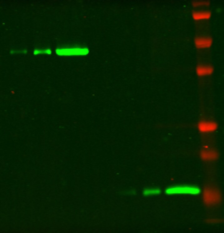
Application: Western BlotSample Tested: recombinant human proteinSpecies: HumanVerified Customer | Posted 03/31/2021Western blot: left to right: sumo3 conjugated-protein,5ng,10ng,50ng,Sumo3 protein(unconjugated) 5ng,10ng,50ng, protein size marker1/1000 primary antibody dilution

There are no reviews that match your criteria.
Protocols
Find general support by application which include: protocols, troubleshooting, illustrated assays, videos and webinars.
- Appropriate Fixation of IHC/ICC Samples
- Cellular Response to Hypoxia Protocols
- ClariTSA™ Fluorophore Kits
- Detection & Visualization of Antibody Binding
- ICC Cell Smear Protocol for Suspension Cells
- ICC Immunocytochemistry Protocol Videos
- ICC for Adherent Cells
- Immunocytochemistry (ICC) Protocol
- Immunocytochemistry Troubleshooting
- Immunofluorescence of Organoids Embedded in Cultrex Basement Membrane Extract
- Immunohistochemistry (IHC) and Immunocytochemistry (ICC) Protocols
- Preparing Samples for IHC/ICC Experiments
- Preventing Non-Specific Staining (Non-Specific Binding)
- Primary Antibody Selection & Optimization
- Protocol for VisUCyte™ HRP Polymer Detection Reagent
- Protocol for the Fluorescent ICC Staining of Cell Smears - Graphic
- Protocol for the Fluorescent ICC Staining of Cultured Cells on Coverslips - Graphic
- Protocol for the Preparation and Fluorescent ICC Staining of Cells on Coverslips
- Protocol for the Preparation and Fluorescent ICC Staining of Non-adherent Cells
- Protocol for the Preparation and Fluorescent ICC Staining of Stem Cells on Coverslips
- Protocol for the Preparation of a Cell Smear for Non-adherent Cell ICC - Graphic
- TUNEL and Active Caspase-3 Detection by IHC/ICC Protocol
- The Importance of IHC/ICC Controls
- View all Protocols, Troubleshooting, Illustrated assays and Webinars
Loading...
Associated Pathways

