
Human Tie-1 Antibody

R&D Systems | Catalog # AF619

R&D Systems

Key Product Details

Species Reactivity

Validated:

Human

Cited:

Human, Mouse

Applications

Validated:

Immunohistochemistry, Western Blot, Flow Cytometry, CyTOF-ready

Cited:

Immunohistochemistry, Western Blot, Flow Cytometry, Immunoprecipitation

Label

Unconjugated

Antibody Source

Polyclonal Goat IgG
Loading...

Product Specifications

Immunogen

Mouse myeloma cell line NS0-derived recombinant human Tie-1
Ala22-Gln760
Accession # P35590

Specificity

Detects human Tie-1 in direct ELISAs and Western blots. In direct ELISAs, approximately 50% cross-reactivity with recombinant mouse Tie-1 is observed and less than 1% cross-reactivity with recombinant human Tie‑2 is observed.

Clonality

Polyclonal

Host

Goat

Isotype

IgG

Scientific Data Images for Human Tie-1 Antibody

Detection of Tie‑1 antibody in HUVEC Human Cells antibody by Flow Cytometry.

Detection of Tie‑1 in HUVEC Human Cells by Flow Cytometry.

HUVEC human umbilical vein endothelial cells were stained with Goat Anti-Human Tie-1 Antigen Affinity-purified Polyclonal Antibody (Catalog # AF619, filled histogram) or control antibody (Catalog # AB-108-C, open histogram), followed by Phycoerythrin-conjugated Anti-Goat IgG Secondary Antibody (Catalog # F0107).
Tie‑1 antibody in Human Placenta by Immunohistochemistry (IHC-P).

Tie‑1 in Human Placenta.

Tie-1 was detected in immersion fixed paraffin-embedded sections of human placenta using 8 µg/mL Goat Anti-Human Tie-1 Antigen Affinity-purified Polyclonal Antibody (Catalog # AF619) overnight at 4 °C. Tissue was stained with the Anti-Goat HRP-DAB Cell & Tissue Staining Kit (brown; Catalog # CTS008) and counter-stained with hematoxylin (blue). View our protocol for Chromogenic IHC Staining of Paraffin-embedded Tissue Sections.
Detection of Tie-1 by Immunocytochemistry/ Immunofluorescence

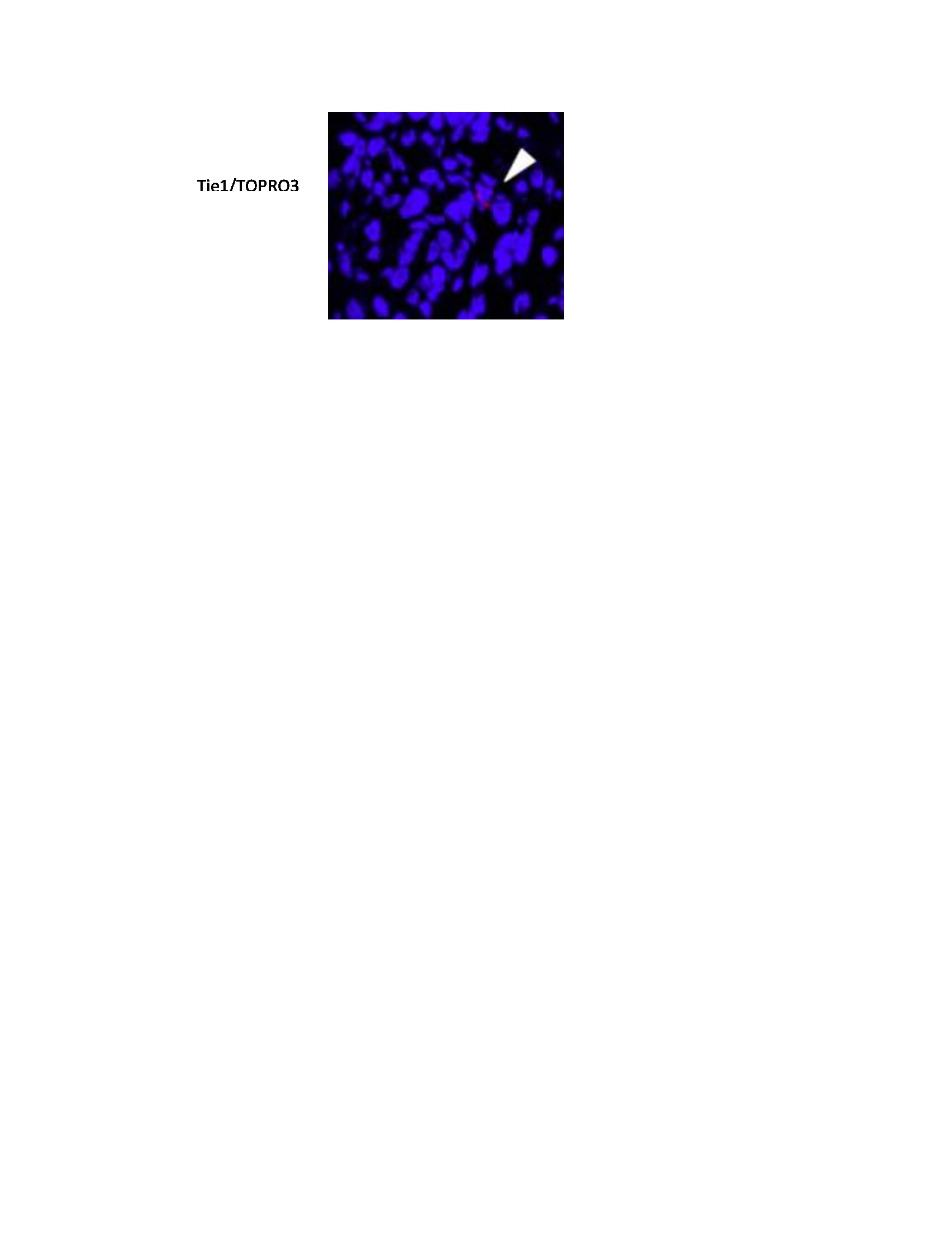
Detection of Tie-1 by Immunocytochemistry/ Immunofluorescence

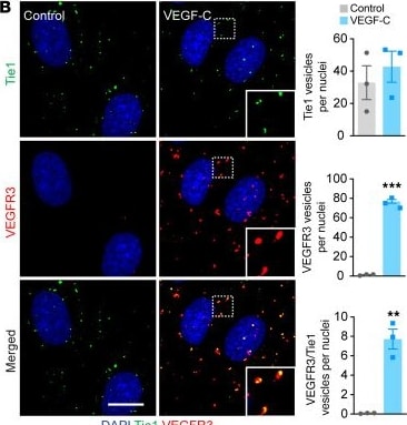
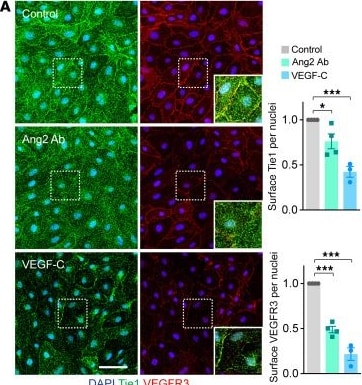
Effect of Ang2 Ab and VEGF-C on the localization of VEGFR3 and Tie1 in cultured LECs.(A) Cell-surface staining of VEGFR3 and Tie1 in LECs treated with a control IgG (n = 4) or Ang2 Ab (n = 4) for 30 minutes or stimulated with VEGF-C (n = 3) for 20 minutes. Scale bar: 50 μm. Magnification: 1.66. Graphs show quantification of VEGFR3 and Tie1 staining on the cell surface normalized to the IgG control. (B) Images showing colocalization of VEGFR3 and Tie1 in intracellular vesicles of LECs stimulated with VEGF-C for 20 minutes. Scale bar: 10 μm. Graph shows quantification of the number of VEGFR3- and Tie1-positive vesicles per nuclei (n = 3). Magnification: 2.36. (C and D) Colocalization of VEGFR3 with Tie1 and EEA1 (sorting endosomes) after 30 minutes of VEGF-C exposure (C) or RAB7 (late endosomes) after 120 minutes of VEGF-C exposure (D). The overview image shows a maximum projection. The boxed regions are shown as zoomed-in images of 1 optical slice per channel. White arrows point to some of the triple-positive (VEGFR3, Tie1, and EEA1 or RAB7) vesicles. Scale bars: 20 μm. Magnification: 3.24. (E and F) PLA immunofluorescence and quantification of PLA spots for Tie1 and VEGFR3 (E) and Ang2 and VEGFR3 (F) in permeabilized LECs (n = 5 fields of view; experiments were repeated 3 times with similar results). Scale bars: 10 μm. Data represent the mean ± SEM. *P < 0.05, **P < 0.01, and ***P < 0.001, by 1-way ANOVA with Dunnett’s post hoc test for multiple comparisons (A, E, and F) and 2-tailed Student’s t test (B). Image collected and cropped by CiteAb from the following open publication (https://www.jci.org/articles/view/155478), licensed under a CC-BY license. Not internally tested by R&D Systems.
Detection of Tie-1 by Western Blot

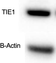
Detection of Tie-1 by Western Blot

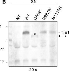
Analysis of TIE1 protein in HEK293T expressing the different TIE1 variants.(A and B) Western blotting of WT-TIE1 and the 3 variants in HEK293T cells transfected using a lentiviral vector. The truncated TIE1-Q682* variant is detected in cell lysate (A) only after 5 minutes of exposure, and is weak in the supernatant (B) after a 20-second exposure. Asterisks indicate the truncated Q682* polypeptide. The lanes were run on the same gel but were noncontiguous. (C) Relative TIE1 protein signals in the lysates and culture supernatants from n = 3 experiments like in A and B. (D) Flow cytometry analysis of TIE1 in transfected HEK293T cells. (E) Quantification of D from n = 3 experiments. Statistical significance in C and E was determined with Brown-Forsythe ANOVA with Dunnett’s post hoc test for multiple comparisons. Data shown as mean ± SEM. **P < 0.01, ***P < 0.001, ****P < 0.0001. Image collected and cropped by CiteAb from the following open publication (https://pubmed.ncbi.nlm.nih.gov/38820174), licensed under a CC-BY license. Not internally tested by R&D Systems.
Detection of Tie-1 by Immunohistochemistry

Detection of Tie-1 by Immunohistochemistry

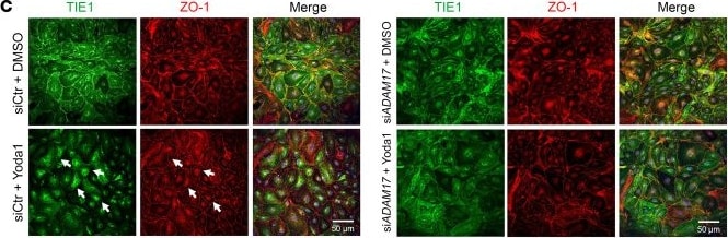
PIEZO1 activation promotes ADAM17-mediated TIE1 shedding in HDLECs.(A) Following a 30-minute treatment with Yoda1, HDLECs displayed a reduced distribution of TIE1 at cell-cell junctions (indicated by arrows), as observed in TIE1 and tight-junction protein 1 (ZO-1) immunostaining. Scale bar: 20 μm. (B) Yoda1-treated HDLECs exhibited increased TIE1 shedding, as confirmed by Western blot analysis (left panel: cell lysate protein samples; right panel: protein samples obtained from TCA precipitation of the culture medium). Each band represents a biological replicate sample (n = 3). (C) HDLECs treated with either siCtr or siADAM17 were stimulated with Yoda1 or vehicle control, followed by staining for TIE1 and ZO-1. Arrows highlight the areas at cell-cell junctions where TIE1 shedding occurred. Scale bars: 50 μm. (D) TIE1 shedding was assessed by Western blot analysis using medium samples subjected to TCA precipitation from siCtr-, siADAM17-, or siADAM10-treated HDLECs after vehicle or Yoda1 treatment. sTIE1, soluble TIE1. Each band represents a biological replicate sample (n = 3). Data are expressed as the mean ± SD. *P < 0.05, **P < 0.01, and ***P < 0.001, by 2-tailed, unpaired Student’s t test (B) or 2-way ANOVA followed by Tukey’s multiple-comparison test (D). Image collected and cropped by CiteAb from the following open publication (https://pubmed.ncbi.nlm.nih.gov/38747287), licensed under a CC-BY license. Not internally tested by R&D Systems.
Detection of Tie-1 by Immunocytochemistry/ Immunofluorescence

Detection of Tie-1 by Immunocytochemistry/ Immunofluorescence

Effect of Ang2 Ab and VEGF-C on the localization of VEGFR3 and Tie1 in cultured LECs.(A) Cell-surface staining of VEGFR3 and Tie1 in LECs treated with a control IgG (n = 4) or Ang2 Ab (n = 4) for 30 minutes or stimulated with VEGF-C (n = 3) for 20 minutes. Scale bar: 50 μm. Magnification: 1.66. Graphs show quantification of VEGFR3 and Tie1 staining on the cell surface normalized to the IgG control. (B) Images showing colocalization of VEGFR3 and Tie1 in intracellular vesicles of LECs stimulated with VEGF-C for 20 minutes. Scale bar: 10 μm. Graph shows quantification of the number of VEGFR3- and Tie1-positive vesicles per nuclei (n = 3). Magnification: 2.36. (C and D) Colocalization of VEGFR3 with Tie1 and EEA1 (sorting endosomes) after 30 minutes of VEGF-C exposure (C) or RAB7 (late endosomes) after 120 minutes of VEGF-C exposure (D). The overview image shows a maximum projection. The boxed regions are shown as zoomed-in images of 1 optical slice per channel. White arrows point to some of the triple-positive (VEGFR3, Tie1, and EEA1 or RAB7) vesicles. Scale bars: 20 μm. Magnification: 3.24. (E and F) PLA immunofluorescence and quantification of PLA spots for Tie1 and VEGFR3 (E) and Ang2 and VEGFR3 (F) in permeabilized LECs (n = 5 fields of view; experiments were repeated 3 times with similar results). Scale bars: 10 μm. Data represent the mean ± SEM. *P < 0.05, **P < 0.01, and ***P < 0.001, by 1-way ANOVA with Dunnett’s post hoc test for multiple comparisons (A, E, and F) and 2-tailed Student’s t test (B). Image collected and cropped by CiteAb from the following open publication (https://www.jci.org/articles/view/155478), licensed under a CC-BY license. Not internally tested by R&D Systems.
Detection of Tie-1 by Western Blot

Detection of Tie-1 by Western Blot

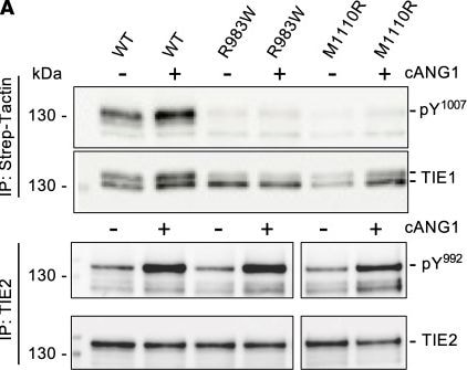
Effect of TIE1 variants R983W and M1110R on TIE1 activation and downstream signaling in endothelial cells.(A) Western blot analysis of TIE1 and TIE2 phosphorylation at Y1007 or Y992 residue (both pY1007 and pY992 detected by anti-pY992 antibody AF2720, R&D Systems) in Comp-ANGPT1–stimulated (cANG1-stimulated) LECs transduced with the WT-, R983W-, or M1110R-TIE1 variant. (B) Quantification of the pY1007/total TIE1 ratios in the samples from experiments like in A. Mean ± SEM, n = 3–5. (C) Western blot analysis of cANG1-stimulated TIE2-PAE cells transduced with the WT-, R983W-, or M1110R-TIE1 variant. (D) Quantification of phospho-AKT-S475/total AKT and phospho-ERK/total ERK ratios from experiments like in C. Data shown as mean ± SEM, n = 4 experiments. Statistical significance in B and D was analyzed by 1-way ANOVA with Tukey’s post hoc test for multiple comparisons. *P < 0.05, **P < 0.01, ***P < 0.001. Image collected and cropped by CiteAb from the following open publication (https://pubmed.ncbi.nlm.nih.gov/38820174), licensed under a CC-BY license. Not internally tested by R&D Systems.

Applications for Human Tie-1 Antibody

Application
Recommended Usage

CyTOF-ready

Ready to be labeled using established conjugation methods. No BSA or other carrier proteins that could interfere with conjugation.

Flow Cytometry

2.5 µg/106 cells
Sample: HUVEC human umbilical vein endothelial cells

Immunohistochemistry

5-15 µg/mL
Sample: Immersion fixed paraffin-embedded sections of human placenta

Western Blot

0.1 µg/mL
Sample: Recombinant Human Tie‑1 Fc Chimera (Catalog # 619-TI)

Reviewed Applications

Read 2 reviews rated 3.5 using AF619 in the following applications:

Flow Cytometry Panel Builder

Bio-Techne Knows Flow Cytometry

Save time and reduce costly mistakes by quickly finding compatible reagents using the Panel Builder Tool.

Advanced Features

  • Spectra Viewer - Custom analysis of spectra from multiple fluorochromes
  • Spillover Popups - Visualize the spectra of individual fluorochromes
  • Antigen Density Selector - Match fluorochrome brightness with antigen density
Build Your Panel Now

Formulation, Preparation, and Storage

Purification

Antigen Affinity-purified

Reconstitution

Reconstitute at 0.2 mg/mL in sterile PBS. For liquid material, refer to CoA for concentration.


Loading...

Formulation

Lyophilized from a 0.2 μm filtered solution in PBS with Trehalose. *Small pack size (SP) is supplied either lyophilized or as a 0.2 µm filtered solution in PBS.

Shipping

Lyophilized product is shipped at ambient temperature. Liquid small pack size (-SP) is shipped with polar packs. Upon receipt, store immediately at the temperature recommended below.

Stability & Storage

Use a manual defrost freezer and avoid repeated freeze-thaw cycles.
  • 12 months from date of receipt, -20 to -70 °C as supplied.
  • 1 month, 2 to 8 °C under sterile conditions after reconstitution.
  • 6 months, -20 to -70 °C under sterile conditions after reconstitution.

Calculators

The reconstitution calculator allows you to quickly calculate the volume of a reagent to reconstitute your vial. Simply enter the mass of reagent and the target concentration and the calculator will determine the rest.

=
÷

Background: Tie-1

Tie-1/Tie (tyrosine kinase with Ig and EGF homology domains 1) and Tie-2/Tek comprise a receptor tyrosine kinase (RTK) subfamily with unique structural characteristics: two immunoglobulin-like domains flanking three epidermal growth factor (EGF)-like domains and followed by three fibronectin type III-like repeats in the extracellular region and a split tyrosine kinase domain in the cytoplasmic region. These receptors are expressed primarily on endothelial and hematopoietic progenitor cells and play critical roles in angiogenesis, vasculogenesis and hematopoiesis.

Human Tie-1 cDNA encodes a 1138 amino acid (aa) residue precursor protein with a 24 residue putative signal peptide, a 735 residue extracellular domain and a 354 residue cytoplasmic domain. Ligands which bind and activate Tie-1 have not been identified. Based on gene-targeting studies, the in vivo functions of Tie-1 have been shown to be related to endothelial cell differentiation and the maintenance of integrity of the endothelium.

References

  1. Partanen, J. and D.J. Dumont (1999) Curr. Top. Microbiol. Immunol. 237:159
  2. Sato, T.N. et al. (1995) Nature 376:70

Long Name

Tyrosine Kinase with Immunoglobulin and Epidermal Growth Factor Homology Domains 1

Alternate Names

Tie1

Entrez Gene IDs

7075 (Human); 21846 (Mouse)

Gene Symbol

TIE1

UniProt

Additional Tie-1 Products

Product Documents for Human Tie-1 Antibody

Certificate of Analysis

To download a Certificate of Analysis, please enter a lot or batch number in the search box below.

Note: Certificate of Analysis not available for kit components.

Product Specific Notices for Human Tie-1 Antibody

For research use only

Citations for Human Tie-1 Antibody

Customer Reviews for Human Tie-1 Antibody (2)

3.5 out of 5
2 Customer Ratings
5 Stars
0%
4 Stars
50%
3 Stars
50%
2 Stars
0%
1 Stars
0%

Have you used Human Tie-1 Antibody?

Submit a review and receive an Amazon gift card!

$25/€18/£15/$25CAN/¥2500 for a review with an image

$10/€7/£6/$10CAN/¥1110 for a review without an image

Submit a review
Amazon Gift Card

Customer Images


Showing  1 - 2 of 2 reviews Showing All
Filter By:
  • Human Tie-1 Antibody
    Name: Anonymous
    Application: Functional Assay
    Sample Tested: HUVEC human umbilical vein endothelial cells
    Species: Human
    Verified Customer | Posted 02/04/2025
    I detected 619-TI with this antibody. This was to confirm the combination can work for my next assay
    Human Tie-1 Antibody AF619
  • Human Tie-1 Antibody
    Name: Mohammad Daud Khan
    Application: Immunohistochemistry
    Sample Tested: Aortic smooth muscle cells
    Species: Human
    Verified Customer | Posted 10/17/2018
    Human Tie-1 Antibody AF619

There are no reviews that match your criteria.

Showing  1 - 2 of 2 reviews Showing All

Protocols

Find general support by application which include: protocols, troubleshooting, illustrated assays, videos and webinars.

FAQs

No product specific FAQs exist for this product.

View all FAQs for Antibodies